Media Veteriner 1996. Vol. 111 (1) Artikel Asli
IN VITRO DEVELOPMENT OF
HOLSTEIN AND JAPANESE BLACK BREEDS EMBRYO
PERKEMBANGAN
IN VITRO EMBRIO SAPI-SAP1
HOLSTEIN DAN JAPANESE BLACK
A.
BOEDIONO')and
T. SUZUK~)
ABSTRACT
Two experiments were conducted with 3,457 oocytes, aspirated from the ovaries of slaughtered Holstein and Japanese Black breeds cows to compare the
use
of superovulated cow serum (SCS) and fetal calf serum (FCS) supplementation into medium for in vjtro development (experiment I) and the effect of different source of oocytes (experiment 11). The endpoint was development to cleavage on day 2 and to blastocyst up to day 9 after msermnabon. In experiment I, oocytes were matured and cultured m vltro (after fertilization) in TCM-199 supplemented with 5% SCS or 5% FCS. The cleavage rate of zygotes cultured in medium supplemented with 5% SCS was higher (P<0,05)than
5% FCS (5621860, 65,3% vs 4451742, 60,0%). The blastocyst production ratewas
sigdicantly higher (PC0,Ol) in SCS treatmentthan
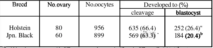
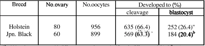
FCS (2321860, 27,0% vs 1241742, 16,7%). In experiment 11, 956 oocytes collected from Holstein breed and 899 oocytes from Japanese Black breed were matured, fertilized and cultured in vitro in medium supplemented with 5% SCS. The mean oocyte number and blastocyst production per ovary were 12,O (956180) and 3,2 (252180) for Holstein breed, 15,0 (899160) and 3,l (184160) for Japanese Black breed. The cleavage rate of zygotes derived from Holstein and Japanese Black breeds ovarieswas
not significantly different (6351956. 66,4% vs 5691899, 63,3%, respectively). However, the blastocyst production rate was sigdicantly higher (P<0,01 in Holstein than Japanese Black breeds (252/956, 26,4% vs 1841899, 20%). These results indicated that the developmental rate to biastocysts of embryos cultured m vitro differ from breed to breed." Deplltmnt of Anatomy, Faculty of Vetninrry Medicine, Bogor Agricultural Uaiversity, ll. T- Kcamnu 3, Boga 1615 1, INDONESIA
arr,
Telah dilakukan dua eksperimen dengan memakai 3.457 oosit yang diperoleh melalui aspirasi ovari sapi Japanese Black, untuk mengetahui pengaruh serum sapi superovulasi (SSS) Calf Serum (FCS) yang disuplementasikan ke dalam medium perkembangan m V I rimen I) dan pengaruh sumber oosit yang berbeda (Eksperimen 11). Pengarnatan dilakukan terhadap perkembangan sampai tingkat pembelahan pada
hari
ke- 2 dan tingkat blastosis padahan
ke-9 setelah inseminasi. Pada eksperimen 1, oosit Qmatangkan dan dikultur in vitro (setelah dibuahi) dalam medium TCM 199 yang ditarnbah dengan 5% SSS atau 5% FCS. Rata-rata tingkat pembelahan sigot yang dikultur dalam medium diberi tambahan SSS 5% lebih tinggi (P<0,05) dibanding FCS 5% (5621860, 65,3% vs 4451742, 60.0%). Sedangkan rata-rata blastosis yang dihasilkan secara bermakna lebih tinggi (P<0,01) dalam SSS dibandingkan dengan FCS (2321860, 27,0?! vs 1241742, 16,7%). Dalam' eksperimen 11, sebanyak 956 oosit yang diarnbil dari sapi Holstein dan 899 oositdari
Japanese Black dimatangkan, dibuahi dan dikultur secara in vitro dalarn medium yang diberi tambahan SSS 5%. Rata-rata jumlah oosit dan blastosis yang dihasilkan per ovari dari sapi Holstein adalah 12.0 (956180) dan 3,2 (252180): sedangkan dari Japanese Black adalah 15,O (899160) dan 3.1 (184160). Rata-rata tingkat pembelahan zigot yang berasal dari ovari sapi Holstein dan Japanese Black tidak berrnakna yaitu masing-masing 6351956, 66.4% dan 5691899, 63,3%. Namun demiluan. rata-rata jumlah blastosis yang dihasilkan oleh ovari sapi Holstein secara nyata lebih tinggi (P<0,01) dibanding yang dihasilkan oleh ovari Japanese Black (252D56, 26,4% vs 1841899, 20,0%). Hal ini menunjukkan bahwa tingkat perkembangan embrio yang cfikultur secara in vitro untuk mencapai tingkat blastosis berbeda dari satu jenis dengan yang laimya.INTRODUCTION
The developmental capacity of bovine oocytes after in vitro fertilization are influenced by factors such as the morphology of cumulus cells attached to oocytes, morphology of the ooplasm, the bull providing the semen and culture conditions (Shioya et al., 1988; Younis and Brackett, 1991; Kroetsch and Stubbings, 1992; Kajihara et al., 1990; Boediono et a)., 1994). Nonnal embryonic development of in vitro matured and fertilized bovine oocytes has
been achieved. Bovine oocytes matured with cumulus cells led to the production of zygote, and it develop, with subsequent pregnancies after transfer to recipient (Kajihara et al., 1990; Xu et al.. 1990).
characteristic changes in gonadotropic hormone profile. The presence of serum in culture medium was necessary for oocyte maturation, fertilization and culture since
sera
could provide cumulus oocyte complexes with protein requirements for in vitro maturation.The purpose of
the
current study was to investigate the pre-implementation development of oocytes collected from Holstein and Japanese Black breeds after mature, fertilize and culture m vitro in medium supplemented with superovulated cow serum (SCS).MATERIALS AND
METHODS
Oocyte collection and in vitro maturation :Cow's ovaries were obtained at a local slaughterhouse and were brought to laboratory within 3 h in physiological saline solution [0.9% (wh) NaCl] supplemented with Penicillin-G (100 IUJml) and streptomycin s d h t e (0.2 mg/ml) at 30"-32OC. Ooqtes in follicles 2-5
mm
in diameter, were collected by aspiration with an 18-G needle filled on a 5 ml syringe in Modified-PBS. Only oocytes surrounded by cumulus cells over more than one-third of their surface and an evenly granulated cytoplasm were used in this experiment (Fig. 1A). Oocyteswere then washed 2-3 times using maturation medium (TCM-199, Earle's salt. Gibco, Grand Island, NY. USA) supplemented with 0.01 mg/ml follicle stimulating hormone (FSH, Denka Pharmaceutical Co., Kawasaki. Japan), 50 p g/ml gentamycin sulfate (Sigma Chemical Co., St. Louis MO.. USA) and 5% superovulated cow serum (SCS. collected on day-7 of estrus) or 5% fetal calf semm (FCS, Sigma Chemical Co., St. Louis MO., USA). The oocytes (100-200
oocytes) were then introduced into the maturation medium (2.5ml) in a polystyrene culture dish (35
mm
diameter,Falcon
1008, Becton Dicknson Co. Ltd., Oxnara CA., USA) coveredwith mineral oil (E.R Squibb & Son, Inc., Princeton, NJ, USA) and cultured for 20 to 22 h at 38.5"C under 5% CO2 in humidified air.
In vitro fertilization
and
culture :Frozen-thawed semen from a single bull (Japanese Black breed) and the same lot
was
used for fertilization in vitro. Frozen sperm was thawed in water (30"-35"C), and the spermatozoa were diluted to about 6 ml with 2.5m M
d e i n added toBrackett
and Oliphant's medium (Caff-BO,Bmkett
and Oliphant, 1975) without bovine serumalbumin
M e r 5 h of inseraination oocytes with adherent cumulus cells were washed by repeated pipetting in culture medium md transferred to the culture medium (TCM-199) supplemented with 5 p g/ml insulin (W&o
Pure
Chemical Industries. Osaka, Japan), 50 p g/ml gentamycin sulfate and 5% SCS or 5% FCS for further development. The culture medium (0.5 ml) in a polystyrene dish (4-we11 multidish; Nuclon, Roskilde. Denmark) was covered with mineral oil (0.5 ml).The cumulus cells surrounding the embryos were removed by pipetting after 48 h of insemination, but the cumulus cells layer (forrmng
a
monolayer at that time) attached to the bottom of culture dish were not disrupted and the embryos were cultured on this layer. The culture mediumwas
replaced
with new medium 4 days after insemination.Experiment I :
huing
m a h n t b n and culture of bovine oocytes fertilized in vitro, the medium was supplemented with 5% SCS or 5% FCS. Each treatment was repeated four times, the sample of SCS and FCS remaining constant throughout.Erpcrierent
I#
:Oocytes of Holstein and Japanese Black breeds were collected separately. According to the results d experiment I. the maturation and culture medium in this experiment was supplemented with 5Yo SCS. The examinations were replicated 7 times using 956 Holstein oocytes and 899 Japanese Black oocytes
Embryo evdwation :
The proportions of embryos that had cleaved (2-. 4- and 8-cell stage) were recorded 48 h
after insemination (when oocytes were taken from cumulus cells). Blastocyst development
was
assessed
on day'-7, -8 and -9, when counted, blastocysts were removed from the culture dish to avoid double counting. The data were analyzed by Chi-square test. A probability of P<0.05 and Pc0.0 1 were considered to be statistically s i m c a n t .Superovulated cow serum (SCS) collection :
The SCS was collected from superovulated cows treated with a total 20 mg FSH. Prostaglandin was administrated 48 h after FSH injection to induce luteolysis. Blood was collected on day-7 (on the time of embryo collection). Whole blood samples were centrifuged twice (3500 rpm, 10 min at 5°C). The serum obtained was then heat-inactivated (56"C, 30
rnin), distributed in small tube (2 ml) and kept in deep freezer (-20°C). The frozen serum
RESULTS Experiment I :
The cultured oocytes displayed a marked expansion of cumulus cells
as
characteristic formatured ova (Fig. 1B). Oocytes were evaluated 48 h after insemination, when developed to cleavage (2-, 4-, 8-cell stage, Fig. 2A), and when developed to blastocyst on day-7, -8 and -9 (Fig. 2C, 2D). The development rate of embryos cultured in medium supplemented with different sources of serum to cleavage rate of zygotes cultured in medium supplemented with 5% SCS was higher (P<0.05)
than
5% FCS (5621860, 65.3% vs 4451742, 60.0%). The blastocyst productions rate was sigxuficantly higher (W0.01) in SCS treatmentthan
FCS (2321860, 27.0% vs 1241742, 16.7%.Experiment 11 :
Total 956 oocytes collected from Holstein breed and 899 oocytes from Japanese Black breed were
used
for in vitro maturation, fertilization and culture. The development rate of Holstein and Japanese Black breeds in vitro produced embryo are shown in Table 2. The mean oocyte number and blastocyst production per ovary were 15.0 (956180) and 3.2 (252180) for Holstein breed, 15.0 (899160) and 3.1 (184160) for Japanese Black breed. The cleavage rate of zygotes derived from Holstein and Japanese Black breeds ovaries was not significantly Merent (6351956, 66.4% vs 5691899. 63.3%, respectively). However, the blastocyst production rate was sigmficantly higher (P~0.01) in Holsteinthan
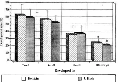
Japanese Black breeds (2561956,26.4% vs 1841899,20%).The deve!opment in vitro of Holstein and Japanese Black breeds embryos on day-2 (when ~t developed to 2-, 4-, and 8-cell stage), -7. -8, and -9 (when it developed to blastocyst) after insemination are shown in Figure 3.
DISCUSSION
Suzulu et. al. (1992) and Shioya et. al. (1988) reported that follicular cells, especially
the junction gap between the ooplasm and cumulus cells used for in vitro fertilization a high number of the fertilized bovine oocyte cleaved and developed into blastocysts.
C d t u r e with somatic cells is necessary for in vltro oocyte development to the blastocyst stage following fertilization. Fertilized oocytes have been co-cultured with bovine
0;Viductal epithelial cells (BOEC; Durnford and Stubbings, 1992; Rorie et al., 1992),
gmulosa cells (Mochzuki et al., 1991), and cumulus cells (Kajihara et al., 1990, Boediono etal., 1994). In the present study, the embryos were co-cultured with cumulus cells for further &velopment. As shown by Suzuki et. al. (1992) and Kajihara et. al. (1990), the co-culture of in vttro fertilized oocytes with cumulus cells provides an appropriate environment for embryonic development, and they obtained pregnancies and offspring by transfemng embryos malting from co-culture with cumulus cells.
la
early work hormonal and follicular factors were found to affect maturation of sheep OBCJ~& in vim and their subsequent developmental capacity (Moor and Trounson, 1977). Inv l h &rtlred oocytes in hormone
free
medium show a limited developmental competence (lk&@ and Hunter. 1985). Several researcherused
the medium supplemented with fetalWserptm (FCS; Saeki et al.. 1990), estrus cow serum (ECS; Schellander et al., 1990), and
sPrperowIated cow serum (SCS: Boedzono et al.. 1994: Suz& et al.. 1992). When maturation
i
d
&&memd~uin
was supplemented with serum. a high proportion of the fertilized bovineoocytes &&pal, into blastocysts. Suzuki et. al. (1982) reported that the
medium
sllpplemented with
serum of the same species was having a beneficial effect for the survival of
the embryocultured
in vitro. The results reported here demonstrated that SCS contains s&@nm which iwrease the cleavage competence and developmental capacity into blastayti4'll in v i h matured and fertilized bovine oocytes. This conclusion draws support fromresult vf
Bwdiono et al. (1994) whoused
SCS for oocyte maturation, fertilization and culture ofbovine
oocytes, which developed into blastocysts.The
further development in vitro of Holstein and Japanese Black breeds fertilized oocytes to cleavage (2 to 8cell stage) were not differ sigruficantly. However, the blastocyst production ratewas
signihntly higher in Holstein and Japanese Black breeds. McLaren andBowman
(1973)f
d
tfItrt
ttte
difference in the start of cleavage inmouse
was determined by maternal genotypeIn
pmmt
study,
the maternal genotype hadno affect
onthe
cleavagerate
of bevineooq@2:
l3uwms,
Mastocyst rate producedfrom
Holstein breed was bgber thanfiom Japanese
Black
W. T k e results indicated that the developmental rate into blastocysts of embryos wWed in vitm differ from breed to breed.ACKNOWLEDGMENTS
We would like to thank Dr. Nrryeean,
H.A.K.
and
Dr. Khan, M.A. for critical reading ofthe
REFERENCES
Boediono. A., Takagi, M., Saha, S. and Suzuki, S. 1994, The influence of day-0 and day-7 superovulated cow
serum
during in vitro development of bovine oocytes. Reprod. Fertil. Dev., 6:261-264.Brackett, B.G. and Oliphant, G. 1975. Capacitation of rabbit spermatozoa in vitro. Biol. Reprod., 12:260-274.
Durnford. R. and Stubbings. R.B.. 1992. The influence of serum and oviductal cells during In vitro maturation on blastocyst development. Theriogenology. 37:205 abst.
Hensleigh. H.C. and Hunter, A.G., 1985. In vitro maturation of bovine cumulus enclosed primary oocytes and their subsequent in vitro fertilization and cleavage. J. Dairy Sci., 68: 1456-1562.
Kajihara, Y.. Kometani, N., Kobayashi,
S..
Shtanaka. Y.. Koshiba. Y. Hishyama, K.,Shiraiwa. K. and Goto, K., 1990. Pregnancy rates and births after co-culture of cumulus cells with bovine embryos derived from in vitro fertilization of In vitro matured follicular oocytes. Theriogenology. 33:202 abst.
Katska, L., Kauffold, P., Smorag, Z., Duschnski. U.. Torner. H. and Kanitz, W.. 1989. Influence of hardening of the zona pellucida on in vitro fertilization of bovine oocytes. Theriogenology, 32:767-777.
Kroetsch, T.G. and Stubbings, R.B.. 1992. Sire and insemination dose does effect in vitro fertilization on bovine oocytes. Theriogenology, 37:240 abst.
McLaren, A. and Bowman, P., 1973. Genetic effects on the timing of early development in the mouse. J. Embriol. Exp. Morphol. 30:491-498.
Mochizulu. H.. Fukui, Y. and Ono, H., 1991. Effect of number of granulosa cells added to culture media for in vitro maturation, fertilization and development of bovine oocytes. Theriogenology, 36:973-986.
Moor, R.M. and Trounson, A.O., 1977. Hormonal and follicular factors affecting maturation of sheep oocytes in vitro and their subsequent developmental capacity. J. Reprod. Fert., 49: 101-109.
Saeki. K.. Leibfried. M.L. and First, N.L., 1990. Are fetal calf serum and hormone necessary during in vitro maturation of cattle oocytes for subsequent development?. Theriogenology, 33 :3 16 abst.
Schellander, K., Fuhrer, R., Brackett, B.G., Korb, H. and ScNeger, W., 1990. In vitro fertilization and cleavage of bovine oocytes matured in medium supplemented with estrous cow serum. Theriogenology, 33:477-485.
Shioya, Y., Kuwayama. M.. Fukushima, M. and Iwasaki, S., 1988. In vitro fertilization and cleavage capability of bovine follicular oocytes classified by cumulus cells and matured in vitro. Theriogenology, 30:489-496.
Suzuki. T., Takahashi, Y., Shimohira, I., Oketani, R and Saito, N., 1982. Nonsurgically transfer and culture of bovine embryos in vitro in the medium supplemented with bovine serum collected from superovulated heifers 7 to 8 days after estrus. Jpn. J. Anim. Reprod.. 28(3): 1 19- 123.
Suzuki, T., Singla, S.K., Sujata, J. and Madan,
M.L..
1992. In vitro fertilization of water M a l o follicular oocytes and their ability to cleave in vitro. Theriogenology, 38: 1187- 1194.Younis. A.I. and Brackett, B.G., 199 1. Importance of cumulus cells and insemination interval for development of bovine oocytes into morulae and biastocysts in vitro. Theriogenology,
Table 1. Development of IVF embryos cultured in vitro using different sera.
Table 2. Development of Holstein and Japanese Black embryos in culture medium supplemented with superovulated cow serum. ,
Serum
SCS
FCS
SCS - supc.rovulatcd cow serum. FCS - fctal calf serum
"'
within colunms. means with different superscripts arc significantly diKerent (" P 0.05; c.d P 0.01, x'-test).No.Oocytes assessed 860 742 Brecd Holstein Jpn. Black
"b within columns. mans with different supvscripts arc significantly diff~rent (P. 0.01. XZ-test).
No.ovary 80 60 Blastocyst
(%I
232" (27.0)1 2 4 ~ (16.7) No.of embryo cleaved
2-cell 42 62 No. oocytes 956 899 4cell 146 72
Developed to (%)
cleavage 635 (66.4) 569
(&.$
' 8cell374
311
blastocyst 252 (26.4)"
184 (20.4)~ Total (%)
562" (65.3)
[image:9.394.24.367.46.175.2] [image:9.394.27.367.276.353.2]Firmre 1. Maturation and Fartilization
-
in vrtro of Bovine Oocvtes (A) Immature oocytes, oocltes surrounded by cimmulus cells andevenly granulatted c>toplasm
(B) Matured ooqtes, oocytes with mucinate expansion of cumn~ulus cells after 2 1 h cultured m vltro
Figure 2. Development in vitro of Bovine Embryos
(A) On day 2 when it developed to cleavage (2-, 4- and 8-11 stage) (B) On day 4 when it developed to morula
(C) On day 8 when it developed to blastocyst (B=blastocyst, EB=expanded blastocyst) (D) On day 9 when it develop to hatching and hatched blastocyst
I I
2-cell 4-cell 8-cell Blastocyst
Developed to
Holstein
a
J. BlackDevelopment rate of Holstein and Japanese Black breeds in vitro produced embryos
Developed to
2-cell 4-cell &ell Blastocyst
J.Black-sd -
11.220 9.970 11.080
3.060 Holstein (96)
64.330 57.670 37.000 25.170
J. Black (%)
61.110 53.670 38.330 22.1 10
Holstein-sd
Media Veteriner 1996. Vol. 111 (1) Artikel Asli
IN VITRO DEVELOPMENT OF
HOLSTEIN AND JAPANESE BLACK BREEDS EMBRYO
PERKEMBANGAN
IN VITRO EMBRIO SAPI-SAP1
HOLSTEIN DAN JAPANESE BLACK
A.
BOEDIONO')and
T. SUZUK~)
ABSTRACT
Two experiments were conducted with 3,457 oocytes, aspirated from the ovaries of slaughtered Holstein and Japanese Black breeds cows to compare the
use
of superovulated cow serum (SCS) and fetal calf serum (FCS) supplementation into medium for in vjtro development (experiment I) and the effect of different source of oocytes (experiment 11). The endpoint was development to cleavage on day 2 and to blastocyst up to day 9 after msermnabon. In experiment I, oocytes were matured and cultured m vltro (after fertilization) in TCM-199 supplemented with 5% SCS or 5% FCS. The cleavage rate of zygotes cultured in medium supplemented with 5% SCS was higher (P<0,05)than
5% FCS (5621860, 65,3% vs 4451742, 60,0%). The blastocyst production ratewas
sigdicantly higher (PC0,Ol) in SCS treatmentthan
FCS (2321860, 27,0% vs 1241742, 16,7%). In experiment 11, 956 oocytes collected from Holstein breed and 899 oocytes from Japanese Black breed were matured, fertilized and cultured in vitro in medium supplemented with 5% SCS. The mean oocyte number and blastocyst production per ovary were 12,O (956180) and 3,2 (252180) for Holstein breed, 15,0 (899160) and 3,l (184160) for Japanese Black breed. The cleavage rate of zygotes derived from Holstein and Japanese Black breeds ovarieswas
not significantly different (6351956. 66,4% vs 5691899, 63,3%, respectively). However, the blastocyst production rate was sigdicantly higher (P<0,01 in Holstein than Japanese Black breeds (252/956, 26,4% vs 1841899, 20%). These results indicated that the developmental rate to biastocysts of embryos cultured m vitro differ from breed to breed." Deplltmnt of Anatomy, Faculty of Vetninrry Medicine, Bogor Agricultural Uaiversity, ll. T- Kcamnu 3, Boga 1615 1, INDONESIA
arr,
Telah dilakukan dua eksperimen dengan memakai 3.457 oosit yang diperoleh melalui aspirasi ovari sapi Japanese Black, untuk mengetahui pengaruh serum sapi superovulasi (SSS) Calf Serum (FCS) yang disuplementasikan ke dalam medium perkembangan m V I rimen I) dan pengaruh sumber oosit yang berbeda (Eksperimen 11). Pengarnatan dilakukan terhadap perkembangan sampai tingkat pembelahan pada
hari
ke- 2 dan tingkat blastosis padahan
ke-9 setelah inseminasi. Pada eksperimen 1, oosit Qmatangkan dan dikultur in vitro (setelah dibuahi) dalam medium TCM 199 yang ditarnbah dengan 5% SSS atau 5% FCS. Rata-rata tingkat pembelahan sigot yang dikultur dalam medium diberi tambahan SSS 5% lebih tinggi (P<0,05) dibanding FCS 5% (5621860, 65,3% vs 4451742, 60.0%). Sedangkan rata-rata blastosis yang dihasilkan secara bermakna lebih tinggi (P<0,01) dalam SSS dibandingkan dengan FCS (2321860, 27,0?! vs 1241742, 16,7%). Dalam' eksperimen 11, sebanyak 956 oosit yang diarnbil dari sapi Holstein dan 899 oositdari
Japanese Black dimatangkan, dibuahi dan dikultur secara in vitro dalarn medium yang diberi tambahan SSS 5%. Rata-rata jumlah oosit dan blastosis yang dihasilkan per ovari dari sapi Holstein adalah 12.0 (956180) dan 3,2 (252180): sedangkan dari Japanese Black adalah 15,O (899160) dan 3.1 (184160). Rata-rata tingkat pembelahan zigot yang berasal dari ovari sapi Holstein dan Japanese Black tidak berrnakna yaitu masing-masing 6351956, 66.4% dan 5691899, 63,3%. Namun demiluan. rata-rata jumlah blastosis yang dihasilkan oleh ovari sapi Holstein secara nyata lebih tinggi (P<0,01) dibanding yang dihasilkan oleh ovari Japanese Black (252D56, 26,4% vs 1841899, 20,0%). Hal ini menunjukkan bahwa tingkat perkembangan embrio yang cfikultur secara in vitro untuk mencapai tingkat blastosis berbeda dari satu jenis dengan yang laimya.INTRODUCTION
The developmental capacity of bovine oocytes after in vitro fertilization are influenced by factors such as the morphology of cumulus cells attached to oocytes, morphology of the ooplasm, the bull providing the semen and culture conditions (Shioya et al., 1988; Younis and Brackett, 1991; Kroetsch and Stubbings, 1992; Kajihara et al., 1990; Boediono et a)., 1994). Nonnal embryonic development of in vitro matured and fertilized bovine oocytes has
been achieved. Bovine oocytes matured with cumulus cells led to the production of zygote, and it develop, with subsequent pregnancies after transfer to recipient (Kajihara et al., 1990; Xu et al.. 1990).
characteristic changes in gonadotropic hormone profile. The presence of serum in culture medium was necessary for oocyte maturation, fertilization and culture since
sera
could provide cumulus oocyte complexes with protein requirements for in vitro maturation.The purpose of
the
current study was to investigate the pre-implementation development of oocytes collected from Holstein and Japanese Black breeds after mature, fertilize and culture m vitro in medium supplemented with superovulated cow serum (SCS).MATERIALS AND
METHODS
Oocyte collection and in vitro maturation :Cow's ovaries were obtained at a local slaughterhouse and were brought to laboratory within 3 h in physiological saline solution [0.9% (wh) NaCl] supplemented with Penicillin-G (100 IUJml) and streptomycin s d h t e (0.2 mg/ml) at 30"-32OC. Ooqtes in follicles 2-5
mm
in diameter, were collected by aspiration with an 18-G needle filled on a 5 ml syringe in Modified-PBS. Only oocytes surrounded by cumulus cells over more than one-third of their surface and an evenly granulated cytoplasm were used in this experiment (Fig. 1A). Oocyteswere then washed 2-3 times using maturation medium (TCM-199, Earle's salt. Gibco, Grand Island, NY. USA) supplemented with 0.01 mg/ml follicle stimulating hormone (FSH, Denka Pharmaceutical Co., Kawasaki. Japan), 50 p g/ml gentamycin sulfate (Sigma Chemical Co., St. Louis MO.. USA) and 5% superovulated cow serum (SCS. collected on day-7 of estrus) or 5% fetal calf semm (FCS, Sigma Chemical Co., St. Louis MO., USA). The oocytes (100-200
oocytes) were then introduced into the maturation medium (2.5ml) in a polystyrene culture dish (35
mm
diameter,Falcon
1008, Becton Dicknson Co. Ltd., Oxnara CA., USA) coveredwith mineral oil (E.R Squibb & Son, Inc., Princeton, NJ, USA) and cultured for 20 to 22 h at 38.5"C under 5% CO2 in humidified air.
In vitro fertilization
and
culture :Frozen-thawed semen from a single bull (Japanese Black breed) and the same lot
was
used for fertilization in vitro. Frozen sperm was thawed in water (30"-35"C), and the spermatozoa were diluted to about 6 ml with 2.5m M
d e i n added toBrackett
and Oliphant's medium (Caff-BO,Bmkett
and Oliphant, 1975) without bovine serumalbumin
M e r 5 h of inseraination oocytes with adherent cumulus cells were washed by repeated pipetting in culture medium md transferred to the culture medium (TCM-199) supplemented with 5 p g/ml insulin (W&o
Pure
Chemical Industries. Osaka, Japan), 50 p g/ml gentamycin sulfate and 5% SCS or 5% FCS for further development. The culture medium (0.5 ml) in a polystyrene dish (4-we11 multidish; Nuclon, Roskilde. Denmark) was covered with mineral oil (0.5 ml).The cumulus cells surrounding the embryos were removed by pipetting after 48 h of insemination, but the cumulus cells layer (forrmng
a
monolayer at that time) attached to the bottom of culture dish were not disrupted and the embryos were cultured on this layer. The culture mediumwas
replaced
with new medium 4 days after insemination.Experiment I :
huing
m a h n t b n and culture of bovine oocytes fertilized in vitro, the medium was supplemented with 5% SCS or 5% FCS. Each treatment was repeated four times, the sample of SCS and FCS remaining constant throughout.Erpcrierent
I#
:Oocytes of Holstein and Japanese Black breeds were collected separately. According to the results d experiment I. the maturation and culture medium in this experiment was supplemented with 5Yo SCS. The examinations were replicated 7 times using 956 Holstein oocytes and 899 Japanese Black oocytes
Embryo evdwation :
The proportions of embryos that had cleaved (2-. 4- and 8-cell stage) were recorded 48 h
after insemination (when oocytes were taken from cumulus cells). Blastocyst development
was
assessed
on day'-7, -8 and -9, when counted, blastocysts were removed from the culture dish to avoid double counting. The data were analyzed by Chi-square test. A probability of P<0.05 and Pc0.0 1 were considered to be statistically s i m c a n t .Superovulated cow serum (SCS) collection :
The SCS was collected from superovulated cows treated with a total 20 mg FSH. Prostaglandin was administrated 48 h after FSH injection to induce luteolysis. Blood was collected on day-7 (on the time of embryo collection). Whole blood samples were centrifuged twice (3500 rpm, 10 min at 5°C). The serum obtained was then heat-inactivated (56"C, 30
rnin), distributed in small tube (2 ml) and kept in deep freezer (-20°C). The frozen serum
RESULTS Experiment I :
The cultured oocytes displayed a marked expansion of cumulus cells
as
characteristic formatured ova (Fig. 1B). Oocytes were evaluated 48 h after insemination, when developed to cleavage (2-, 4-, 8-cell stage, Fig. 2A), and when developed to blastocyst on day-7, -8 and -9 (Fig. 2C, 2D). The development rate of embryos cultured in medium supplemented with different sources of serum to cleavage rate of zygotes cultured in medium supplemented with 5% SCS was higher (P<0.05)
than
5% FCS (5621860, 65.3% vs 4451742, 60.0%). The blastocyst productions rate was sigxuficantly higher (W0.01) in SCS treatmentthan
FCS (2321860, 27.0% vs 1241742, 16.7%.Experiment 11 :
Total 956 oocytes collected from Holstein breed and 899 oocytes from Japanese Black breed were
used
for in vitro maturation, fertilization and culture. The development rate of Holstein and Japanese Black breeds in vitro produced embryo are shown in Table 2. The mean oocyte number and blastocyst production per ovary were 15.0 (956180) and 3.2 (252180) for Holstein breed, 15.0 (899160) and 3.1 (184160) for Japanese Black breed. The cleavage rate of zygotes derived from Holstein and Japanese Black breeds ovaries was not significantly Merent (6351956, 66.4% vs 5691899. 63.3%, respectively). However, the blastocyst production rate was sigmficantly higher (P~0.01) in Holsteinthan
Japanese Black breeds (2561956,26.4% vs 1841899,20%).The deve!opment in vitro of Holstein and Japanese Black breeds embryos on day-2 (when ~t developed to 2-, 4-, and 8-cell stage), -7. -8, and -9 (when it developed to blastocyst) after insemination are shown in Figure 3.
DISCUSSION
Suzulu et. al. (1992) and Shioya et. al. (1988) reported that follicular cells, especially
the junction gap between the ooplasm and cumulus cells used for in vitro fertilization a high number of the fertilized bovine oocyte cleaved and developed into blastocysts.
C d t u r e with somatic cells is necessary for in vltro oocyte development to the blastocyst stage following fertilization. Fertilized oocytes have been co-cultured with bovine
0;Viductal epithelial cells (BOEC; Durnford and Stubbings, 1992; Rorie et al., 1992),
gmulosa cells (Mochzuki et al., 1991), and cumulus cells (Kajihara et al., 1990, Boediono etal., 1994). In the present study, the embryos were co-cultured with cumulus cells for further &velopment. As shown by Suzuki et. al. (1992) and Kajihara et. al. (1990), the co-culture of in vttro fertilized oocytes with cumulus cells provides an appropriate environment for embryonic development, and they obtained pregnancies and offspring by transfemng embryos malting from co-culture with cumulus cells.
la
early work hormonal and follicular factors were found to affect maturation of sheep OBCJ~& in vim and their subsequent developmental capacity (Moor and Trounson, 1977). Inv l h &rtlred oocytes in hormone
free
medium show a limited developmental competence (lk&@ and Hunter. 1985). Several researcherused
the medium supplemented with fetalWserptm (FCS; Saeki et al.. 1990), estrus cow serum (ECS; Schellander et al., 1990), and
sPrperowIated cow serum (SCS: Boedzono et al.. 1994: Suz& et al.. 1992). When maturation
i
d
&&memd~uin
was supplemented with serum. a high proportion of the fertilized bovineoocytes &&pal, into blastocysts. Suzuki et. al. (1982) reported that the
medium
sllpplemented with
serum of the same species was having a beneficial effect for the survival of
the embryocultured
in vitro. The results reported here demonstrated that SCS contains s&@nm which iwrease the cleavage competence and developmental capacity into blastayti4'll in v i h matured and fertilized bovine oocytes. This conclusion draws support fromresult vf
Bwdiono et al. (1994) whoused
SCS for oocyte maturation, fertilization and culture ofbovine
oocytes, which developed into blastocysts.The
further development in vitro of Holstein and Japanese Black breeds fertilized oocytes to cleavage (2 to 8cell stage) were not differ sigruficantly. However, the blastocyst production ratewas
signihntly higher in Holstein and Japanese Black breeds. McLaren andBowman
(1973)f
d
tfItrt
ttte
difference in the start of cleavage inmouse
was determined by maternal genotypeIn
pmmt
study,
the maternal genotype hadno affect
onthe
cleavagerate
of bevineooq@2:
l3uwms,
Mastocyst rate producedfrom
Holstein breed was bgber thanfiom Japanese
Black
W. T k e results indicated that the developmental rate into blastocysts of embryos wWed in vitm differ from breed to breed.ACKNOWLEDGMENTS
We would like to thank Dr. Nrryeean,
H.A.K.
and
Dr. Khan, M.A. for critical reading ofthe
REFERENCES
Boediono. A., Takagi, M., Saha, S. and Suzuki, S. 1994, The influence of day-0 and day-7 superovulated cow
serum
during in vitro development of bovine oocytes. Reprod. Fertil. Dev., 6:261-264.Brackett, B.G. and Oliphant, G. 1975. Capacitation of rabbit spermatozoa in vitro. Biol. Reprod., 12:260-274.
Durnford. R. and Stubbings. R.B.. 1992. The influence of serum and oviductal cells during In vitro maturation on blastocyst development. Theriogenology. 37:205 abst.
Hensleigh. H.C. and Hunter, A.G., 1985. In vitro maturation of bovine cumulus enclosed primary oocytes and their subsequent in vitro fertilization and cleavage. J. Dairy Sci., 68: 1456-1562.
Kajihara, Y.. Kometani, N., Kobayashi,
S..
Shtanaka. Y.. Koshiba. Y. Hishyama, K.,Shiraiwa. K. and Goto, K., 1990. Pregnancy rates and births after co-culture of cumulus cells with bovine embryos derived from in vitro fertilization of In vitro matured follicular oocytes. Theriogenology. 33:202 abst.
Katska, L., Kauffold, P., Smorag, Z., Duschnski. U.. Torner. H. and Kanitz, W.. 1989. Influence of hardening of the zona pellucida on in vitro fertilization of bovine oocytes. Theriogenology, 32:767-777.
Kroetsch, T.G. and Stubbings, R.B.. 1992. Sire and insemination dose does effect in vitro fertilization on bovine oocytes. Theriogenology, 37:240 abst.
McLaren, A. and Bowman, P., 1973. Genetic effects on the timing of early development in the mouse. J. Embriol. Exp. Morphol. 30:491-498.
Mochizulu. H.. Fukui, Y. and Ono, H., 1991. Effect of number of granulosa cells added to culture media for in vitro maturation, fertilization and development of bovine oocytes. Theriogenology, 36:973-986.
Moor, R.M. and Trounson, A.O., 1977. Hormonal and follicular factors affecting maturation of sheep oocytes in vitro and their subsequent developmental capacity. J. Reprod. Fert., 49: 101-109.
Saeki. K.. Leibfried. M.L. and First, N.L., 1990. Are fetal calf serum and hormone necessary during in vitro maturation of cattle oocytes for subsequent development?. Theriogenology, 33 :3 16 abst.
Schellander, K., Fuhrer, R., Brackett, B.G., Korb, H. and ScNeger, W., 1990. In vitro fertilization and cleavage of bovine oocytes matured in medium supplemented with estrous cow serum. Theriogenology, 33:477-485.
Shioya, Y., Kuwayama. M.. Fukushima, M. and Iwasaki, S., 1988. In vitro fertilization and cleavage capability of bovine follicular oocytes classified by cumulus cells and matured in vitro. Theriogenology, 30:489-496.
Suzuki. T., Takahashi, Y., Shimohira, I., Oketani, R and Saito, N., 1982. Nonsurgically transfer and culture of bovine embryos in vitro in the medium supplemented with bovine serum collected from superovulated heifers 7 to 8 days after estrus. Jpn. J. Anim. Reprod.. 28(3): 1 19- 123.
Suzuki, T., Singla, S.K., Sujata, J. and Madan,
M.L..
1992. In vitro fertilization of water M a l o follicular oocytes and their ability to cleave in vitro. Theriogenology, 38: 1187- 1194.Younis. A.I. and Brackett, B.G., 199 1. Importance of cumulus cells and insemination interval for development of bovine oocytes into morulae and biastocysts in vitro. Theriogenology,
Table 1. Development of IVF embryos cultured in vitro using different sera.
Table 2. Development of Holstein and Japanese Black embryos in culture medium supplemented with superovulated cow serum. ,
Serum
SCS
FCS
SCS - supc.rovulatcd cow serum. FCS - fctal calf serum
"'
within colunms. means with different superscripts arc significantly diKerent (" P 0.05; c.d P 0.01, x'-test).No.Oocytes assessed 860 742 Brecd Holstein Jpn. Black
"b within columns. mans with different supvscripts arc significantly diff~rent (P. 0.01. XZ-test).
No.ovary 80 60 Blastocyst
(%I
232" (27.0)1 2 4 ~ (16.7) No.of embryo cleaved
2-cell 42 62 No. oocytes 956 899 4cell 146 72
Developed to (%)
cleavage 635 (66.4) 569
(&.$
' 8cell374
311
blastocyst 252 (26.4)"
184 (20.4)~ Total (%)
562" (65.3)
[image:22.394.24.367.46.175.2] [image:22.394.27.367.276.353.2]Firmre 1. Maturation and Fartilization
-
in vrtro of Bovine Oocvtes (A) Immature oocytes, oocltes surrounded by cimmulus cells andevenly granulatted c>toplasm
(B) Matured ooqtes, oocytes with mucinate expansion of cumn~ulus cells after 2 1 h cultured m vltro