ISSN: 1978-3019
Development of Domestic Cat Embryo Produced by Preserved Sperms
KARTINI ERIANI1∗∗∗∗∗, ARIEF BOEDIONO2, ITA DJUWITA2, SONY HERU SUMARSONO3, AL-AZHAR4
1Faculty of Mathematics and Science, Syiah Kuala University, Jalan Syeh Abdurrauf 3, Banda Aceh 23111, Indonesia 2Faculty of Veterinary Medicine, Bogor Agricultural University, Jalan Rasamala, Darmaga Campus, Bogor 16680, Indonesia
3Faculty of Mathematics and Science, Institut Teknologi Bandung, Jalan Ganesha 10, Bandung 40132, Indonesia 4Faculty of Veterinary Medicine, Syiah Kuala University, Banda Aceh 23111, Indonesia
Received May 27, 2008/Accepted December 18, 2008
The ability to mature and fertilize oocytes of endangered species may allow us to sustain genetic and global biodiversity. Epididymis sperms may be the last chance to ensure preservation of genetic materials after injury or death of a valuable animal. Studies have been conducted to determine wether both epididymis sperms and oocytes can be used to produce viable embryos and offspring. The purpose of this study was to determine how long cats sperms contained in epididymis were remain motile and had intact membranes when preserved at 4 oC, and to determine whether such those preserved sperms are able to fertilize oocytes. Epididymis was preserved immediately in phosphate buffer saline at 4 oC for 1, 3, and 6 days. The observation of sperm quality and viability after preservation was performed by vital staining acrosom and
Hoechst-Propidium Iodine. Biological functions of sperms were evaluated by in vitro culture technique for fertilization, micro fertilization and embryonic development rate in CR1aa medium. The results showed that average motility of sperms collected from ductus deferens, cauda and corpus epididymis decreased not significantly (P > 0.05) from 0, 1, 3, and 6 days of preservation times (from 83.0%, 80.2%, 79.0%; 80.9%, 75.0%, 75.5%; 52.0%, 63.2%, 55.0% to 34.6%, 34.6%, 33.3%, respectively). The general results showed that sperms from epididymis preserved for 1,3,and 6 dayscan be used for IVF. The rate of embryonal cleavage produced by IVF technique using sperms collected from epididymis preserved for 1-,3-and 6-days were 33.3, 26.7, and 20.0%, respectively and significantly different (P < 0.05) from that of controll (50.0%). In conclusion, sperms contained in epididyimis preserved at 4 oC in PBS (Phospate Buffer Saline) for 1-6 days can be used to IVF and in vitro production ofcat embryos.
Key words: gamet, preservation, in vitro fertilization
___________________________________________________________________________
_________________ ∗
∗∗
∗∗Corresponding author. Phone: +62-8159655785, Fax: +62-651-7428212, E-mail: [email protected]
INTRODUCTION
The death of endangered animals in the marginal areas located far away from diagnostic laboratories would potentially result in the lost of genetic material sources in the form of sperms and oocytes. In this condition preservation becomes one of alternative ways could be used to save the genetic materials. It was assumed that preservation would minimize metabolism rate maintaining cell vialibity necessary for many biological functions.
One of animals considered as endangered species is Felidae family, both big felids (big cats from the group of
Panthera) and small felids (small cats from the group of Felis). Almost all species of wild cats in Indonesia are now reduce in their population and some of them are in endanger condition including Panthera tigris sumatrae (Sumatran tigers),
Panthera pardus (panthers), Felis mormorata (wild cats),
Felis temminkii (gold cat), Pardofelis nebulosa (plant tiger), Prionailurus planiceps (dampak cat), Felis bengalensis (rock cat) (data from Leuser Management Unit). According to Pope (2000), more than 36 spesies of wild cat can be categorized as vulnerable or endanger animals from the hunting and the lost of natural habitats.
In order to support programs have been set up by the government to conserve protected endanger animals, some research have been conducted by using reproductive biotechnology for increasing population growth rate. These efforts need the use of animal models closely related both in family and physical characters. Wright and Walters (1980) divide species of cats into three genera namely Panthera
(Great cats), Acinonyx (Cheetah), and Felis (Small cats). For this reason, domesticated local cat (Felis catus) can be used as an alternative representative animal model due to its close relationship. According to Pope (2000), domestic cat can be used not only as an animal model for developing in vitro
technique, but also more impontantly as recipient mother for embryos from small wild cats.
In this study epididymis preservation was carried out in phopate buffer saline (PBS) solution in order to learn adaptation capability of epididymis in maintaining sperm viability. It is hoped that results of the study can be used as part of alternative solution for optimizing the use of genetic potential of wild animals, in particular those endangered species.
MATERIALS AND METHODS
Physiology and Pharmacology of Veterinary Faculty of Bogor Agricultural University. Testis and ovarium used as samples were collected from 12 male and female cats aged 2-3 years old and 2-3.5 kg weight. Both testis and ovarium were collected by castration and ovariectomy surgery at the Surgery Laboratory, the Department of Clinic, Reproduction, and Pathology of Veterinary Faculty of Bogor Agricultural University.
Preservation and Sperm Collection of Ductus Deferens and Epididymis. Epididymis and ductus deferens were preserved in PBS medium and kept in a refrigerator of 4 oC.
The left epididymis and ductus deferens were observed in the castration day (day 0), while the right epididymis and ductus deferens were preserved and then observed in the day one (day 1), three (day 3), and six (day 6) after preservation to evaluate the sperm consentration and motility. Observation of sperm viability was carried out until the 21th day. Sperm was
aspirated from cauda and corpus epididymis 10X using 26 G of gauge connected with 1 ml of syringe containing of 0.2 ml of mPBS. The solution was then transferred into a vial and centrifuge at 500 g for 5 minutes. Ductus deferens was cut into 2 cm of fragments and flushed by using syringe containing of 0.2 ml of mPBS. Flushing solution was transferred into a vial and centrifuge again at 500 g for 5 minutes. The concentration, motility, and viability of sperms were then evaluated by using standard methods.
The percentage of motil sperms was counted by substracting the number counted sperms with the number of dead sperms. Sperm viability was analyzed from the percentage of intact acrosomes observed microscopically by using eosin-B fast green vital staining (Aalseth & Saacke 1986).
Oocyte Collection. Oocytes were collected from a fresh ovarium obtained from ovariectomy surgery. After separating the ovary from connective tissues, oocytes was collected by slicing the cortex of ovary using a knife and put the slice in petridisk containing of mPBS. The number and quality of oocytes were observed by inverted microscope.
The oocyte quality was determined by visually evaluating the compactness and number of granulosa and homogenity of cytoplas condition as performed by Carolan et al. (1994). The grade A oocytes have homogenous cytoplasm surrounded by 5-10 layers of intact granulosa. The grade B oocytes have homogenous cytoplasm surrounded by 3-5 layers of intact granulosa. The grade C oocytes have homogenous cytoplasm surrounded by 1-2 layers of cumulus cells granulosa. The grade D oocytes have homogenous cytoplasm without cumulus cells.
Oocyte Maturation. Oocytes collected from the ovary were washed three times with CR1aa media (Rosenkrans and First
1994), added with fetal bovine serum (FBS) 10% (v/v), bovine
serum albumin (BSA) 0.03%, follicle stimulating hormone
(FSH) 10 mg/ml, luteinizing hormone (LH) 20 mg/ml, estradiol
1 mg/ml and antibiotic (10,000 IU of penicilline/ml and 10 mg of streptomysin/ml). Fifteen to ten grades A-B oocytes were placed in 50 ml of CR1aa dropping medium and covered with oil mineral. The oocytes were then cultured in an incubator
containing of 5% CO2 at 37 oC for 24 hours.
In Vitro Fertilization (IVF) and Embryo Production.
Sperms collected from ductus deferens, cauda- and corpus epidydimis were then processed for IVF. Each obtained sperm suspension was poured into a vial and centrifuged at 500 g for 5 minutes in CR1aa media enriched with 2.5 mM of caffeine, 36 ml of heparin and antibiotic. Sperm motility used for IVF
was selected by swim-up technique (Younglai et al. 2001).
Final concentration of sperms used for IVF was 2 x 10⁶/ml.
Suspension containing of sperms was prepared in droping medium of 100 ml and covered with mineral oil. The dropping medium was filled with 15 of oocytes and cultured in incubator
containing of 5% CO2 at 37 oC for 24 hours. After 18 hours
incubated in fertilization media, oocytes were transferred into CR1aa culturing media. Observation of embryos development was performed after 96 hours of incubation. Parameters observed were the number of embryos developed into 2-8 of cells and morulla. Evaluation of data was carried out based on the percentage of developing embryos i.e. the number of developed embryos per the number of oocytes cultured.
Experiment Design and Data Analysis. This study has used complete random design with 6 and 3 replications for observation of sperm quality and development of embryos, respectively. Data of percentage of fertilization rate and embryo development obtained were analyzed by Analysis of Variance. Statistical analysis was continued by Tukey-Kramer HSD test if there was differences in any treatment groups (Steel & Torrie 1991).
RESULTS
Sperm Concentration and Motility. Concentration of sperms collected from ductus deferens, and cauda and corpus epidydimis at the day 1, 3, and 6 after preservation was not significantly different (P > 0.05) from that control (Table 1). These results were different from those found by other researchers. According to Hafez and Hafez (2000), sperm
concentration of cat was range from 1.5 x 10⁸/ml upto 28.0
x
108/ml with the average of 13.0
x 108/ml. Even though results
obtained were different from that of Hafez, they were still representative enough to be used in the IVF since Risopatron
et al. (1996) claimed that minimal concentration of sperms
Table 1. Concentration and motility of sperms collected from ductus deferens and epididymis of cat after preservation
needed for successful IVF was 1.0 x 10⁶/ml, and that optimum
concentration was 10.00 x 10⁶/ml.
The motility of sperms collected from ductus deferens, cauda and corpus epidydimis on the day 6 of preservation time were 34.6, 34.6, and 33.3%, respectively. These results were significantly different (P < 0.05) from that control (0H) that reaching of 80%, in the average. According to Hafez and Hafez (2000) frozen sperms should have at least 40% of motility to be used in artificial insemination program.
Sperm Viability. The success of fertilization was markedly influenced by sperm viability, especially the intact acrosom. In this study, observation of acrosom reaction was perfomed
microscopically by using vitalstaining with eosin-B fast green
(Figure 1). It showed that storage of epididymis and ductus
deferens in a refrigerate temperature of 4 oC until day 6 could
keep the quality and viability of sperms of having intact
acrosom in minimal limit requiring for succesfull in vitro embryo
production. The percentage of living sperm with intact acrossom from different sources as shown in Table 2.
Although there was a decrease in sperm viability, storage
of epididymis in low temperature (4 oC) could give better
protection on chemical compound contained in epididymical fluid as well as good support for a longer viability of sperms in epididymis at low temperature, could protect chemicals contained in epididymical fluid and support sperm viability longer than that in higher temperature. It was appeared that low temperature (preservation) could minimize unacceptant change in comparation to preservation in room temperature. This, in turn, has a longer chance to protect sperms. According
to Soler et al. (2003) quality of sperms collected from cauda
epididymis kept in 5 oC for four days was significantly higher
than that kept in room temperature.
Embryo Production of In Vitro Fertilization. Sperm capability of performing fertilization should be supported by fluidity and intact of its plasma membrane. These factors affected capacitation process, acrosom reaction and interaction of membrane of sperms with membrane of ovum (zona pellucida). Plasma membrane of sperms also played a role in maintaining their permeability, the capacity of membrane to transport compounds from inside and outside of sperms. In addition, plasma membrane was also needed in metabolic processes resulting energy for supporting sperm motility.
Motility was very important for good in vitro and in vivo
fertilization.
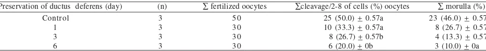
Oocytes fertilized by sperms collected from caudal epididymis preserved for day 1, 3, and 6 still have capability to reach cleavage stage (2-8 of cells) of 33.3, 26.7, and 20.0%,
respectively (Table 3). Statistical analysis showed that
capability of oocytes fertilized by sperms preserved for 1H to reach cleavage stage was not significantly different from that
of control (P > 0.05).Capability of oocytes fertilized by sperms
collected from that of control (P < 0.05). There was indication of reduction in cleavage rate of fertilized oocytes to reach cleavage stage of 2-8 cells. This phenomenon indicated that acrosom cap of sperms collected from ductus deferens
a b
c d
Figure 1. Sperms appearance in vital staining, a. live sperms with intact acrosom, b. live sperms with lysis acrosom, c. dead sperms with intact acrosom, d. dead sperms with broken acrosom. Bar = 1 µm.
Table 2. Percentage of living sperms with intact acrosom or acrosom reaction after preservation from different sources
Live sperm/intact acrosom (%)
Number followed by the same letters showed no significant difference at 5% Tukey-Kramer HSD probability. DD: ductus defferens, CA: cauda epididymis, CR: corpus epididymis.
Table 3. Development of embryos produced by IVF method using preserved sperms
Preservation of ductus deferens (day) (n) ∑ fertilized oocytes ∑cleavage/2-8 of cells (%) oocytes ∑ morulla (%)
preserved in 4 oC was still could be kept. However, longer
preservation time would result in more reduction in quality and viability of sperms to perform fertilization. Intact acrosom
caps was very important for the success of in vitro fertilization
because vesicles contained in acrosom caps have some enzymes played significant roles fertilization process. If acrosoms were broken, these enzymes will be lost so that sperms capacity to pass barrier surrounding oocytes will be lost and there was no fertilizitaion would be occurred.
DISCUSSION
Sperm Concentration, Motility, and Viability. It was assumed that the reduction in sperm motility occurred during preservation period resulted from accumulation of metabolic wastes. According to Nazlie (2004) during preservation sperms were still alive and performed their activities by using energy produced by metabolic processes. However, this energy gradually decreased, and by product of the metabolism such us lactic acids were in the high proportion. The lower pH due to from this lactic acid accumulation was toxic and had negative effect to some metabolic enzymes. As a consequence, all metabolic processes will be disrupted.
In addition to due to the metabolic wastes, the decrease in sperm motility, was due to reactive oxygen species (ROS) (Soeradi 2004). ROS could induce DNA damage (Fraser 2004). In this study, it was predicted that the low sperm motiliy was caused by the high proportion of DNA damage. According to
Moustafa et al. (2004) there were three hypotesa could explain
DNA damage: (i) DNA damage from the packaging and
ligation processes during sperm maturation, (ii) oxydative stress resulted in DNA destruction and the increase of ROS
formation such as 8-hydroxydeoxyguanosine in DNA of
sperms, and (iii) DNA fragmentation caused by apoptosis. Factors causing DNA damage, however, were not deeply investigated in this study.
According to Fraser and Strzezek (2004) factors influencing DNA damage were species spesific and difficult to be explained. In pig, DNA destruction was caused by ROS formation during sperm ageing ultimately disturbing the integrity of other sperms. In this study, it was difficult to determine whether the long preservation time at low
temperature (4 oC) can bring higher oxidative stress resulting
in overproduction of ROS. This assumption will also contradict to the principle of oxidation-reduction. However, long preservation could create destructive effects. There was such indication that ROS was potentially formed during preservation because majority of Fellidae were reported having poor quality of sperms (Farstad 2000) and abnormality
of ejaculated sperms > 60% (Penfold et al. 2003). Nazlie (2004)
reported that abnormality of sperms collected from corpus epididymis of domesticated cats on day 0 (control) was as high as 25.75%. This will potentially trigger formation of ROS during preservation.
Moreover, it was suggested that chain reaction of lipid peroxidation and other free radical compounds could be
inhibited their formation by adding antioxidants such as gluthathion, cystein, catalase, gluthathion peroxidase,
β-caroten, vitamin E (tocoferol), and vitamin C (ascorbic acid)
in the medium. In this study, washing and capacitation media used were CR1aa containing of amino acid of MEM Non Essential Amino Acid (one of its component was cystein) and Minimum Essential Amino Acid (MEM-BME) that probably could play a role as antioxidant coumpounds as well as fixing plasm membrane of damaged sperms. As the results the reduction of sperm motility could be minimized during the washing of sperms.
Production of Embryos by IVF. By using IVF technique we obtained 26.67, 13.33, and 10% morulla for the preservation
of day 1, 3,and 6, respectively. These results were not
significantly different (P > 0.05) from control (46.00%). There is a suspition that parthenogenesis has occurred in this study. From totally 302 oocytes, 15 (4.9%) oocytes cleavaged before fertilization. However, fertilization rates obtained in this study was higher than that of parthenogenesis, 33.3, 26.7, and 20.0% for day 1, 3, and 6 of preservation time respectively. The
developed oocytes caused by swim-up sperms were used for
in vitro fertilization. According to Younglai et al. (2001) DNA
fragmentation in sperms prepared by swim-up method was
lower than that of non swim-up. Howart et al. (1993) referred
in Goodrowe et al. (2000) suggested that fertilization rate in
domesticated cats by using freshly swim-up sperms was
markedly higher (94.6%) than that of without swim-up (53.2%).
Muratori et al. (2000) claimed that the high DNA fragmentation
occurred in the unselected sperms and in swim-up sperms
has positive correlation with abnormal morphology as well as
with weak sperm tails. Barroso et al. (2000) performed sperm
analysis to infertile men by using terminal deoxynucleotidyl
transferase-mediated dUDP nick-end labelling (Tunnel)
assay, suggested that there was relationship between low
sperm motility and the high DNA damage.
In this study, it was assumed that sperm preservation may caused DNA damage as reflected by the low sperm motility. This assumption was also supported by data of sperm
observation by using Hoechst-Propidium Iodine staining.
The data showed that swim-up became one of important
treatments needed for fertilization rate even though in fact there were immotil sperms with intake DNAs.
for preservation time of day 3 and 6, sperms collected from caudal epididymis have relatively higher percentage of development (2-8 cells) than that from ductus deferens and corpus epididymis. The occurrence of developing pattern above was probable caused by the higher capability of nutrition content of cauda epididymis to maintain better
quality and viability of sperms. According to Tomes et al.
(1979) fluid of caudal epididymis has lower sodium and chloride contents but higher potassium, phosphor, and protein than that of retetestis fluid. Epididymis also synthesizes carnitin, glycerilphosphorilcholin (GPC), fructosa, and glycoprotein (Johnson & Everitt 1995). Bearden and Fuquay (1997) said that the fluid of cauda epididymis has an optimum condition for sperms due to its low PH, high viscosity, high
CO2 concentration, high ratio of K+ and Na+, positive influence
of testosterone on epididymis function, and combination of other factors resulting in low metabolism rate of this organ so that can prolong viability of sperms.
For the percentage of morulla stage development (Figure 3e,f), sperms collected from the three sources showed no differences. There was no embryo develop into blastocys since
according to Skrzyszowska et al. (2002) in cat growth inhibition
occurred in morulla stage. The proportion of cat embryos
developed into blastocys produced by in vitro culture from
the 2- to 4- cells was higher than that from the 1-cell (Figure 3a). This showed that embryonic growth stage started from
in vitro culture has high impact to the increase in the
development delay of cat embryo into morulla stage (Figure 3).
ACKNOWLEDGEMENT
The author would like to say great appreciate and thank to Yuhara Sukra for his help, guidance and support. Our great thank you and appreciation were also directed to Ivan Krisna (Management Unit of Leuser) for his valuabe data and information.
REFERENCES
Aalseth EP, Saacke RG. 1986. Vital staining and acrosomal evaluation of bovine sperm. Gamet Research 15:73-81.
Barroso G, Morshedi M, Oehninger S. 2000. Analysis of DNA fragmentation, plasma membrane translocation of phosphatidylserine and oksidative stress in human sperm. Hum Reprod 15:1338-1344.
Bearden HJ, Fuquay JW. 1997. The male reproduction system. In:
Applied Animal Reproduction (4th Ed). New Jersey: Prentice Hall.
p 27.
Carolan C, Monaghan P, Gallagher M, Gordon I. 1994. Effect of recovery method on yield of bovine oocytes per ovary and their developmental competence after maturation, fertilization, and culture in vitro. Theriogenology 41:1061-1068.
Farstad W. 2000. Current state in biotechnology in Canine and Feline reproduction. Anim Reprod Sci 60:375-387.
Fraser L. 2004. Structural damage to nuclear DNA in mammalian spermatozoa: its evaluation techniques and relationship with male infertility. Polish J Vet Sci 7:311-321.
Fraser L, Strzezek. 2004. The use of comet assay to assess DNA integrity of boar spermatozoa following liquid preservation at 5 and 16 oC. Folia Histochemica et cytobiologica 42:49-55.
Goodrowe KL et al. 2000. Piecing together the puzzle of carnivore reproduction. Anim Reprod Sci 60:389-403.
Hafez ESE, Hafez B. 2000. Reproduction in Farm Animals. Seventh Edition. Baltimore: Lippincott Wiliams & Wilkin.
Johnson MH, Everitt BJ. 1995. Essential Reproduction 4Ed. Oxford:
Blackwell Science. p 152-158.
Moustafa MH et al. 2004. Relationship between ROS production, apoptosis and DNA denaturation in spermatozoa from patients examined for infertility. Hum Reprod 19:129-138.
Muratori M, Piomboni P, Baldi E. 2000. Functional and ultrastructural features of DNA-fragmented human sperm. J Androl 21:903-912. Nazlie CS. 2004. Kajian Kualitas Spermatozoa Kucing Asal Eididymis dan Duktus Deferens Setelah Proses Preservasi pada Suhu 4 oC
[Tesis]. Bogor: Sekolah Pascasarjana, Institut Pertanian Bogor. Penfold LM, Jost L, Evenson DP, Wildt DE. 2003. Normo- versuv
teratospermic domestic cat sperm chromatin integrity evaluated by flow cytometry and intracytoplasmic sperm injection. Biol Reprod 103-108.
Pope CE. 2000. Embryo technology in conservation efforts for endangered felids. Theriogenology 53:163-174.
Risopatron J et al. 1996. Migration/sedimentation sperm selection method use in bovine in vitro fertilization comparison with washing/ centrifugation. Theriogenology 46:65-73.
Figure 3. Development of embryo in vitro fertilized. a. phase 2-cells,
b. phase 3-cells, c. phase 4-cells, d. phase 8-cells, e-f. phase compact morulla. Bar = 20 µm.
a b c
d e f Cleavage DD
Cleavage CR Cleavage CA
Morula DD Morula CA Morula CR 7 0
6 0
5 0
4 0
3 0
2 0
1 0
0
Embryo development (%)
0 1 3 6 Preservation time (day)
Rosenkrans CF, First NL. 1994. Effect of free amino acids and vitamin on cleavage and development rate of bovine zygotes in vitro.
Anim Sci 72:434-437.
Skrzyszowska M et al. 2002. In vitro developmental competence of domestic cat embryos after somatic cloning: a preliminary report.
Theriogenology 58:1615-1621.
Soeradi O. 2004. Radikal bebas pada pria infertil. Kumpulan Makalah/ Abstrak Paradigma Terkini Genetika dan Reproduksi. Departemen Biologi Kedokteran FKUI. p 95-101
Soler AJ, Perez-Guzman MD, Garde JJ. 2003. Storage of red deer epididymides for four days at 5 oC: effect on sperm motility,
viability, and morphology integrity. Exp Zool 295:188-199.
Steel RGD, Torrie JH. 1991. Prinsip dan Dasar Statistik. Ed ke-2. Jakarta: Gramedia.
Tomes GL, Robertson DE, Lightfoot. 1979. Sheep Breeding Second Edition. London: Butteworth.
Wright M, Walters S. 1980. The Book of the Cat. 14-15, 213. London: Pan Books.
Younglai EV, Holt D, Brown P, Jurisicova A, Casper RF. 2001. Sperm swim-up techniques and DNA fragmentation. Hum Reprod