Technique of Overlap Extension by Polymerase Chain Reaction for Splicing
Cauliflower Mosaic Virus (CaMV) 35S Promoter and
DhPEX11-Like
Silvi Ikawati
1,2*, Yung-fu Yen
21
Faculty of Agriculture, University of Brawijaya, Malang, Indonesia
2
Department of Bioagricultural Science, National Chiayi University, Chiayi, Taiwan
Abstract
The promoter plays an important role in the regulation of gene expression. The problem is some of binary vector that absence from promoter at cloning site. The cauliflower mosaic virus (CaMV) 35S promoter is a strong and constitutive promoter that widely used to produce transgenic organisms. In this experiment the cauliflower mosaic virus (CaMV) 35S
promoter was spliced at upstream of DhPEX11-like for driving downstream transgenes DhPEX11-like expression used the technique of Overlap Extension by The Polymerase Chain Reaction. In gene splicing, internal primers are used to amplify some overlapping regions of both genes and then these internal primers are combined with the external primers in PCR process which allows amplification of the entire region. In the experiment, the recombinant PCR successfully spliced the 35S-DhPEX11 gene. This method is simple, rapid and reduced reagents used because it does not need many vector constructions
.
Keywords:DhPEX11-Like, Gene splicing, Oligonucleotide, PCR, Promoter
INTRODUCTION
Promoters are one of the essential constitute of gene, as they are required to drive expression of both the selectable marker gene and the gene of interest in transgenes [1]. The problem is some of binary vector is absence from promoter at cloning site. Combining genes or regulatory elements to make hybrid genes is a widely used methodology throughout the biological sciences [2]. One of the techniques for synthesis of arti-ficial genes is called gene splicing, in which seg-ments of DNA are joined together to create a new genetic combination [3]. Gene splicing by overlap extension is a technique for combining the DNA molecule from two genes on the short nucleotide sequences that have been recom-bined in precise junctions without using re-striction endonucleases or ligase [4]. The polymerase chain reaction (PCR) has greatly en-hanced the field of molecular biology by making numerous regions of the genome (coding and noncoding), in both extant and extinct taxa, accessible for detailed analysis [5]. Previous research demonstrated that the PCR recom-binant can be used to remove selectable markers or other introduced transgenes that are no
Correspondence author:
Silvi Ikawati
Email : [email protected]
Address : Department of Plant Protection, Faculty of Agriculture, University of Brawijaya, Jl. Veteran Malang, 65145
longer desired and therefore can be a useful tool for genome engineering in plants [6]. The ability to fuse two DNA fragments by overlap extension can be exploited further to splice two or more DNA fragments from different genes [7]. Overlap extension-PCR also can be used as a means for site directed mutagenesis, introducing desired mutations to the final hybrid gene [2]. Under PCR conditions, the common sequence allows strands from two different fragments to hybridize to one another, forming an overlap [4]. Initial PCRs generate overlapping gene segments that are then used as template DNA for another PCR to create a full-length product [8].
MATERIALS AND METHODS ubiquitin 1 and actin promoters [11]. Cauliflower Mosaic Virus (CaMV) and the closely related Figwort Mosaic Virus are circular duplex DNA viruses which replicate via transcription of a full-length (35S) genomic RNA intermediate [12]. The cauliflower mosaic virus (CaMV) 35S promoter is a strong and constitutive promoter that widely used for production purposes [13, 14].
Plasmid preparation
To get the promoter and the gene for splice process, first was needed to extract plasmid that contained the genes from E. coli. The trans-formed white single colony E. coli was picked out from a petri dish and was transferred to small volume of LB-medium 5 mL containing the am-picillin antibiotic (0.2 mg/ml) and shaken at 37°C and 200 rpm for 16-24 h. Plasmid Miniprep Purification Kit (GeneMark) was used to extract the plasmid from E. coli culture, according to the manufacturer’s manual. Cell pellet was collected in a 1.5 mL Eppendorf tube (1-3 ml of cells) by centrifugation of E. coli culture at 14,000 rpm for 1 min.
The supernatant was decanted and the pellet was resuspended in 200 μl Solution I by pipetting or vortexing. And 200 μl Solution II was added into the tube and the mixture was inverted 5 times. Next, 200 μl Solution III was also added and the tube was inverted again (5 times) to lyse cells. The lysate was centrifuged at 14,000 rpm for 5 min and the supernatant was transferred into a spin column combined with collection tube. The flow-through in the collection tube was discarded. The spin column was added 700 μl Wash Solution and centrifuged at 14,000 rpm for 1 min. The washing step was repeated for one more time. The spin column was centrifuged for 5-10 min at top speed to remove residual trace of ethanol. After ethanol removed completely, the spin column was then transferred into a new 1.5 mL Eppendorf tube and was added 30-100 μl of
Gene splicing by overlap extension consists of three times PCRs process. In the first stage reactions produced the two DNA fragments, and the first products to be used as template in the ribonucleotide triphosphate (dNTP), 0.25 μl of Taq Phusion High-Fidelity DNA Polymerase, 5 μl of 5X Phusion HF reaction buffer and 1 μl primer mix (10 μM each). PCR amplification was performed initialized at 94°C for 3 min, followed by 28 cycles (94°C for 45 s, 60°C for 30 s, and 72°C for 30 s), with a final extension of 5 min at 72°C.
Gel agarose preparation
For preparing 1% gel, 0.25 g of agarose powder (Seakem, Marine Colloids, Inc.) dissolved in 25 ml 0.5X Tris acetate EDTA buffers (20 mM Tris acetate, 0.5 mM EDTA). The mixture was heated in a microwave oven for about 1-2 min to dissolve agarose. The solution was cooled for 4 min and poured into electrophoresis apparatus. After gel has solidified, 0.5X TAE buffer was poured into gel box, and comb was removed after solidified. A dye solution was added at DNA samples prior to electrophoresis. DNA molecular weight marker was used in each size marker lane of the electrophoresis gel. Electrophoresis was performed at 100 V for 35 minutes or until the dye neared the bottom of the gel. The gel was then placed in a solution of ethidium bromide in water and stained for 2 minutes. About 10 minutes the gel was rinsed in water. The gel is illuminated with UV transilluminator then photo-graphed.
PCR recombinant [16]
fragments. The gel was sliced using a clean razor blade and was excised with the Wizard DNA Clean-Up system kit (Promega). Gel slice was transferred in a 1.5-ml microcentrifuge tube (Eppendorf) and 10 μl Membrane Binding Solution was added per 10 mg of gel slice. The gel was incubated at 50-65°C until gel slice is completely dissolved.
Dissolved gel mixture was transferred to the Minicolumn assembly and then centrifuged at 14,000 rpm for 1 min. Following centrifugation flow-through was discarded and the minicolumn was washed with 700 μl Membrane Wash Solution (ethanol added) then centrifuged at 14,000 rpm for 1 minute. The washing step was repeated with 500 μl Membrane Wash Solution and centrifuged at 14,000 rpm for 5 minutes. The column was recentrifuged for 1 min with the microcentrifuge lid open (or off) to allow evaporation of any residual ethanol. Fifty μl Nuclease-Free Water was added to the minicolumn and centrifuged at 14,000 rpm for 1 min. For further confirmation these PCR products were sequenced.
Confirmation of DNA fragments
Confirming DNA fragments of CaMV35S promoter, DhPEX11-like and 35S/DhPEX11 we did PCR, enzyme digestion and sequence analysis.
For the digestion reaction includes 1.5 μg of
plasmid DNA, 2 μl of 10X buffer, 0.2 μl of 100x BSA, 0.5 μl of restriction enzyme, and sterile water to 20 μl. The reaction was incubated for 1 h at the temperature specific for the enzyme
used. Digested products were electrophorated on 1 % agarose gels.
DNA Sequencing
After PCR and enzyme digestion the transformed strains of plasmids were delivered to Mission Biotech Company (Taipei, Taiwan) for sequencing. DNA sequence data were analyzed using the National Centre of Biotechnology (NCBI) web site (http://www.ncbi.nlm.nih.gov) for alignment by the program nucleotide blast (BLASTn).
RESULT AND DISCUSSION First Stage PCR
In the first stage PCRs produces two DNA fragment with the sequence 5′ and 3′to the splice point (1)–(2) in Fig. 1. However, since the hybrid oligonucleotides have the splice regions, pro-ducts in the first stage will splice at the short sequences derived from the other (2) in Fig. 1. Therefore when the two products are combined they can partially anneal and used two external primers in the second stage PCR to get the final result (3–4) in Fig. 1 [9].
This technique uses 2 steps PCR, first step is to get two kind of template that the promoter and gene sequence are joined use internal Oligonucleotides. Second step used two kind of template with external Oligonucleotides (Table 1). The oligonucleotides design (Table 1) were based on the full-lengthed nucleotide sequence of 35S promoter in NCBI (http://www.ncbi.-nlm.nih.gov) and DhPEX11-like [17].
Figure 1. Gene splicing by overlap extension by polymerase chain reaction [9]
Table 1. Oligonucleotides for cloning CaMV 35S Promoter and DhPEX11-like and splicing CaMV35S Promoter and DhPEX11-like
to generate 35S/DhPEX11
Gene Oligonucleotide sequences 5'-3' (A) First
35S Pro
35S-F GGGAATTCCATGGAGTCAAAGATTCAAATAGAGGACCTAACAG 35S-R GCTATTTCATCCTGGGTCATGGTCAAGAGTCCCCCGTGTT
DhPEX11
PEX11-F AACACGGGGGACTCTTGACCATGACCCAGGATGAAATAGC PEX11-R CCGGATCCTCATAATCATTAGGAGGAGGAGCAGCTGCTTC
(B) Second
35S-F GGGAATTCCATGGAGTCAAAGATTCAAATAGAGGACCTAACAG PEX-R CCGGATCCTCATAATCATTAGGAGGAGGAGCAGCTGCTTC
The extracted plasmid DNA from pCAMBIA 1302 was used as template in PCR with prime pair of 35S-F and 35S-R for cloning CaMV 35S Promoter, as well as the DhPEX11-like fragment was cloned from the transformed pMETB Plasmid using the prime pair of PEX11-F and PEX11-R [17]. The secondary PCR spliced the two DNA fragmented of CaMV 35S promoter and DhPEX11-like to become to the recombinant of 35S/DhPEX11 gene, using outprimer pair of 35S-F and PEX11-R. The hybrid oligonucleotides are designed from the known nucleotide sequences to generate fragments that will have overlapping sequence [9]. They had 20 bp overlap which sequences were underlay at 5’end of 35S
promoter and 3’end of PEX11-like gene (Fig. 2).
The availability of thermostable DNA polyme-rases with a much lower tendency to add a non-templated nucleotide to DNA fragments [18] may decrease the rate of mutation further and the need to blunt end intermediate products [9].
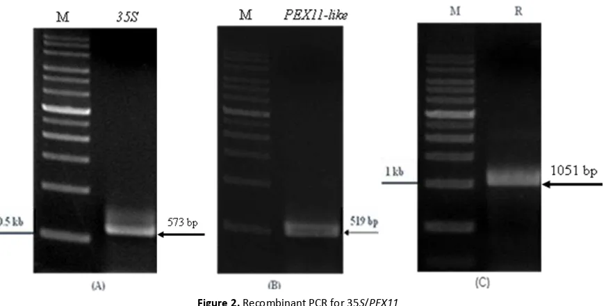
The first PCR cloned the 35S promoter and the second PCR cloned the DhPEX11-like gene, their sizes were confirmed by analysis on 1% gel electrophoresis with expected with expected DNA fragments of 573 bp (Fig. 2A) and 579 bp (Fig. 2B), respectively. The size of the recom-bination had expected band and sequenced being 1051 bp. (Fig. 2C).
Figure 2. Recombinant PCR for 35S/PEX11
(A) PCR analysis of 35S promoter. The figure showed the expected size of 35S promoter was 573 bp. (B) PCR analysis of PEX11-like gene. the expected size for PEX11-like was 519 bp.
(C) PCR analysis of 35S/PEX11 recombinant, the 1051 bp band was shown. M: 1-kb DNA markers. 35S: 35S promoter.
PCR recombinant product
PCR recombinant product was ligated to be cloned into pGEM-T Easy vector and infected to E. coli which was spanned on LB-agar medium containing X-gal, IPTG and ampicillin. The white colonies was the host of transformant vector which was confirmed by PCR. Its reactant were run on 1% agarose gel and distinct band of 1051 bp revealed under the UV light (Fig. 3A). It presented the DNA fragment of 35S/PEX11 in the transformant plasmid.
Confirmed DNA fragments
The expression cassette of 35S/PEX11 that released from the pGEM plasmid DNA digested with BamHI and HindIII enzymes, the reactant was run on 1% agarose gel which appeared both 1051 bp fragment and 3 Kb of the vector backbone (Fig. 3B). The final product that has been amplified by flanking primers also can be obtained restriction enzymes sites to insert it into an expression vector for cloning step [8].
DNA sequencing
The identity value of alignment results of the original sequence of 35S/PEX11-like and the sequence of 35S/PEX11-like fragment cloned from p-GEM T-easy vector was 98%.
In the experiment, the recombinant PCR successfully spliced the 35S-DhPEX11 gene which appeared strong expression of transformant. SOE by PCR is rapid because it does not need many vector constructions which are a time consuming process. This method is simple and widely applicable approach has significante advantages over standard recombinant DNA techniques [4]. Moreover, the recombinant might be amplified and cloned into expression vector, and it is readily applied in plant transformation. The 35S promoter of cauliflower mosaic virus (CaMV) able to confer high-level gene expression in most organs of transgenic plants [19].
Fig. 3. Gel checking of the DNA fragments inserted into pGEM-T Easy vector. (A) Polymerase chain reaction (PCR) analysis. The figure showed the expected size of 35S/PEX11 cloned into vector. (B) The constructed 35S/PEX11/pGEM-T was digested by EcoRI and SpeI enzymes for double checked before sequencing, the 1051 bp band and the 3.0 kb band were shown. M: 1-kb DNA markers. C: Constructed 35S/PEX11/pGEM-T.
CONCLUSION
Recombinant PCR in this study successfully spliced the 35S-DhPEX11 gene. SOE by PCR did not need many vector constructions thus less time consuming. This simple and widely applicable method of approach has significante advantages for standard recombinant DNA. The use on 35S promoter of CaMV confer high-level gene expression for transgenic plants as in this study.
REFERENCES
[1] Deo, P.C., A.P. Tyagi, M. Taylor, R. Harding, D. Becker. 2010. Factors affecting somatic embryogenesis and transformation in mo-dern plant breeding. Nat. Appl. Sci. 28. 27-40.
[3] Li,X., Y. Qiu, Y. Shen,C. Ding, P. Liu, J. Zhou, Z. Ma. 2008. Splicing together different regions of a gene by modified polymerase chain reaction-based site-directed mutage-nesis. Anal. Biochem. 373. 398–400.
[4] Horton, R.M., H.D. Hunt, S.N. Ho, J.K. Pullen, L.R. Pease. 1989. Engineering hybrid genes without the use of restriction enzymes: gene splicing by overlap extension. Gene. 77. 61–68.
[5] Bradley, R.D., D.M. Hillis. 1997. Recom-binant DNA sequences generated by PCR amplification. Mol. Biol. Evol. 14. 592-593. [6] Thomson, J.G., R. Chan, R. Thilmony, Y.
Y. Yau, D.W. Ow. 2010. PhiC31 recombi-nation system demonstrates heritable germinal transmission of site-specific excision from the Arabidopsis genome. BMC Biotech. 10. 1-12.
[7] Vallejo, A.N., R.J. Pogulis, L.R. Pease. 2003. Mutagenesis and synthesis of novel recom-binant genes using PCR. In: Dieffenbach, C.W., G.S. Dveksler (Eds). PCR Primer A Laboratory Manual. Cold Spring Harbor Laboratory Press. New York. 467-468. [8] Heckman, K.L., L.R. Pease. 2007. Gene
splicing and mutagenesis by PCR-driven overlap extension. Nature Prot. 2. 924-932. [9] Warrens,A.N, M.D. Jones, R.I. Lechle. 1997.
Splicing by overlap extension by PCR using asymmetric amplification: an improved technique for the generation of hybrid proteins of immunological interest. Gene. 186. 29–35.
[10] Higuchi, R. 1990. Recombinant PCR. In: M.A. Innis, D.H. Gelfand, J.J. Sninsky, T.J. White (Eds). PCR Protocols A Guide to Methods and Applications. Academic Press. San Diego.
[11] Bhatnagar-Mathur, P., V. Vadez, K.K. Sharma. 2007. Transgenic approaches for abiotic stress tolerance in plants: retrospect and prospects. Plant Cell Reports. 27. 411-424.
[12] Gruber, M.Y., W.L. Crosby. 1993. Vectors for plant transformation. In: Glick, B.R., J.E. Thompson (Eds). Methods in Plant Molecu-lar Biology and Biotechnology. CRC Press. Florida. 89-119.
[13] Alves, A.C., V.M. Quecini, M.L.C. Vieira. 1999. Plant transformation: advances and perspectives Scientia. Agricola. 56. 1-8. [14] de Wilde, C., H. van Houdt, S. de Buck, G.
Angenon, G. de Jaeger, A. Depicker. 2000. Plants as bioreactors for protein production:
avoiding the problem of transgene silencing. Plant Mol. Biol. 43. 347-359.
[15] Ling, M.F., B.H. Robinson. 1995. A one-step polymerase chain reaction site-directed mutagenesis method for large gene-cassettes with high efficiency, yield, and fidelity. Anal. Biochem. 230 (1). 167-172. [16] Blattner, F.R. 1999. Direct amplification of
the entire ITS region from poorly preserved plant material using recombinant PCR. Biotechniques. 27. 1180-1186.
[17] Hue, L.T. 2010.Characterization of a salt-induced the mitochondrial import inner membrane translocase subunit DhTIM22 gene from the extreme halophilic yeast Debaryomyces hansenii and its overex-pression to enhance yeast tolerance to stresses. Master Thesis. Departement of Bioagricultural, National Chiayi University Science.
[18] Hu, G. 1993. DNA polymerase-catalyzed addition of nontemplated extra nucleotides
to the 3′ end of a DNA fragment. DNA Cell.
Biol. 12. 763–770.
![Figure 1. Gene splicing by overlap extension by polymerase chain reaction [9] Promoter and gene illustrated in the form of bars](https://thumb-ap.123doks.com/thumbv2/123dok/2881484.1696800/3.595.94.523.515.705/figure-splicing-overlap-extension-polymerase-reaction-promoter-illustrated.webp)