Indo. J. Chem., 2005, 5 (3), 274 - 277
Suharso
274
* Email address : [email protected]
CHARACTERIZATION OF SURFACE OF THE (010) FACE OF BORAX CRYSTALS
USING
EX SITU
ATOMIC FORCE MICROSCOPY (AFM):
CLEAVAGE AND CLEAVAGE STEPS
Suharso
*Department of Chemistry, Faculty of Mathematics and Natural Sciences, University of Lampung Jl. Sumantri Brojonegoro 1, Gedung Meneng-Bandarlampung
Received 16 August 2005; Accepted 29 August 2005
ABSTRACT
The surface topology of borax crystals grown at a relative supersaturation of 0.21 has been investigated using ex situ atomic force microscopy (AFM). It was found that the cleavage of borax crystals along the (010) face planes has features of the cleavage of layered compounds, exhibiting cleavage steps of low heights. The step heights of the cleavage of the (010) face of borax crystal are from one unit cell to three unit cells of this face.
Keywords:AFM, cleavage, borax.
INTRODUCTION
Scanning Probe Microscope (SPM) is a family of instrument that is used to measure properties of surface, including Atomic Force Microscope (AFM) and Scanning Tunneling Microscope (STM). The Atomic Force Microscope was developed in 1986 by Binnig, Quate, and Gerber in a collaboration between IBM and Stanford University [1]. In early 1990, digital Instrument Inc. introduced the first commercial AFM.
Like the STM, the AFM also uses a very sharp tip to probe and map the morphology of a surface. However, in AFM there is no requirement to measure a current between tip and sample. In this case, the tip is positioned on the end of a long cantilever with a low spring constant (of the order of 1 Newton/m). In contact mode AFM, the first scanning technique, the tip-sample force is held fixed by maintaining a constant and very low deflection of the cantilever, pushing the tip against the sample. This force can be in the range of interatomic forces in solids (about 10-9Newton).
The vertical resolution of the AFM is mainly determined by the resolution of the vertical scanner movement which is lower than 1 Å, and the number of data points in the vertical direction, while the lateral resolution is determined by the tip shape and the number of data points. Since its invention, AFM has given to be a very powerful technique for investigating surfaces at low to high resolution, and for studying, in situ, the dynamic processes of crystal growth and dissolution and some
researchers used this technique to investigate a surface topography of various crystals.
Krasinki and Rolandi reported ex situ AFM investigation of surface topographies of the (100) and (101) faces of as-grown potassium dihydrogen phosphate (KDP) crystals. Growth hillocks, step bunching, growth spirals, two- dimensional nucleation and hollow cores were detected [2].
Land with his colleague observed an in situ
AFM on the canavalin crystals. The results show that, depending on the supersaturation, growth occurs on steps of one growth unit in height generated either by simple and complex screw dislocation sources, two dimensional nucleating islands, or macroclusters which sediment onto the surface before spreading laterally as step bunches [3].
The Investigation of the growth on the (001) face of barite in situ at room temperature using contact mode AFM was carried out by some researchers. They observed that growth under moderate supersaturation conditions follows the birth and spread model; two-dimensional nuclei form simultaneously with the advancement of molecular height cleavage steps. They concluded that the surface crystallography strongly controls nucleation and growth [4].
Indo. J. Chem., 2005, 5 (3), 274 - 277
Suharso
275
(010) face occurs by combined spiral and two-dimensional nucleation mechanism [5].
To our knowledge, the surface topography of borax crystals has not been observed by atomic force microscopy to date. Most of the researches gave poor visual data. In this present research, the (010) faces of borax crystals will be characterized usingex situatomic force microscopy.
EXPERIMENTAL SECTION
Solubility of borax in water
The driving force for crystallization is usually expressed as a supersaturation ratio, defined as [6]:
*
A
A
S
where A and A* are the initial and the equilibrium solute concentrations. The solubility of borax in water used has been reported by Nies and Hulbert [7], and Sprague [8].
Seed Preparation
The seed solution was prepared from 30 g of Univar AR grade sodium tetraborate dissolved in 100 mL of Milli-Q water by heating up until 60 °C and filtering through filter paper. The solution was quickly cooled down into petri dish that covered by a transparent plastic, producing 40-200 m well crystals. Only single seed crystals between 40 -120m in size were used to investigate the growth rate.
Experimental Set-up
AFM investigations were carried out in air with a Digital Instruments Nanoscope E AFM, using a 12 or 150 m scanner. All the AFM images (height) were collected in the contact mode. Wide triangular shaped 200 m cantilevers made of gold-coated Si3N4, with force constant of 0.12 N/m were used.
Seed crystals were grown in 100 mL of the borax solution filtered through a 0.45m membrane filter in the covered plastic bottle at temperature 20 °C. As soon as the crystals grew into an acceptable size for the AFM experiment, individual crystal were taken out carefully and dried with a tissue. Then, the seed crystals were gently placed on the AFM sample holder using a double-sided sticky tape or 'blue tack'. Ex situAFM measurements were then immediately carried out.
RESULT AND DISCUSSION
The investigation of the surface topography on a cleavage borax crystal has been carried out on the surface of the (010) face at relative supersaturation of 0.21 and room temperature. The
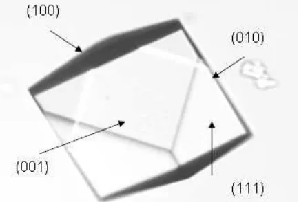
unit cell height in this direction is 1.061 nm. This face at the low supersaturation has small area. Therefore it is very hard to take the image under atomic force microscopy. The surface of this face can be seen in Figure 1.
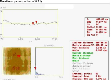
Figure 2 (a, b) illustrates an example of an array of steps observed on the (010) cleavage plane of borax crystal. Most of the cleavage steps seen in this figure are of elementary height, but some of the steps of height are approximately two and three unit cell of this face (Figure 3). The highest cleavage step observed on the (010) face of the borax crystal had a three units cell. These steps are parallel to the (100) face.
Investigations of mono-, bi- and trilayer steps as well as multilayer steps of heights up to several tens of nm by AFM on the cleavage surfaces of ionic crystals have often been reported in the literature. Monolayer steps emerging on the (100) cleavages faces of MgO single crystal was observed by Sangwal et al and they also found monolayer and multilayer on the (100) cleavage face of L-arginine phosphate monohydrate (LAP) single crystals. The highest cleavage step observed on a LAP crystal had five units cell [9]. The result presented here on cleavage steps on the (010) face of borax crystal grown from aqueous solutions is very similar to the cleavage LAP crystal reported by Sangwal [9]. However, in contrast to the very large height of multilayer cleavage steps of another ionic crystals, most of the cleavage steps observed on the (010) cleavage face of borax crystals are elementary steps and the maximum height of the highest step observed is much lower than observed in the case of ionic crystals [10].
It may be concluded that the cleavages of borax crystals along the (010) planes has features of the cleavage of layered compounds like LAP, mica and hydrated potassium niobates, which exhibit cleavage steps of low heights. In contrast to
Indo. J. Chem., 2005, 5 (3), 274 - 277
Suharso
276
(a) (b)
Figure 2 (a,b). Height and deflection images on the (010) face of cleavage borax crystal (Relative supersaturation of 0.21)
Figure 3 Section analysis of the steps on the (010) face of cleavage borax crystal from Figure 2(a)
the cleavage of these, the cleavages of another ionic crystals usually show a large number of cleavage steps of different height. This difference in the behaviour of cleavage steps is assosiated with the nature and strenght of bonds involved during cleavage of the crystals [10].
CONCLUSION
AFM investigation produced very useful information about the surface characteristics of the (010) faces of borax crystals. The cleavage of borax crystals along the (010) face planes has features of the cleavage of layered compounds, exhibiting cleavage steps of low heights. The step
heights of the cleavage of the (010) face of borax crystal are from one unit cell to three unit cells of this face (unit cell of this face = 1.061 nm).
ACKNOWLEDGEMENT
Indo. J. Chem., 2005, 5 (3), 274 - 277
Suharso
277
REFFERENCES
1. Binning, G., Quate, C.F. and Gerber, C., 1986,
Phys. Rev. Lett., 56, 9, 930-933.
2. Kransinski, M.J. and Rolandi, R., 1996, J. Crystal Growth,169, 548-556.
3. Land, T.A., De Yoreo, J.J., and Lee, J.D. Yu.G., 1997,Surf. Sci.384, 136-155.
4. Pina, C.M., Bosbach, D., Prieto, M. and Putnis, A., 1998,J. Crystal Growth,187, 119-125. 5. Kuznetsov, V.A., Samotoin, N.D., Okhrimenko,
T.M. and Rak, M., 2000,Phys. Stat. Sol., 179, 349.
6. Mullin, J.W., 1993, Crystallization, third edition, Butterworth-Heinemann Ltd, Oxford.
7. Nies, N.P., and Hulbert, R.W., 1967, J. Chem. Eng. Data, Vol. 12 (3), 303-313.
8. Sprague, R.W., 1980, In Mellor’s Comprehensive Treatise on Inorganic and Theoritical Chemistry, Vol. 5, 254, London and New York, Longman.
9. Sangwal, K., Torrent-Burgues, J., Sanz, F., and Servat, J, 1997,Surface Science374, 387-396. 10. Sangwal, K., Torrent-Burgues, J., Sanz, F., and