Osteoradionecrosis and cholesteatoma of external auditory canal in
post-radiotherapy nasopharyngeal carcinoma patient
Devira Zahara, Agus Multazar
Departement of Ear, Nose and Throat - Head and Neck Surgery Faculty of Medicine University of Sumatera Utara
H. Adam Malik Hospital Medan
ABSTRACT
Background: Osteoradionecrosis and cholesteatoma of the external auditory canal following external-beam radiotherapy as the treatment of nasopharyngeal carcinoma is a rarely found complication. Patients with external auditory canal cholesteatoma (EACC) typically present with chronic otorrhea and dull pain due to the local invasion of squamous tissue into the bony external audioty canal (EAC). Purpose: To remind ENT specialists and general practitioners about the risk osteoradionecrosis and cholesteatoma of external auditory canal in nasopharyngeal carcinoma patient after radioteraphy treatment. Case: We report a case of osteoradionecrosis and cholesteatoma of EAC in nasopharyngeal carcinoma (NPC) patient with complaint of a foul-smelling discharge from her right and left ears. Two years previously she had undergone external-beam radiotherapy to the neck as the treatment for nasopharyngeal carcinoma. Management: The cholesteatoma was removed microscopicaly on local anasthesia. After the cholesteatoma had been removed the right ear result of pure tone audiometry showed mild degree conductive hearing loss (27,7 dB), while the left ear within normal hearing threshold. Conclusion: Osteoradionecrosis and cholesteatoma of external auditory canal could develop as a complication of radioteraphy in nasopharyngeal carcinoma patient.
Keywords: osteoradionecrosis, cholesteatoma, radiotherapy, chronic otorrhea.
ABSTRAK
Latar belakang: Komplikasi osteoradionekrosis dan kolesteatoma pada liang telinga luar akibat radioterapi pada pengobatan karsinoma nasofaring sangat jarang terjadi. Pasien dengan kolesteatoma liang telinga luar biasanya datang dengan keluhan otore kronis dan nyeri akibat invasi dari jaringan skuamus ke tulang liang telinga luar. Tujuan: Memberi wawasan bagi dokter umum dan spesialis THT-KL tentang adanya risiko osteoradionekrosis dan kolesteatoma pada liang telinga luar akibat radioterapi pada penderita karsinoma nasofaring. Kasus: Dilaporkan satu kasus osteoradionekrosis dan kolesteatoma di liang telinga luar pada penderita karsinoma nasofaring dengan keluhan sekret telinga berbau busuk pada liang telinga kanan dan kiri. Dua tahun sebelumnya pasien tersebut mendapat radioterapi untuk pengobatan karsinoma nasofaring. Penatalaksanaan: Kolesteatoma diangkat secara mikroskopis dengan anestesi lokal. Pemeriksaan audiometri nada murni pascatindakan didapati tuli konduktif derajat ringan (27,7dB) pada telinga kanan sedangkan telinga kiri dalam batas normal. Kesimpulan: Radionekrosis dan kolestatoma liang telinga luar merupakan komplikasi terapi radiasi pada kasus karsinoma nasofaring.
Kata kunci: osteoradionekrosis, kolesteatom, radioterapi, otore kronis.
Alamat korespondensi: Devira Zahara, e-mail: d3_za@yahoo.com atau multazar_md@yahoo.com
INTRODUCTION
Osteoradionecrosis (ORN) is one of the
most serious complication caused by
radio-therapy treatment in nasopharyngeal
carci-noma (NPC) patients. ORN rarely occurs in
the mainstay treatment of nasopharyngeal
carcinoma (NPC) with the classic 3 H
principle of Hypoxia, Hypovascularity and
Hypocellularity, but it may actually impairs
normal collagen synthesis and cell production,
leading to tissue breakdown and eventual
radionecrosis.1 ORN, though not commonly
seen nowadays, is one of the most serious
complications of radiotherapy in NPC.2 It
may occur in the skull base,1,2 mandible,1,3
maxillaand external auditory canal in NPC
patients. Signs of ORN include unhealed
ulcer, exposed bone and accompanying
granuloma, all of which closelymimic signs
of malignancy and pose a significant
challenge for differentiation.1,3
External auditory canal ORN is
seldomly reported in the literature.1,4-7 It is probably overlooked by many clinicians
because the initial symptom of crust, otorrhea,
and otalgia may lead to the diagnosis of
chronic otitis media instead. However,
potentially disastrous complications, such as
malignant otitis externa, meningitis or brain
abscess, may occur and resulted in death.7-12 We report a nasopharyngeal carcinoma
case with osteoradionecrosis and
cholestea-toma of the external auditory canal after
external-beam radiotherapy treatment in a
woman who had experienced this
compli-cation 2 years after she had undergone 32nd times radiotherapy. Her condition resolved
after removal of crust from the external
auditory canal, followed by antibiotic therapy
and periodic aural toilet with local anasthesia.
CASE REPORT
A 39-year-old woman complained of a
foul-smelling discharge from her right and left
ears since 4 months previously and she had
used ear drop antibiotics but got no
impro-vement. Two years before she had undergone
external-beam radiotherapy to the neck as the
treatment for nasopharyngeal carcinoma.
Otoscopic examination of the right ear
showed hyperaemic with crust of the
external auditory canal. The left ear was
hyperaemic and the external auditory canal
was filled with crust, which after removal
of the crust, the tympanic membrane was
seen intact. The external auditory canal was
evaluated and found that the posterior part
of the skin was destroyed until the bone
was exposed. Histologic examination of the
aural crust revealed keratinizing squamous
cell of the epithelium (choleasteatoma).
After the cholesteatoma had been
removed, a strip of gauze with topical
anti-biotics was deposited in external ear canal
for three days and after the removal of the
gauze, she got topical antibiotic treatment
and regular aural toilet.
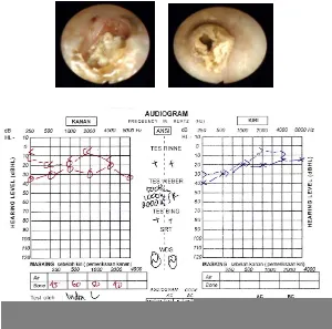
As for the right ear, the result of pure
tone audiometry showed mild degree
conduc-tive hearing loss (27,7 db), and both of the tympanometry was normal, while the left
ear within normal hearing threshold.
Computed tomography of the temporal bone did not suggest middle ear pathology,
no erosion in the temporal bone that was
compatible with osteoradionecrosis and showed an EACC as a soft-tissue mass in
the inferior EAC, with associated erosion of
Figure 1: Coronal temporal bone CT image shows an EACC as a soft-tissue mass in the inferior EAC, with associated erosion of the subajacent bone.
Figure 2: The right and the left external auditory canal
Figure 3: Pure tone audiometry
DISCUSSION
Osteoradionecrosis and cholesteatoma of
the external auditory canal following
external-beam radiotherapy as the treatment
of nasopharyngeal carcinoma is a rarely found
complication following a long post treatment
interval, in this case 2 years. Patients usually
present with chronic, offensive otorrhea and
occasionally otalgia.1-4,12
External auditory canal cholesteatoma
(EACC) is a rare entity with an estimated
occurrence of one in 1000 new patients at
otolaryngology clinics. Patients with EACC
typically present with chronic otorrhea and
dull pain due to the local invasion of
squamous tissue into the bony EAC. Most
cases are spontaneous or occur after trauma
ear-canal stenosis or obstruction has also
been reported to produce EACC. The clinical
differential diagnosis including neoplasms of
the EAC and inflammatory or infective
conditions such as keratosis obturans,
postinflammatory medial canal fibrosis, and
malignant otitis externa.7,8,12
A cholesteatoma is a cystic structure
lined by keratinizing stratified squamous
epithelium with associated periostitis and
bone erosion, which is most commonly
found in the middle ear cavity. Middle ear
cholesteatomas may be congenital or acquired,
but approximately 98% are acquired. Although
cholesteatomas are found almost
exclusi-vely in the middle ear and mastoid, in rare
cases they occur in the EAC. The estimated
incidence of EACC is 0.1–0.5% of all otologic
patients. Although EACC is rare, recognizing
it as a distinct entity is important because its
management is notably different from that of
its clinical differential diagnoses.7,9,10
The exact etiology of EACC is unclear.
Most cases are spontaneous or occur after
trauma in the auditory canal, although ear
canal stenosis or obstruction has also been
described as a causative factor. Trauma,
either canal skin lacerations, may isolate the
squamous epithelium or cause stenosis of the
canal; either of these events could lead to
EACC.1,7-11
Normal epithelial migration from the
tympanic membrane and EAC is an
impor-tant self-cleansing property of the outer ear.
Epithelial migration carries the keratin
debris laterally outward from the tympanic
membrane for removal. Spontaneous EACC
has been described as a disease of the
elderly due to the loss of normal migration
in aging epithelium of the canal wall.
Another pathophysiologic hypothesis suggests
that accumulation of keratin debris induces
changes of cellular proliferation in the
EAC.1,7
Other literatures described a lower than
average migratory rate of epithelium in the
inferior wall of the ear canal in cases of
EACC. The cause of EACC may be partly
related to abnormal epithelial migration,
which leads to the local accumulation of
squamous epithelium that can evolve into
an EACC. In addition, the decreased
migratory rate is thought to be related to a
poor blood supply.1,7
Imaging can be valuable in evaluation of
EACC. In temporal bone CT examination,
EACC is most commonly seen as an EAC
soft-tissue mass with associated bone erosion
and intramural bone fragments. The bone
erosion adjacent to the soft-tissue mass may
be smooth, similar to a middle ear
chole-steatoma; however, the erosion may be
irregular secondary to the necrotic bone and
periostitis.7,10
Patients with EACC usually present
with chronic otorrhea, dull pain and less
commonly, they present with hearing loss.
Gross pathologic analysis of EACC
demons-trates extensive erosion of the bony EAC by a
keratini-zing epithelial sac with a localized periostitis
and sequestration of bone. The tympanic
membrane is typically normal. The interface
between the EACC and the bone is erosive.
This erosion is thought to be related to
proteolytic enzymes along the margin of the
lesion produced within the cyst lining; these
weaken the bone and result in periostitis
and sequestration of bone. The erosion could
also be partly related to the accumulation of
keratin debris, which traps moisture and
results in a bacterial infection that can cause
ulceration of the epithelial layer and
granulation tissue formation in patients who
have a superimposed infection.7
In the diffuse type, a widespread
ischemic osteonecrosis involves the skull
base and adjacent structures. These patients
have usually received higher doses of
external irradiation to the temporal bone.
Severe otalgia and pulsatile, offensive
otorrhea are common. Cranial nerve palsies
might also be present. Diffuse
osteoradio-necrosis is associated with a recognized
incidence of local or regional complications,
such as suppurative labyrinthitis, trismus,
meningitis, cerebrospinal fluid leakage, and
internal carotid aneurysm.3-6
On otoscopic examination, EACC can be
difficult to distinguish from other
inflam-matory, infective, or neoplastic processes of
the EAC; examples of these include keratosis
obturans, postinflammatory medial canal
fibrosis, malignant otitis externa, and
squamous cell carcinoma (SCC). Keratosis
obturans is the most closely related condition
and the one most difficult to distinguish. In
fact, keratosis obturans was previously
considered to represent the same disease
process as EACC.7
The diagnosis is established on the
basis of the history and clinical examination
and audiometric findings. Imaging is usually
not performed for diagnosis. Surgical
mana-gement is the treatment of choice.
Interes-tingly, EACC has been described in
association with postinflammatory medial
canal fibrosis in rare cases.7,9,10
The management of osteoradionecrosis in
the external auditory canal by conservative
treatment with frequent aural toilet and
topical antibiotics is often administered for
localized osteoradionecrosis.4,6,12
Radiation exerts the 3 Hs of hypoxia,
hypovascularity and hypocellularity, impairs
normal collagen synthesis and cell production,
and leads to tissue breakdown and eventual
ORN. ORN of the external auditory canal
typically occurs in the lower portion of the
canal, which can be explained by the
down-ward pressure exerted by wearing a hearing
aid or picking at ear wax. The pressure may
also involve the perichondrium that carries
the main blood supply to the cartilage,
resulting in pressure necrosis. Then,
micro-organisms maypenetrate through the pressure
ulcer, leading to serious perichondritis and
chondritis.1,4-9
The principle of treating ORN is
cannot be revitalized and has to be surgically
removed.3 The lack of vascularized tissue
around the ORN site further complicates
treatment. Prevention of infection with
antibiotics and maintaining local hygiene by
frequent cleansing are also mandatory in
controlling the disease.3,4,8
The symptomatology of external auditory
canal ORN of purulent otorrhea, otalgia,
exposed necrotic bone and granuloma, may
mimic that of chronic otitis media, external
auditory canal cholesteatoma or ear
malig-nancy. The differentiation between ORN
and recurrent cancer might be difficult.2,5-9 NPC patients with external auditory
canal ORN typically present with foul odor
because of the sequestrum and thus may be
socially repelled. The symptoms of otorrhea
or otalgia, which mimic those of chronic
otitis media, may be neglected by patients.
The disease is also ignored by clinicians,
probably because of unawareness of the
disease entity.2,4-8
ORN involving the external auditory
canal is quite unusual in NPC patients. The
clinical presentations of otorrhea, otalgia and
crust formation are very similar to those of
chronic otitis media, external auditory canal
cholesteatoma or malignancy. A high
suspi-cion index is mandatory for early diagnosis.
The disease may lead to disastrous
complica-tions and should never be neglected by
clinicians. CT evaluation could help the
diagnosis by showing soft-tissue attenuating
mass in the EAC with erosion of adjacent
bone, and bone fragments are often present
within the mass. The management should be
by surgical evacuation of the pathological
entities.
REFERENCES
1. Po Hao S, Tsang Ming N, Chang Ping K, Chen Kuo C, Chao Chieh W. Osteoradionecrosis of external auditory canal in nasofaring carcinoma. Teipe: Departement of Otolaryngology, Chang Gung University College of Medicine, 2006. p. 116-20.
2. Patient information. Oncology directorate and haematology SDU radiotherapy side effects for benign head and neck conditions. Addenbrooke’s Hospital HNS, Cambrige University Hospitals NHS foundations Trust. 2006. p. 1-6.
3. Bhandare N, Antonelli PJ, Morris CG, Malayapa RS, Mendenhall WM. Ototoxicity after radio-therapy for head and neck tumors. Departement of Radiation Oncology, and Departement Otolaryngology University of Florida College of Medicine, Gainesville, FL. Elsevier Inc. USA. 2007. p. 469-78.
4. Low Kein W, Toh TS, Wee J, Chong SF, Wang DY. Sensorineural hearing loss after radio-therapy and chemoradio-therapy; A single, blinded, randomized study. J Clin Oncol 2006: 1904-8. 5. Amin MS, Meyers AD. External ear inflamatory
deseases. [Accesed 30 Maret 2012]. Available from:
http://emedicine.medscape.com/article/845990-overview#showall.
6. Owen HH, Rosborg J, Gaihede M. Choles-teatoma of the external ear canal: etiological factors, symptoms and clinical findings in a series of 48 cases. BMC Ear, Nose and Throat Disorders 2006. [Accesed 10 April 2012].
Available from: http://www.biomedcentral.com/1472-6815/6/16.
7. Heilbrun ME, Salzman KL, Glastonbury MG, Harsnberger HR, Kennedy RJ, Shelton C. External auditory canal cholesteatoma. Am J Neuroradiol 2003; 751–6.
8. Chihani M, Aljalil A, Touati M, Bouaity B, Ammar H. Post-traumatic cholesteatoma complicated by a facial paralysis: a case report. Department of Oto-Rhino-Laryngology, Avicenna Military Hospital, Marrakesh, Kingdom of Morocco. [Accesed 12 April 2012].
Available from: http://www/ajol.info/index.php/sajr/article/view
9. Lin SD, Pai CY, Nieh S. External auditory canal cholesteatoma. tri-service general hospital. National Defense Medical Center, Taipei, Taiwan, Republic of China. J Med Sci 2006: 231-4.
10. Rianto BU. Imunobiomolekuler kolesteatoma timpani. Bagian Ilmu Kesehatan Telinga Hidung Tenggorok, Fakultas Kedokteran Universitas Gadjah Mada Rumah Sakit Dr. Sardjito Yogyakarta. J ORLI 2009; 39(1):47-54
11. Ghazi B, Salima K, Salem T, Lamia O. Spontaneous external auditory canal cholestea-toma: Report of 3 cases. ENT Department, La Rabta Hospital, Tunis – Tunisi 2009. p. 383. 12. Loock JW. Osteoradionecrosis of the temporal
bone. In: Scott-Brown’s Otorhinolaryngology Head and Neck Surgery. 7th ed. Great Britain 2008. p. 3376-8.