J.Food Pharm.Sci. 4 (2016) 1 – 4
Avalaible online at www. jfoodpharmsci.com
Research Article
Effect of Green Tea Extract (Camellia Sinensis) with Nanocitosan
Capsulation on Gingivitis Healing
Annisa Hidaratri Uningojati
1, Dilla Asriyani
1, Urfa Tabtila
1, Fathul Muin
2, B. Nadya Kausara
2, Indra
Bramanti
31 Faculty of Dentistry, Gadjah Mada University, Yogyakarta 2 Faculty of Pharmacy, Gadjah Mada University, Yogyakarta
3 Pediatric Dentistry, Faculty of Dentistry, Gadjah Mada University, Yogyakarta
ARTICLE INFO ABSTRACT
Received 16/04/2016
Received in revised form 17/05/2016 Accepted 20/06/2016
Available online 02/01/2017
1. Introduction
Green tea (Camellia sinensis) is one of the plants with the highest flavonoids[1]. The flavonoids proven to have antiinflamatory, antibacterial and antioxidant activities. Flavonoids in green tea evidently can inhibit the growth of bacterial pathogens common periodontal disease like Porphyromonas gingivalis, Provotella
intermedia and Rovotella nigrescens. Flavonoids green
tea also shown bactericidal effect against gram negative anaerobic rod bacterial pigment black species P.
gingivalis and Provotella. Green tea has proven to have
anti-Streptococcus mutans activity. Gingivitis treatment using green tea polyphenols (EGCG) reduces the ability
of P. gingivalis to attach oral epithelial cells[2].
Chitosan is one of polisaccharides derived from Crustacean shells that mostly used as matrix of various drugs and plant extracts. Chitosan began to applied widely in the pharmaceutical, food and health industries. Chitosan have some characteristics include anti-microbial, wound healing, non-toxic, biocompatibility, degradable and also dissolves in water. Chitosan in micro/nanoparticel form have so many benefit that is non-toxic, stable during the use of high surface area, and can be the matrix of various drugs and plant extracts[3].
Inflamation on gingivitis was an acute inflamation. Early stage of gingivitis will increase vascular permeability that can lead to migration of leukocyt polymorphonuclear from vascular then caused edema. During inflamation, monocyt will migrates from Gingivitis is one of the high oral manifestations in Indonesia. Gingivitis can be cured by using green tea extract containing flavonoids. This flavonoid can be encapsulated with nanochitosan. This study aims to determine the effect of topical application of green tea extract gel with chitosan capsulation on gingivitis healing. The subjects of this study were 36 rats wistar white male rats. Rat was divided into 4 groups. Group I was given chicaflo gel (chitosan encapsulated flavonoid), group II was given green tea extract gel as comparison, group III was given base gel as negative control and group IV was given standard anti-inflammatory gel as positive control. The gel was applied to wounds in gingival mice that had previously been injured using a biopsy punch. On the 2nd, 5th and 7th days after gel application, 3 rats from each group were decapitated and jawed for histologic preparations. Furthermore, the number of inflammatory cells on the preparations of each rat group is computed and compared in number. The results showed a significant decrease in the number of inflammatory cells in chicaflo gel applications compared to controls. Our conclusion is that the use of chicaflo gel can improve the effectiveness of gingivitis healing.
peripheral blood vessel, then penetrates blood vessel wall to reach inflamatory tissue. This migrate monocyt will differentiate into macrophage in inflamatory tissue. Lesions of gingivitis later can be changed from initially only the presence of PMN leukocytes to the increase of limfocyt and macrophage[4]. Gingivitis prevalence in Indonesia ranks second, reach 96,58%[5].
Research using green tea extracts with nano-chitosan drug carrier for gingivitis treatment never be done before. This study used white male rats Wistar strain. This study was intended to determine the effect of green tea extract with nano-chitosan drug carrier in gingivitis therapy on proliferation stage. In addition, the results of this study was compared by Difflam Mouth Gel as positive control.
2. Materials and Methods
2.1. Material and Samples
Materials: grinder, rotary evaporator, magnetic stirrer, homogenizer, microscope, spectrophotometer, pH stick, scatter test kits, sticking power test kit, mortar, and stamper
Samples: dry green tea, chitosan, sodium tripolyphosphate, acetic acid, 70% ethanol, 95% NaCl, formalin buffer, CMC-Na, nipagin, nipasol, and aquadest.
2.2. Making Tea Extract
Preparation of tea extract was done using maseration technique. The dried green tea is smoothed using a grinder. The purpose of the treatment is to increase the particle surface area to increase the powder mixing with the solvent. Then, green tea powder is macerated with 70% ethanol solvent for 24 hours with stirring. The maserate is then filtered and the obtained filtrate is concentrated with a rotary evaporator to obtain a thick green tea extract.
2.3. Nanocitosan Capsule
The capsule of green tea extract into nanocitosan was done by ionic gelation method. Preparation of chitosan nanoparticles was done by dissolving chitosan 200 mg into 100 mL 1% acetic acid solution by using magnetic stirrer. Then, 2 grams of green tea extract dissolved in 100 mL chitosan solution with magnetic stirrer. Preparation of sodium tripolyphosphate solution was done by dissolving 40 mg of sodium tripolyphosphate with 40 mg of aquadest. After that, 1% sodium tripolyphosphate solution was poured gradually with 5000 rpm stirring using homogenizer for 30 minutes to form a nanoparticle suspension. Sodium tripolyphosphate will serve as a cross-linker. Nanoparticles chitosan-extract green tea then separated by way of centrifugation.
2.4. Gel Formulation
The green extract gel and CHICAFLO were prepared using some additives. The standard gel formula with CMC-Na base is as follows:
R / CMC-Na 2 ingredients are weighed, then inserted CMC-Na into the mortar, then added water and crushed until homogeneous and fluffy. After mixing with homogeny inserted nipagin and nipasol then re-crushed. The above treatments were replicated 2 times with the addition of the active ingredient, the first mortar inserted the green condensed extract, the second mortar incorporated the chitosan-encapsulated green tea extract (Chicaflo), and the third mortar was not added the active ingredient. Then each mortar is crushed to homogeny to form an elastic gel mass.
Furthermore, gel quality test in the form of homogeneity test, organoleptic test, spreading test, sticky power test, Ph.
2.5. Wound Healing Test
2.5.1.Preparing Animal
Try: The experimental animals used in this study were male white rats wistar strain aged 3 months heavily 200 grams. Total of 36 male white rats were divided into four treatment groups, each group consisted of 9 tails.
2.5.2.Animal Injury
Try: First try the animal injected ketamine. The gingivitis openings were performed with a 2 mm biopsy punch. The treatment group was divided into 4 groups:
1. Group I: Given application of green tea extract gel with nanocitosan capsulation (Chicaflo Gel)
2. Group II: Application of green tea extract gel 3. Group III: Application of gel without active
substance as a negative control
4. Group IV: Application of Difflam gel as a positive control.
Drug and Gel administration in experimental animals: Dosage is applied evenly on the wound area every 2 times a day for seven days.
Observation of wounds. Wound observations were performed on days 2, 5, and 7 by observing changes in the number of inflammatory cells and the proliferation process in wound healing including angiogenesis, epithelialization, and fibrosis.
Observation of wounds. Wound observations were performed on the 2nd, 5th, and 7th days by observing changes in the number of inflammatory cells and the proliferation process in wound healing including angiogenesis, epithelization, and fibrosis.
3. Results and Discussion
3.1. Research Result
In this study the data obtained will be tabulated and analyzed statistically with SPSS using two-way ANOVA method.
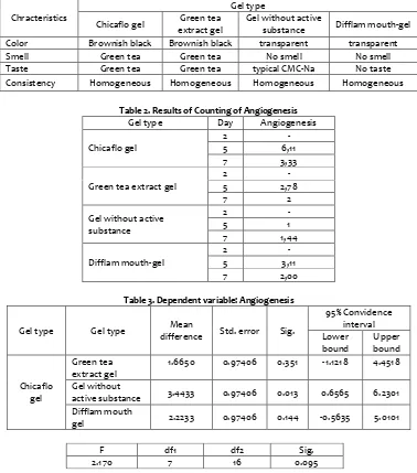
Table 1. Result of Gel Quality Test Color Brownish black Brownish black transparent transparent
Smell Green tea Green tea No smell No smell
Taste Green tea Green tea typical CMC-Na No taste
Consistency Homogeneous Homogeneous Homogeneous Homogeneous
Table 2. Results of Counting of Angiogenesis Gel type Day Angiogenesis
Chicaflo gel
difference Std. error Sig.
95% Convidence
active substance 3.4433 0.97406 0.013 0.6565 6.2301 Difflam mouth
gel 2.2233 0.97406 0.144 -0.5635 5.0101
3.2. Discussion
Based on the results of the study, it was shown that green tea extract gel with nanocitosan capsulation (Chicaflo) was able to stimulate the proliferative phase in wound healing process shown by the increasing amount of fibroblast, angiogenesis and epithelization. The proliferative phase consists of fibroplasia, granulation, epithelialization and angiogenesis that begins 24 hours after the wound[6]. During this process proliferation of fibroblasts and endothelial cells plays an important role in the occurrence of angiogenesis. This process is controlled by FGF, TGF-β and VEGF[7].
The process of healing the wound on the oral mucosa runs faster than the wound on the skin. This is due to injury to the oral mucosa containing immune mediators, blood vessels and less profibrotic mediators but has higher rates of re-epithelization and faster fibroblast proliferation than in skin[8].
Wound healing process is a process of restoring the structure of cells and tissue layers damaged in a complex and dynamic[9]. When injured, the physiological response of the body will automatically perform the process of
wound healing and cell regeneration automatically[10, 11]. Although this process runs natural and natural, but Required the existence of certain conditions that can accelerate the healing process, one of which is nutrition[12, 13]. Treatment of gingivitis using Chicaflo gel showed increased proliferative phase stimulation caused by nutrients contained in this gel have an influence on wound healing process.
Chicaflo gel made from green tea extract containing polyphenols flavonoids have the ability as antiinflamasi[14]. There are two ways of flavonoids in inhibiting inflammation, by inhibiting capillary permeability and inhibit acid metabolism arakidonat[15, 16]. Gingivitis treatment using the chicaflo gel proved able to stimulate wound more effectively. The results showed the increase of thickness of the epithelium in gingivitis treatment using a gel Chicaflo significant compared to the control treatment of gingivitis. This shows that Chicaflo gel can increase epithelialization.
Epithelialization thickness resulting in gingivitis therapy using Chicaflo gel showed epithelialization and wound closure process that is faster and shorter
F df1 df2 Sig.
2.170 7 16 0.095
inflammatory phase. Chicaflo gel contains flavonoids from green tea which works to improve the process of mitogenesis, cell interaction and adhesion molecules play an important role in the proliferative phase and epithelialization terhada healing process of scar tissue.
4. Conclusion
The conclusion of this study is that green tea extract with nanocitosan capsulation / Chicaflo gel (Chitosan encapsulated flavonoids) can improve the effectiveness of gingivitis healing.
Reference
[1] Oktaria, R., & Rahmanisa, S. 2016. Pengaruh
Epigallocatechin-3-Gallate (EGCG) pada Teh Hijau terhadap
Acne vulgaris. Jurnal Kedokteran Universitas Lampung. 7(2):
33.
[2] Shinde, G., Sheikh, S., Gupta, S., & Muglikar, S. 2015. Determination of the effect of white tea consumption in patients with generalized chronic gingivitis. Universal
Research Journal of Dentistry. 5(1):22.
[3] Rismana, E., Kusumaningrum, S., Bunga, O., Nizar, dan Marhamah. 2014. Pengujian Aktivitas Antiacne Nanopartikel Kitosan-Ekstrak Kulit Buah Manggis (Garcinia
mangostana). Media Litbangkes. 24 (1) : 19-27.
[4] Panagakos, F. S., dan Davies, R. M. 2011. Gingival Diseases -
Their Aetiology, Prevention and Treatment. Rijeka : InTech.
Hal. 107.
[5] Warongan, G., Wagey, F., & Mintjelungan, C. 2015. Gambaran Status Gingiva Pada Ibu Hamil Di Puskesmas Bahu Manado. Jurnal e-GiGi. 3(1) : 143-148.
[6] Morton,Laurel M. danPhillip, Tania J. 2016. Wound Healing and Treating Wounds. Differential diagnose and evaluation
of chronic wounds. University School of Medicine, Boston.
American Academy of Dermatology
[7] Larjava, Hannu. 2012. Oral Wound Healing : Cell Biology and
Clinical Management, Faculty of Dentistry, University of
British Columbia, Vancouver – Canada.
[8] Glim JE, Egmond MV, Niessen FB, Everts V, Beelen RH. Detrimental dermal wound healing: What can we learn from the oral mucosa. International Journal of tissue repair
and regeneration. 2013; 21(5): 648 660.
[9] Dewi IALP, Damriyasa IM, Dada IKA. 2013. Bioaktivitas Ekstrak Daun Tapak Dara(Catharanthus roseus) terhadap Periode Epitelisasi dalam Proses Penyembuhan Luka pada Tikus Wistar. Indonesia Medicus Veterinus. 2(1): 58 – 75. [10]Ferdinandez MK, Dada IKA, Damriyasa IM. Bioaktivitas
Ekstrak Daun Tapak Dara (Catharantus roseus) Terhadap Kecepatan Angiogenesis dalam Proses Penyembuhan Luka pada Tikus Wistar. Indonesia Medicus Veterinus 2013; 2(2): 180-190.
[11] Nurcahaya MI. Pengaruh ekstrak etanol lidah buaya (Aloe vera) terhadap peningkatan jumlah fibroblas pada proses penyembuhan luka mukosa rongga mulut tikus (Rattus
norvegicus) strain wistar. Skripsi. Surakarta: 2015. p. 35.
[12] Guo S. Dipietro LA. Factors Affecting Wound Healing.
Journal of Dental Research. 2010; 89(3): 219-229.
[13] Oroh CG, Pangemanan DHC, Mintjelungan CN. Efektivitas lendir bekicot (Achatina fulica) terhadap jumlah sel fibroblas pada luka pasca pencabutan gigi tikus wistar.
Jurnal e-GiGi (eG) 2015; 3(2): 515-519.
[14] Chairunnisa A. Pengaruh aplikasi ekstrak daun ceremai (Phyllanthus acidus (L.)) terhadap jumlah fibroblas pada hari ke-7. Skripsi. Banda aceh: 2015. p. 29.
[15] Fitriyani A, Winarti L, Muslichah S dan Nuri. Uji antiinflamasi ekstrak metanol daun sirih merah (Piper crocatum Ruiz & Pav ) pada tikus putih. Majalah Obat Tradisional 2011; 16(1): 34-42.
[16]Hidayati NA, Listyawati S, Setyawan AD. Kandungan kimia dan uji antiinflamasi ekstrak etanol Lantara camara L. pada tikus putih (Rattus Novergicus L.) jantan. Bioteknologi 2008; 5(1): 10-17.