Research Article
HIGH-THROUGHPUT SCREENING OF 75 EUPHORBIACEAE AND MYRTACEAE PLANT
EXTRACTS FOR IN-VITRO ANTITUMOR AND PRO-APOPTOTIC ACTIVITIES ON HUMAN
TUMOR CELL LINES, AND LETHALITY TO BRINE SHRIMP
MAY A. EL MANAWATY
1, WALID FAYAD
1, NABAWEYA M. EL-FIKY
2, GAMILA M. WASSEL
†1,
BASSEM S. EL-MENSHAWI
*31Drug Bioassay-Cell Culture Laboratory, Pharmacognosy Department, National Research Center, Dokki, Giza, 12622, Egypt, 2Pharmacognosy Department, Faculty of Pharmacy, Cairo University, Cairo 11562, Egypt, 3Drug Research Center, Future University, Fifth
Settlement, 90th Street, New Cairo 11835, Egypt.Email: [email protected]
Received: 11 Feb 2013, Revised and Accepted: 04 Apr 2013
ABSTRACT
Objective: Various approaches are used for discovering cytotoxic anticancer therapeutics from plants. In this study 75 extracts representing 47 plant species belonging to families Euphorbiaceae and Myrtaceae were screened by three in-vitro bioassays, to detect extracts with possible antitumor activity.
Methods: The methanol plant extracts were screened over three bioassays viz., cytotoxicity on HepG2 human hepatocellular carcinoma cell line assessed by MTT method, pro-apoptotic activity on HCT116 human colon carcinoma cell line measured by the increase of M30 neo-epitope apoptosis biomarker through sandwich ELISA, as well as brine shrimp lethality (BSL) bioassay. Results: 54 extracts were active in at least one bioassay. 43 extracts (57%) scored cytotoxic on HepG2, compared to 32 (43%) pro-apoptotic extracts in HCT116-M30 assay and 18 (24%) lethal extracts in BSL test. Out of the 43 cytotoxic extracts 15 (20%) possessed pro-apoptotic activity, which may be considered the ideal candidate agents. Conclusion: The relatively high number of active extracts detected in the 3 bioassays may be attributed to the selected families being rich in reported bioactivities and folk medicinal uses. The extracts of Melaleuca leucadendron L. and Callistemon rigidus R.Br. were considered the most promising ≥9 % toxicity in (epG cytotoxicity and ≥ fold increase in apoptotic background signal at ppm ; while the activity of C. rigidus is firstly to be recorded here and subsequent investigation for its active compounds is recommended. The three bioassay approach described could be useful in screening large numbers of extracts for detecting antitumor agents.
Keywords: Cytotoxicity, M30 apoptosense, HepG2 human hepatocellular carcinoma, HCT116 human colon carcinoma, Cancer therapeutics drug discovery
INTRODUCTION
Natural products have been considered as a valuable source of drug leads for many years[1]. They have been selected to interact with biological targets [2]. The high degree of their chemical diversity makes them attractive for discovery of new drugs[2]. The testing of their extracts has been widely practiced in the pharmaceutical industry[1].
Many species belonging to family Euphorbiaceae have been used as folk medicinal remedies to treat some skin disorders, diarrhea, dysentery, venereal diseases and as anthelmintic [3, 4]. Some of these species were reported to possess anti-proliferative, and antibacterial activities, as well as an effect against genital herpes (HSV-2) [5, 6]. Many of the species belonging to family Euphorbiaceae are known to be toxic and poisonous [5, 6] The main active principles found in this family are terpenoids [7], where specific diterpenes called phorboids are responsible for its toxicity [5, 6]. Family Myrtaceae also comprises several species that are used in folk medicine, mainly as an anti-diarrheal, antimicrobial, antioxidant, anti-rheumatic, anti-inflammatory and anti-hypercholesterolemic agents. This family is rich in essential oils [8].
Based on the above broad bioactivities of Euphorbiaceae and Myrtaceae, the present study aimed to screen some of their available species growing in Egypt for the following bioactivities: antitumor/cytotoxicity and pro-apoptotic effects on human tumor cell lines, in addition to lethality on brine shrimps. The general purpose is to test an approach for a systematic screening of large number of extracts in the search for therapeutics against tumors.
MATERIALS AND METHODS Plant material
Specimens of the available and accessible wild and cultivated plants, belonging to families Euphorbiaceae and Myrtaceae, were collected from different localities in Egypt. Voucher specimens of these plants,
viz. 32 Euphorbiaceae species and 15 Myrtaceae (cf. Table 1) were prepared and deposited in the Herbarium of the Pharmacognosy Department, National Research Center, Egypt. The taxonomical nomenclature is in accordance with Boulos (1999, 2000, 2002, 2005) [9-12] and Huxley et al. (1992) [13].
Preparation of extracts
The 47 plant species under investigation were separated into their organs which yielded 75 parts (cf. Table 1); these were dried in solar ovens at 45°C, powdered and kept in labelled containers. Sufficient weight of each plant powder was exhaustively extracted with methanol. The methanol extracts were filtered, evaporated under vacuum at 350C, freeze dried and stored at -20°C. The extraction and drying procedures during the preparation of the extracts were done cautiously to prevent de-naturation or artifact formation. The freeze dried plant extracts were deposited at the Extract Bank of the In-Vitro Bioassay Laboratory, National Research Center, Egypt.
In vitro antitumor bioassay on human hepatocellular carcinoma cell line (HepG2 cytotoxicity)
Cell culture—HepG2 cell line was maintained in RPMI-1640, 10% foetal bovine serum at 37°C in 5% CO2 and 95% humidity. Cells were sub-cultured using trypsin versene 0.15%.
Viability test—After about 24h of seeding 10000 cells per well (in 96-well plates), when cells have reached 60-70% confluence, the medium was changed to serum-free medium containing a final concentration of the extracts of 100 ppm in triplicates. The cells were treated for 24h. 100ppm adriamycin was used as positive control and 0.5% DMSO was used as a negative control. Cell viability was determined using the MTT [3-(4,5-dimethylthiazol-2-yl)-2,5-diphenyltetrazolium bromide] assay as described by Mosmann 1983 [14].
The equation used for calculation of percentage cytotoxicity:
(1-(av(x)/(av(NC)))*100
Where: Av: average, X: absorbance of sample well measured at 495nm with reference 690nm, NC: absorbance of negative control measured at 495nm with reference 690nm
This method is feasible for high throughput screening
Pro-Apoptosis bioassay (HCT116-M30 pro-apoptosis) Cell
culture—HCT116 colon carcinoma cell line was maintained in
McCoy`s 5A modified medium, 10% foetal calf serum at 37°C in 5% CO2 and 95% humidity. Cells were sub-cultured using trypsin versene 0.15%. Cells were checked for mycoplasma using DAPI staining.
Apoptosis induction test—Pro-apoptotic activity was assessed using the M30-apoptosense® kit according to the instruction of the manufacturer (Peviva AB, Bromma, Sweden). Test: Day 1: 10000 cells per well were seeded in a 96-well plate. Day 2: The cells were exposed to 100 ppm extract for 24 h. 40 µM cisplatin was used as positive control. μL of % NP were added per well. Lysis of the cells was allowed to occur on a rotatory shaker for 5 minutes at room temperature. Gentle mixing was done by pipetting up and down, taking care not to create air bubbles, then μL of the medium/lysate were added to the well of M30 Coated Microstrips [15, 16] 7 μL of the diluted M (RP horseradish peroxidase) Conjugate solution were added to each well (within 20 min from the addition of the cell lysate). The plate was sealed and incubated on shaker (600rpm) for 4 h. The plate was then washed five times with μL diluted wash solution. μL of TMB , ’, , ’ -Tetramethylbenzidine) were added to each well and incubation for min in the dark. μL of the stop solution . M sulphuric acid were added. Shaking for 5-10 seconds was done to ensure complete mixing. The absorbance was determined after 5 minutes at 450 nm in a microplate reader within 30 min.
Results are expressed as fold increase of the negative control (FINC) background caused by spontaneous apoptosis according to the following formula: FINC= OD450 (of test extract)/OD450 (of negative control), Where OD450 is the optical density obtained at 450 nm wavelength. 0.5% DMSO was used as negative control. This method is feasible for high throughput screening
Brine shrimp lethality bioassay (BSL)
A solution of instant ocean sea salt was prepared by dissolving 2.86g in 75ml distilled water. 50mg of Artemia salina eggs were added in a hatching chamber[17]. The hatching chamber was kept under a fluorescent bulb for 48h for the eggs to hatch into shrimp larvae. 20mg of each extract were dissolved in 2ml methanol, from which
50µl were transferred into vials corresponding to 100 ppm. The methanol was allowed to evaporate to dryness in about 24h at room temperature. 2.5ml of instant ocean solution containing one drop of DMSO were added to each vial and 10 nauplii of Artemia salina were added to each vial. The final volume of the solution was adjusted to 5ml with sea salt solution immediately after adding the nauplii. 24 h later, the number of surviving shrimps were counted and recorded [18]. The experiment was done in triplicates. 100ppm of Annona
cherimolia leaves extract was used as a positive control. 0.5% DMSO
was used as negative control.
The equation used for calculation of percentage lethality: Percentage
lethality = [10 – (( S1+S2+S3)/3)] * 100, Where S1, S2, S3 are the number of viable nauplii in the sample vials.
Determination of LC values
In case of highly active extracts possessing ≥90% cytotoxicity on HepG2 cells and those giving ≥90% lethality on BSL, five descending concentrations were prepared and tested. The results were used to calculate the LC50 values of each extract using Probit analysis and utilizing the SPSS computer program (SPSS for windows, statistical analysis software package / version 9 / 1989 SPSS Inc., Chicago, USA).
RESULTS
In this investigation an extract was counted as active when possessing (scoring) >50% in HepG2 cytotoxicity, >50% lethality in BSL, or inducing >1.5 fold increase in the apoptotic signal of the negative control in the HCT116-M30 pro-apoptosis.
The results obtained in the three bioassays are expressed in Table (1) and showed that 54 extracts were active in either one or more of the three bioassays. A summary of the distribution of the number of active extracts in the three bioassays is shown in Figure (1). 43 extracts were cytotoxic to HepG2 compared to 32 pro-apoptotic extracts on HCT116-M30 pro-apoptosis and 18 lethal extracts in BSL assay. Only 2 extracts (viz. numbers 5 and 9) out of the 18 lethal extracts in BSL, were inactive in the other two bioassays. BSL assay detected 14 active extracts out of the 43 cytotoxic extracts on HepG2.
Highly active extracts, which gave ≥90% cytotoxicity or lethality, possessed LC50 values ranged between 10-40 and 41-91 ppm for HepG2 cytotoxicity and BSL, respectively (cf. Tables 2, 3). This might be interpreted by higher sensitivity of the HepG2 model compared to BSL bioassay.
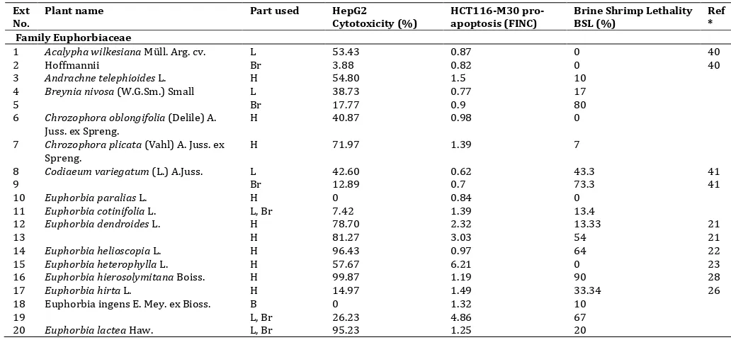
Table 1: Activity of plant extracts (100 ppm) in three bioassays: HepG2 cytotoxicity, HCT116 pro-apoptosis and brine shrimp lethality Ext
1 Acalypha wilkesiana Müll. Arg. cv.
Hoffmannii
6 Chrozophora oblongifolia (Delile) A.
Juss. ex Spreng.
H 40.87 0.98 0
7 Chrozophora plicata (Vahl) A. Juss. ex
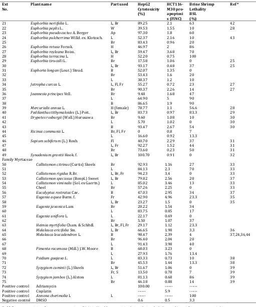
Table 1 (Contd.): Activity of plant extracts (100 ppm) in three bioassays: HepG2 cytotoxicity, HCT116 pro-apoptosis and brine shrimp lethality Ext
No.
Plant name Part used HepG2
Cytotoxicity (%)
HCT116-M30 pro-apoptosi s (FINC)
Brine Shrimp Lethality BSL (%)
Ref *
21 Euphorbia neriifolia L. L, Br 89.25 2.1 63 42
22 Euphorbia peplis L. W 39.53 1.55 10 28
23 Euphorbia pseudocactus A. Berger Ap 97.30 1.8 60
24 Euphorbia pulcherrima Willd. ex. Klotzsch. L 52.37 2.16 10 43
25 Br 83.43 0.96 20
26 Euphorbia retusa Forssk. H 46.97 2 86
27 Euphorbia royleana Boiss. L, Br 59.47 3.68 70
28 Euphorbia terracina L. H 52.20 0.75 100
29 Euphorbia tirucalli L. Br 17.58 1.06 0 25
30 L, Br 93.17 0.48 37 25
31 Euphoria longan (Lour.) Steud. B 52.07 1.35 0
32 Br 53.43 1.6 20
33 L 38.37 1.2 10
34 Jatropha curcas L. L, Fl, Fr 55.27 0.72 23 27
35 Br 90.37 2.26 14 27
36 Joannesia principes Vell. Br 9.48 1.68 47
37 L 64.90 5 90
38 B 86.65 1.9 90
39 Mercurialis annua L. H (female) 78.77 1.1 56.6 28
40 Pedilanthus tithymaloides (L.) Poit. L, Br 83.73 0.97 83.3 29
41 Drypetes roxburgii (Wall.) Hurusawa Br 9.60 1.08 10 30
42 L 5.70 1.02 0 30
43 B 93.47 2.67 54 30
44 Ricinus communis L. Br, Fl, Fr 0 0.8 7
45 L 16.60 0.92 13.3
46 Sapium sebiferum (L.) Roxb. Fl 40.70 2.29 37 31
47 L, Fr 92.27 1.52 44 31
48 Br 73.60 0.23 50 31
49 Synadenium grantii Hook. f. L, Br 100.70 0.91 0 32
Family Myrtaceae
50 Callistemoncitrinus (Curtis) Skeels Br 92.93 1.16 27 33
51 L 84.13 2.3 70 33
52 Callistemon rigidus R.Br. L, Br, Fr 94.23 3.4 0 33
53 Callistemonspeciosus (Bonpl.) Sweet L, Br 79.82 2.56 20 37
54 Callistemon viminalis (Sol. ex Gaertn.)
Cheel
L 85.02 3.46 13 33
55 Br 57.26 2.25 0 33
56 Eucalyptus rostratus Cav. B 47.03 2.95 34 37
57 Eugenia aquea Burm. f. Fr 42.90 4.96 23.3 35
58 L, Br 23.27 1.5 0 35
59 Eugenia javanica Lam Br 20.22 1.54 34
60 L 83.75 0.85 17
61 Eugenia uniflora L. L 22.17 0.69 0
62 Br 5.50 1.07 37
63 Heimia myrtifolia Cham. & Schltdl. L, Br, Fl, Fr 29.17 1.12 23.3
64 Melaleuca ericifolia Sm. L, Br 66.65 1.98 3.3 36
65 Melaleuca leucadendron L. B 98.47 2.39 4 37,24,36,44
66 Br 96.40 2.04 20
67 L 91.43 3.98 40
68 Pimenta racemosa (Mill.) J.W. Moore B 68.03 3.23 0
69 L 27.93 1.76 13.4
70 Psidium guajava L. L 83.33 0.73 10 38
71 Br 45.53 1.44 13.3 38
72 Syzygium cuminii (L.) Skeels L, Br 51.67 1.06 0 39
73 Fr, S 13.50 0.78 7 39
74 Syzygium jambos (L.) Alston L 81.13 0.68 86 39
75 Br 46.18 0.88 14 39
Positive control Adriamycin 100.00 --- ---
Positive control Cisplatin --- 5.9 ---
Positive control Annona cherimolia L L --- --- 100
Negative control DMSO 0.6 0.5 3.3
Bold figures = activity of extracts possessing >50 % in HepG2 cytotoxicity, brine shrimp lethality, and/or >1.5 in pro-apoptosis bioassays. FINC: fold increase of negative control.
Parts: Ap: Aerial part; B: Bark; Br: Branches; Fl: Flowers; Fr: Fruits; H: Herb; L: Leaves; R: Root system; S: Seeds; St: Stem; W: Weed.
Fig. 1: Illustrative scheme showing the distribution of the numbers of active extracts in the three bioassays (cf. Table 1).
Table 2: LC50values of extracts inducing ≥ 9 percent in HepG cytotoxicity
Extract number Plant name Part used *LC50 (ppm)
14 Euphorbia helioscopia L. H 30.7 (±2.25)
16 Euphorbia hierosolymitana Boiss. H 26.8 (±1.34)
20 Euphorbia lactea Haw. L, Br 25(±2.26)
23 Euphorbia pseudocactus A. Berger Ap 19.4 (±1.06)
30 Euphorbia tirucalli L. L, Br 23.4 (±1.57)
35 Jatropha curcas L. Br 35.2 (±4.56)
43 Drypetes roxburgii (Wall.) Hurusawa B 10 (±1.91)
47 Sapium sebiferum Roxb. L, Fr 33.4 (±4.63)
49 Synadenium grantii Hook. F. L, Br 40.3 (±3.35)
50 Callistemoncitrinus (Curtis) Skeels Br 22.2 (±0.52)
52 Callistemon rigidus R.Br. L, Br, Fr 40 (±2.6)
65 Melaleuca leucadendron L. B 32 (±4.56)
66 Br 27 (±1.91)
67 L 14 (±2.05)
Positive control Adriamycin 21.6 (±1.24)
Parts: Ap: Aerial part; B: Bark; Br: Branches, Fr: Fruits, H: Herb, L: Leaves, * Confidence limit = 95%
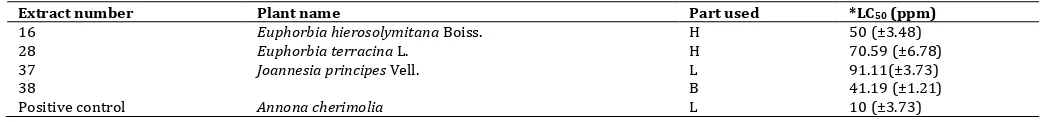
Table 3: LC50values of extracts inducing ≥ 9 percent in brine shrimp lethality
Extract number Plant name Part used *LC50 (ppm)
16 Euphorbia hierosolymitana Boiss. H 50 (±3.48)
28 Euphorbia terracina L. H 70.59 (±6.78)
37 Joannesia principes Vell. L 91.11(±3.73)
38 B 41.19 (±1.21)
Positive control Annona cherimolia L 10 (±3.73)
Parts: B: Bark; H: Herb, L: Leaves. , * Confidence limit = 95% 23 extracts were active in both HepG2 cytotoxicity and HCT116-M30 pro-apoptosis assays, indicating that the cell death mode induced by these extracts is apoptosis. However, 20 extracts were cytotoxic on HepG2 but not inducing apoptosis in HCT116. This could be explainable by the death mode, induced by these extracts at the specified concentration, to be other than apoptosis and most likely necrosis. A similar observation was previously reported [19]. On the other hand, 9 extracts were pro-apoptotic, but not cytotoxic on HepG2. These extracts might be late acting, and if incubated for longer time (e.g. 72-96 hours) they might have shown cytotoxic activity [16].
DISCUSSION
The present study utilized three different in vitro bioassays to screen 75 plant extracts, representing 47 species belonging to families Euphorbiaceae and Myrtaceae, to predict their possible anticancer activity. The three bioassays used were: i- cytotoxicity on HepG2 human hepatocellular carcinoma cell line assessed by MTT method (HepG2 cytotoxicity), ii- pro-apoptotic activity on HCT116 human colon carcinoma cell line, quantified by the M30-apoptosense® kit (HCT116-M30 pro-apoptosis), and iii- brine shrimp lethality assay (BSL). All extracts were screened at 100 ppm in the three bioassays and activity was assessed after 24 hours.
Comparing the results obtained (cf. Table 1) with the literature available on the species examined, it is apparent that many of the bio-active extracts detected were previously reported to possess related bio-activities, cf. references [20-44] (cf. Table 1). The most prominent genera relevant to the literature were Euphorbia,
Callistemon and Melaleuca.
The results obtained in the BSL assay may lead to the conclusion that BSL is a useful simple tool that could be combined with other bioassays to detect cytotoxicity in large number of plant extracts [45]. However, false negative results can occur. Further studies with larger number of extracts and/or other human tumor cell lines may be recommended to reach a validated correlation.
cytoskeleton component cytokeratin-18 by caspases during apoptosis[48].
Out of the 23 extracts which scored positive in both assays (HepG2 cytotoxicity and HCT116-M30 pro-apoptosis), the extracts of
Melaleuca leucadendron L. and Callistemon rigidus R.Br. (cf. extract anticancer and pro-apoptotic activities, and accordingly may be considered the most promising active in the present studyand is recommended for further chemical and biological investigations. The three bioassay approach described in this study appears to be Center Karolinska, Department of Oncology and Pathology, Karolinska Institute and Hospital, Stockholm, Sweden.
The plant species were authenticated by Professor LoutfyBoulos, Faculty of Science, Alexandria University, Egypt and by Mme Therese Labib, Agronomist, Orman Garden, Dokki, Cairo. The ASRT-Egypt funded project Biotechnological methods for drug discovery has provided the bioassay facilities.
Note: Our Co-Author Prof. Dr. Gamila M. Wassel† was deceased in
25 Nov, 2012.
REFERENCES
1. Shu, Y.Z., Recent natural products based drug development: a
pharmaceutical industry perspective. J Nat Prod, 1998. 61(8): p.
1053-71.
2. Koehn, F.E. and G.T. Carter, The evolving role of natural
products in drug discovery. Nat Rev Drug Discov, 2005. 4(3): p.
206-20.
3. Perumal Samy, R., S. Ignacimuthu, and A. Sen, Screening of 34
Indian medicinal plants for antibacterial properties. J
Ethnopharmacol, 1998. 62(2): p. 173-82.
4. Taylor, R.S., F. Edel, N.P. Manandhar, and G.H. Towers,
Antimicrobial activities of southern Nepalese medicinal plants. J Ethnopharmacol, 1996. 50(2): p. 97-102.
5. Hohmann, J. and J. Molnar, [Euphorbiaceae diterpenes: plant toxins or promising molecules for the therapy?]. Acta Pharm Hung, 2004. 74(3): p. 149-57.
6. Betancur-Galvis, L.A., G.E. Morales, J.E. Forero, and J. Roldan,
Cytotoxic and antiviral activities of Colombian medicinal plant
extracts of the Euphorbia genus. Mem Inst Oswaldo Cruz, 2002.
97(4): p. 541-6.
7. Rakshit K. Devappa, H.P.S.M., Klaus Becker, Jatropha Diterpenes: a Review. J Am Oil Chem Soc, 2010. 88(3): p. 301-22.
8. Stefanello, M.E., A.C. Pascoal, and M.J. Salvador, Essential oils from neotropical Myrtaceae: chemical diversity and biological properties. Chem Biodivers, 2011. 8(1): p. 73-94.
9. Boulos, L., Flora of Egypt : Azollaceae-Oxalidaceae. 1999, Cairo, Egypt: Al Hadara Publ. . Society (Great Britain), The new Royal Horticultural Society
dictionary of gardening. 1992, London: Macmillan Press.
14. Mosmann, T., Rapid colorimetric assay for cellular growth and survival: application to proliferation and cytotoxicity assays. J Immunol Methods, 1983. 65(1-2): p. 55-63.
15. Hagg, M., K. Biven, T. Ueno, L. Rydlander, P. Bjorklund, K.G. Wiman, et al., A novel high-through-put assay for screening of
pro-apoptotic drugs. Invest New Drugs, 2002. 20(3): p. 253-9.
16. El-Menshawi, B.S., W. Fayad, K. Mahmoud, S.M. El-Hallouty, M. El-Manawaty, M.H. Olofsson, et al., Screening of natural products for therapeutic activity against solid tumors. Indian J Exp Biol, 2010. 48(3): p. 258-64.
17. Meyer, B.N., N.R. Ferrigni, J.E. Putnam, L.B. Jacobsen, D.E. Nichols, and J.L. McLaughlin, Brine shrimp: a convenient general
bioassay for active plant constituents. Planta Med, 1982. 45(1):
p. 31-4.
18. Fatope, M.O., Ibrahim H. and Takeda, Y. , Screening of Higher Plants Reputed as Pesticides Using the Brine Shrimp Lethality Assay. Int. J. Pharmacog., 1993. 31(4): p. 250-4.
19. Herrmann, R., W. Fayad, S. Schwarz, M. Berndtsson, and S. Linder, Screening for compounds that induce apoptosis of cancer cells grown as multicellular spheroids. J Biomol Screen, 2008. 13(1): p. 1-8.
20. Baloch, I.B. and M.K. Baloch, Isolation and characterisation of new bio-active compounds from Euphorbia cornigera: cytotoxic
ingenol esters. Nat Prod Res, 2012. 26(20): p. 1857-63.
21. Aljancic, I.S., M. Pesic, S.M. Milosavljevic, N.M. Todorovic, M. Jadranin, G. Milosavljevic, et al., Isolation and biological evaluation of jatrophane diterpenoids from Euphorbia dendroides. J Nat Prod, 2011. 74(7): p. 1613-20.
22. Lu, Z.Q., S.H. Guan, X.N. Li, G.T. Chen, J.Q. Zhang, H.L. Huang, et al., Cytotoxic diterpenoids from Euphorbia helioscopia. J Nat Prod, 2008. 71(5): p. 873-6.
23. Flores JS, R.R., The secretions and exudates of plants used in
mayan traditional medicine. J Herbs Spices Med Plants, 1996. 4:
p. 53-9.
24. Evandri, M.G., L. Battinelli, C. Daniele, S. Mastrangelo, P. Bolle, and G. Mazzanti, The antimutagenic activity of Lavandula angustifolia (lavender) essential oil in the bacterial reverse
mutation assay. Food Chem Toxicol, 2005. 43(9): p. 1381-7.
25. Rishikesh S Babar, U.P.K., Nitin N Mali, Sandeep B Patil, Nilofar S Naikwade, In-vitro cytotoxicity activity of Euphorbia hirta, Euphorbia tirucalli and Euphorbia neriifolia extract against
B16F10 melanoma cell line. Inventi Impact:
Ethnopharmacology, 2012. 2012: p. http://www.inventi.in/Article/pep/655/12.aspx. .
26. Aylward, J.H., Anti-cancer compounds 2004, Peplin Biotech Pty. Ltd. (Fortitude Valley, AU) USA.
27. Lin, J., F. Yan, L. Tang, and F. Chen, Antitumor effects of curcin
from seeds of Jatropha curcas. Acta Pharmacol Sin, 2003. 24(3):
p. 241-6.
28. Abu-Dahab, R.a.A., F. , Antiproliferative activity of selected medicinal plants of Jordan against a breast adenocarcinoma cell line (MCF7) Sci. Pharm., 2007. 75: p. 121-36.
29. Mongkolvisut, W. and S. Sutthivaiyakit, Antimalarial and antituberculous poly-O-acylated jatrophane diterpenoids from Pedilanthus tithymaloides. J Nat Prod, 2007. 70(9): p. 1434-8. 30. Raghavendra HL, K.T., Valleesha NC, Sudharshan SJ and
Chinmaya A. , Screening for Cytotoxic activity of Methanol Extract of Putranjiva roxburghii Wall (Euphorbiaceae) Seeds.
Phcog J, 2010. 2(10): p. 335-7.
31. Liu, H.B., H. Zhang, J.H. Yu, C.H. Xu, J. Ding, and J.M. Yue,
Cytotoxic diterpenoids from Sapium insigne. J Nat Prod, 2012. 75(4): p. 722-7.
32. Hassan, M.E.M., M. M. D and Mohamed, S.M., Two New Phorbol-Type Diterpene Esters from Synadenium grantii Hook F. Leaves.
Rec. Nat. Prod., 2012. 6(3): p. 255-62.
33. Niaz, A.G., A.; Syed, W. A. S.; Ismail, S.; Mehreen, G. and Imran, K., Acute toxicity, brine shrimp cytotoxicity and relaxant activity
of fruits of callistemon citrinus curtis. BMC Complementary and
Alternative Medicine, 2011, . 11: p. 99-107.
34. Kajangwe, V., J.C. Chalchat, J. Rutayisire, A. Ndagijimana, M.J. Mukazayire, and P. Duez, Chemical composition and antibacterial activity of the essential oil of Callistemon speciosus (Sims) DC.
growing in Rwanda. Planta Med, 2008. 74(09): p. PI19.
35. Subarnas, A., A. Diantini, R. Abdulah, A. Zuhrotun, C. Yamazaki, M. Nakazawa, et al., Antiproliferative activity of primates-consumed plants against MCF-7 human breast cancer cell lines
36. Abdel Bar, F.M., A.M. Zaghloul, S.V. Bachawal, P.W. Sylvester, K.F. Ahmad, and K.A. El Sayed, Antiproliferative triterpenes from Melaleuca ericifolia. J Nat Prod, 2008. 71(10): p. 1787-90. 37. Aggarwal, B.B., A. Bhardwaj, R.S. Aggarwal, N.P. Seeram, S.
Shishodia, and Y. Takada, Role of resveratrol in prevention and therapy of cancer: preclinical and clinical studies. Anticancer Res, 2004. 24(5A): p. 2783-840.
38. Chen, K.C., C.L. Hsieh, C.C. Peng, H.M. Hsieh-Li, H.S. Chiang, K.D. Huang, et al., Brain derived metastatic prostate cancer DU-145 cells are effectively inhibited in vitro by guava (Psidium gujava L.) leaf extracts. Nutr Cancer, 2007. 58(1): p. 93-106.
39. Cock, I.E., Antimicrobial Activity of Syzygium australe and
Syzygium leuhmannii Leaf Methanolic Extracts. Pharmacognosy
Communications, 2012. 2(2): p. 71-77.
40. Lim, S.W., K.N. Ting, T.D. Bradshaw, N.A. Zeenathul, C. Wiart, T.J. Khoo, et al., Acalypha wilkesiana extracts induce apoptosis by
causing single strand and double strand DNA breaks. J
Ethnopharmacol, 2011. 138(2): p. 616-23.
41. Abdul Manaf Ali, M.M.M., Saleh H. El-Sharkawy, Junainah Abdul Hamid, Nor Hardiani Ismail, Faujan Ahmad and Nordin Lajis,
Antiviral and Cytotoxic Activities of Some Plants Used in Malaysian Indigenous Medicine. Pertanika J. Trop. Agric. Sci, 1996 19(2/3): p. 129-36.
42. Papiya Bigoniya and Rana, A.C., Radioprotective and in-vitro cytotoxic sapogenin from Euphorbia neriifolia (Euphorbiaceae)
leaf. Tropical Journal of Pharmaceutical Research, 2009. 8(6): p. 521-30
43. Smith-Kielland, I., J.M. Dornish, K.E. Malterud, G. Hvistendahl, C. Romming, O.C. Bockman, et al., Cytotoxic triterpenoids from the leaves of Euphorbia pulcherrima. Planta Med, 1996. 62(4): p. 322-5.
44. Wolter, F., A. Clausnitzer, B. Akoglu, and J. Stein, Piceatannol, a natural analog of resveratrol, inhibits progression through the S phase of the cell cycle in colorectal cancer cell lines. J Nutr, 2002. 132(2): p. 298-302.
45. Taksim Ahmed, M.N.U., MD. Kamal Hossain, Nahid Hasan, MD. Sohel Rana, Evaluation of antioxidant and cytotoxic potinetial of Artocarpus chama Buch. seeds using in vitro models.
International Journal of Pharmacy and Pharmaceutical Sciences, 2013. 5(1): p. 283-289.
46. Vijayabaskaran M., V.N., Arif Pasha MD., Babu G, Sivakumar P, Perumal P, Jayakar B, Invitro cytotoxic effect of ethanolic extract
of Pseudarhtria viscida Linn. International Journal of Pharmacy
and Pharmaceutical Sciences, 2010. 2(3): p. 93-94.
47. Kaufmann, S.H. and W.C. Earnshaw, Induction of apoptosis by
cancer chemotherapy. Exp Cell Res, 2000. 256(1): p. 42-9.