Contents lists available atScienceDirect
International Biodeterioration & Biodegradation
journal homepage:www.elsevier.com/locate/ibiod
E
ff
ect of biological colonization on ceramic roo
fi
ng tiles by lichens and a
combined laser and biocide procedure for its removal
J. Pena-Poza
a, C. Ascaso
b,∗∗, M. Sanz
c, S. Pérez-Ortega
d, M. Oujja
c, J. Wierzchos
b,
V. Souza-Egipsy
e, M.V. Cañamares
e, M. Urizal
f, M. Castillejo
c, M. García-Heras
a,∗aInstituto de Historia (CCHS-CSIC), Calle Albasanz 26-28, 28037 Madrid, Spain
bMuseo Nacional de Ciencias Naturales (MNCN-CSIC), Calle Serrano 115bis, 28006 Madrid, Spain cInstituto de Química Física Rocasolano (IQFR-CSIC), Calle Serrano 119, 28006 Madrid, Spain dReal Jardín Botánico (RJB-CSIC), Plaza Murillo 2, 28014 Madrid, Spain
eInstituto de Estructura de la Materia (IEM-CSIC), Calle Serrano 121, 28006 Madrid, Spain fThor Especialidades SA, Barcelona, Spain
A R T I C L E I N F O
Keywords: Biodeterioration Ceramic roofing tiles Lichens
Removal procedures Laser irradiation Biocide Acticide®CF
A B S T R A C T
Biodeterioration damage is an important issue in conservation and restoration of built heritage, especially when ceramic materials are used. Biological colonization of ceramic roofing tiles by lichens is a common phenomenon. However, there are no reports to date of lichens removal from unglazed roofing tiles for conservation purposes.
This paper for thefirst time reveals the results of a combined procedure undertaken to assess the removal of
lichens on different kinds of unglazed ceramic roofing tiles by treatments based on both dual sequential laser irradiation and treatment using Acticide®CF biocide. Three species of lichens were identi
fied:Verrucaria ni-grescens,Calogaya decipiensandPyrenodesmia teicholyta. The chemical and mineralogical composition of roofing
tiles were characterized by X-rayfluorescence (XRF) spectrometry, optical polarized petrographic microscopy,
and X-ray diffraction (XRD). Laser irradiation was accomplished by applying sequences of nanosecond laser
pulses at two wavelengths (1064 and 266 nm). After dual sequential laser irradiation a biocide was applied. To assess the combined effect of both treatments several techniques were used, including stereo andfluorescence
(FM) microscopies, scanning (SEM) and transmission (TEM) electron microscopies, and FT-Raman spectroscopy. Chemical composition of the analyzed roofing tiles was shown as a relevant factor regarding the degree of interaction between the biological colonization and the substrate, and hence, the bioweathering effect. The
combined procedure has proved to be very effective in ablating cortical layers in all species, or even complete
areolae inV. nigrescens, enhancing biocide effect in the thalli ofC. decipiensandP. teicholyta, and producing the complete damage of both bionts.
1. Introduction
A wide biodiversity has been recognized in architectural ceramic materials such as bricks, roofing tiles and glazed wall tiles. This bio-diversity very often causes biodeterioration on these materials which in turn induce not only aesthetic but also chemical and physical damage. Decay by biodeterioration is an important issue in conservation and restoration of built heritage, especially when ceramic materials were used (Warren, 1999).
Different types of biological colonization have been identified in such materials, from microorganisms to plants, including bacteria, cy-anobacteria, algae, lichens and fungi (Coutinho et al., 2015). Regarding
biodeterioration damage, bricks are the most studied type of archi-tectural ceramic material (Wang et al., 2011), while roofing (Gazulla et al., 2011) and glazed wall tiles (Coutinho et al., 2013) are hardly investigated. In addition, biodeterioration of stained glass windows have been also reported (Carmona et al., 2006). Studies focused on the impact of the lichen thalli on a monumental stone substrate have re-vealed a high bioreceptivity of rocks to lichen colonization depending on rock surface roughness, porosity, and capillarity, as well as a strong incidence of lichens in the biodeterioration of stone built heritage (de los Ríos et al., 2004; 2009).
Ceramic roofing tiles have been traditionally elaborated from a composition very similar to that used for bricks, that is, an uncoated or
http://dx.doi.org/10.1016/j.ibiod.2017.10.003
Received 23 December 2016; Received in revised form 6 October 2017; Accepted 10 October 2017 ∗Corresponding author.
∗∗Corresponding author.
E-mail addresses:[email protected](C. Ascaso),[email protected](M. García-Heras).
unglazed body mainly composed of illitic-kaolinitic clays with quartz and a variable amount of carbonates which is subsequently fired at low/medium temperatures. The main function of roofing tiles is to protect the building against rain and sun and, due to this reason they are commonly exposed to weathering and biological agents which later may cause biodeterioration. Fired clay roofing tiles werefirst used in Egypt and other ancient civilizations such as Babylon. However, Romans were probably the ancient people which more extensively roofed their buildings. Although the technique of glazed roofing tiles was already known by Babylonians it was widely spread later by the Islamic influence in medieval times (Campbell and Pryce, 2003).
Biological colonization of ceramic roofing tiles by lichens is a fre-quent and common phenomenon. However, this kind of colonization over the ceramic roofing tiles has been scarcely described (Kiurski et al., 2005). It affects not only the aesthetic aspects of the building roofs but also roof functionality can be affected by lichens cover (Laiz et al., 2006). For this reason any study related to the removal of lichens from roofing tiles may be of significant importance, even for roofing tiles conservation purposes. Removal procedures of biofilms composed by algae, cyanobacteria and fungi with application of four biocides have been evaluated, but in glazed majolica tiles, byCoutinho et al. (2016). However, the process of lichens eradication from ceramic roofing tiles has never been reported up to date.
In this study it is hypothesized that biological weathering (lichen thalli) of ceramic roofing tiles made by traditional methods of ela-boration depends on the characteristics of the substrate (uncoated or unglazed ceramic). The usefulness of combined treatments using dual sequential laser irradiation and biocides is further assessed in order to help decision making in conservation and restoration of historical buildings in which ancient or traditional ceramic roofing tiles need to be necessarily preserved.
Laser cleaning is a well-established technique because it provides fine and selective removal of superficial deposits and encrustations such as biological and black crusts on stone substrates (Cooper, 1998; Maravelaki-Kalaitzaki et al., 2003; Tornari et al., 2006). In the case of lichen elimination, laser cleaning constitutes a promising alternative to more conventional cleaning techniques (de Cruz et al., 2009; Speranza et al., 2013; Osticioli et al., 2015). This technique has been also used for the elimination of dark deposits originated from air pollution on ter-racotta substrates, with close composition to ceramic roofing tiles (Oujja et al., 2005).
2. Materials and methods
2.1. Ceramic roofing tile samples
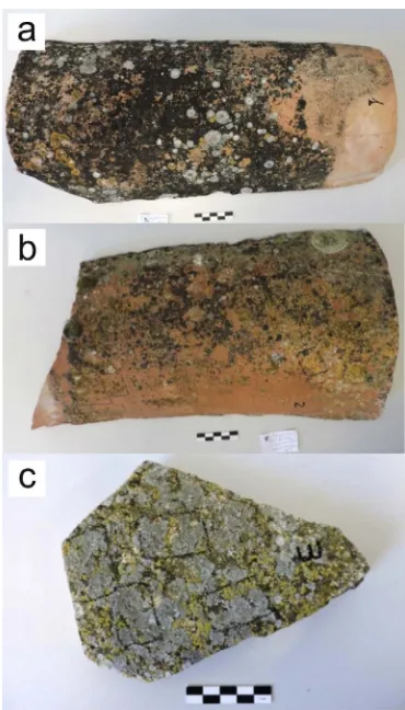
Three unglazed ceramic roofing tiles coming from Segovia (samples 1 and 2) and Guadalajara (sample 3) provinces located in the center of Spain under a mesothermal climate were selected to undertake this research. All of them belonged to traditional rural built heritage and were elaborated following traditional methods. They have been natu-rally exposed to both weathering and biological agents for years and all showed an extensive biological colonization on their surfaces (Fig. 1). Ceramic body samples from roofing tiles were characterized by the following techniques: X-rayfluorescence (XRF) spectrometry, optical polarized petrographic microscopy through thin-section examination, and X-ray diffraction (XRD).
Chemical analyses by XRF were carried out with a PANalytical Axios wavelength dispersed X-ray spectrometer equipped with a rho-dium tube of 4 kW and 60 kV. Analytical determinations were under-taken through the standard-less analytical software IQ+ (PANalytical) from synthetic oxides and natural minerals. Thin-sections for petro-graphic observations were cut perpendicularly to the surface of the roofing tiles. The observations were accomplished with a Kyowa Bio-Pol 2 polarizing light microscope. Micrographs from thin-sections were recorded with a Moticam 2500 camera. XRD analyses were carried out
with a PANalytical X'Pert-MPD unit using Kα of copper radiation
(1.54056 Å), under set conditions of 45 kV and 40 mA. Diffractograms were obtained between 2θ= 5–60°. Both XRD and XRF analyses were carried out on powder samples prepared by grinding ceramic body roofing tile fragments, with their most external surfaces removed to avoid contaminations by biocolonization, in an agate mortar.
2.2. Procedures for the removal of the lichen thalli
Lichen thalli were treated according to two different procedures: dual sequential laser irradiation alone and dual sequential laser irra-diation plus chemical treatment containing an Acticide®CF biocide. The most effective of the two procedures was the second one. Consequently, the results and discussion of this paper will be focused on this combined procedure. However, as control, untreated and only chemically treated thalli were also tested.
Regarding the best laser treatment to remove lichen thalli, some authors have reported dual IR-UV sequential irradiation at 1064 and 355/266 nm as the ideal treatment to eliminate lichen colonization crusts while ensuring preservation of the lithic substrate (Sanz et al., 2015, 2017). In the present study, dual IR-UV sequential irradiation at 1064 and 266 nm has been applied. The wavelength of 1064 nm was selected because it is known that it produces ultrastructural changes and therefore metabolic damage in the mycobiont hyphae penetrating stone substrates (Speranza et al., 2013), whereas the 266 nm wave-length is adequate because lichen specimens usually present larger light absorbance at this wavelength (Nguyen et al., 2013; Sanz et al., 2017). Dual sequential irradiation was carried out with a Q-switched Nd:YAG laser (pulse width 17 ns, repetition frequency 1 Hz) using the funda-mental wavelength of 1064 nm and its fourth harmonic at 266 nm.
beams, with a squared cross section of ca. 0.25 mm2, were directed to the surface of the sample with the help of mirrors. The pulse energy was measured in front of the sample by a joulemeter (Gentec ED-200). The laserfluences used for irradiation of the samples were 1.8 J cm−2for 1064 and 0.2 J cm−2for 266 nm, respectively. These values are just below the corresponding ablation thresholds of the bare roof tiles. During irradiation, the samples were translated perpendicularly to the laser propagation direction to obtain uniformly irradiated areas of up to 1 cm2.
The chemical treatment that includes the biocide Acticide® CF supplied by Thor Specialties S.A. is based on the following components: 1) a mixture of alkyldimethylbenzylammonium chloride and 1-octyl-2H-isothiazol-3-one (Acticide®CL1), which works as cleaner and anti-fungal/antialgal agents, both compounds are miscible with water and stable over the pH range 4–10 and up to temperature of approximately 60 °C; 2) 2-octil-2H-isotiazol-3-ona and terbutryn (Acticide®CF), anti-fungal/antialgal agents which are sparingly soluble in water and so-luble in most organic solvents and stable in presence of light over a pH range 2–10 and up to temperature of 100 °C; and 3) an alkyl poly-methysiloxane (organosilicon polymer, Advansil® PMR) which is a water-repellent resin for waterproofing. The use of water-repellent resin was in agreement with the biocides application procedure elaborated by the company Thor Specialties S.A.
A water-repellent resin is prepared by adding 1% of Acticide®CF to a mixture of 250 g Advansil®PMR plus 4750 g of white spirit. Acticide® CL1 was applied with a brush and let it dry for 24 h. Then, the water-repellent resin containing Acticide®CF was applied.
2.3. Techniques used to assess the effect of the different treatments
Several techniques were applied, including stereomicroscopy to describe morphological changes,fluorescence microscopy (FM) to ob-serve the viability of lichen photobionts after the different treatments, scanning electron microscopy (SEM) to analyze surface change of li-chens, scanning electron microscopy in backscattered mode (SEM-BSE) to study the effects of treatments inside the thalli, transmission electron microscopy (TEM) to observe cytological induced alterations, and FT-Raman spectroscopy to detect possible structural and chemical changes. Lichen thalli were examined under a Leica S8APO dissecting ste-reomicroscope and macroscopic photographs were taken with a Leica EC3 image capture system. Thallus identity was achieved following Smith et al. (2009)andClauzade and Roux (1985). The preparations were observed through a Zeiss AxioImager D1fluorescence microscope with Plan-Apochromat x63/1.40 oil immersion objective. A CCD Ax-iocam HRc Rev 2 Zeiss camera and Carl Zeiss AxioVision 4.7 software were used to capture and record brightfield andfluorescence signals. Fragments of untreated (control) and irradiated lichen thalli were gently separated from the substrate surface using a sterile blade. Hand-made sections of the lichen thallus immersed in distilled water were deposited on glass slides and after covering them by cover glass were examined in brightfield and byfluorescent microscopy using sets of filters: DAPI (Zeiss Filter Set 49; Ex/Em: 323-347/385-455 nm) and rhodamine (Zeiss Filter Set 20; Ex/Em: 540-552/567-647 nm).
The surface of ceramic roofing tile samples colonized by lichen thalli untreated (control) and treated with the two procedures described in 2.2 were examined after air drying at least for 24 h by SEM at low vacuum with 20 kV acceleration potential using a FEI Inspect-S SEM equipment. Samples of the lichen thalli-roofing tile interface were prepared for SEM-BSE using the method developed byWierzchos and Ascaso (1994). The roofing tile samples, cut to expose cross-sections of both the lichen thalli and roofing tile substrate, werefixed in glutar-aldehyde 3.25% in phosphate buffer and osmium tetroxide 1% in the same buffer, dehydrated in an ethanol series and embedded in LRWhite resin. After polymerization at 60 °C for 48 h, surfaces were fi ne-po-lished and carbon coated. The resulting cross-sections were examined through a FEI Inspect-S SEM equipment with a solid-state four-diode,
BSE detector operated at 20–25 kV acceleration potentials.
Fragments of the untreated (control) and chemically treated lichen thalli with the two procedures described in 2.2 were processed ac-cording to the protocol described byde los Ríos and Ascaso (2002). Samples werefixed in glutaraldehyde 3.25% and osmium tetroxide 1%, dehydrated in an ethanol series and embedded in Spurr's resin at 70 °C for 48 h. Ultrathin-sections were cut with an ultramicrotome Reichert Ultracut E, stained with lead citrate and observed in a Leo EM 910 and JEOL JEM 1011 TEM equipments at acceleration potential of 80 kV.
A RFS 100/S-G Bruker spectrometer equipped with a cooled Ge detector was employed for FT-Raman spectroscopic measurements. The excitation source consists of a continuous Nd:YAG laser emitting at 1064 nm. Controlled laser power outputs (20–50 mW) were applied to avoid damage of the samples during measurements. The light scattered from an area of < 0.01 cm2 was collected in backscattering (180°) geometry. Each data point resulted from the accumulation of 500 scans and the wavenumber resolution was 4 cm−1.
A CM-310 Metrotec spectrocolorimeter was used to measure the chromatic properties of the roofing tile samples and specifically the changes that could have been induced by laser irradiation. The ob-servation area was circular and 1 cm in diameter. Five spectra were obtained in each zone and averaged to obtain one data point. The CIE-Lab color space was used to measure color shifts expressed in three variables, namely, ΔL* (positive values indicate lighter and negative values darker hues),Δa* (positive values indicate shift to red and ne-gative values shift to green), andΔb* (positive values indicate shift to yellow and negative values shift to blue). The values ofL*,a* andb* were measured on a chosen reference (cut edge of the roofing tile), on the non-treated bio-encrustation and on the laser irradiated test-areas. The difference of the measured coordinates to the coordinates of the chosen reference for each sample was then calculated (i.e.
ΔL* =L*−L*ref, etc.). The magnitude of the overall color change is given by:
ΔE*= [(ΔL*)2+ (Δa*)2+ (Δb*)2]1/2 (1)
3. Results
3.1. Characterization of ceramic roofing tiles
Chemical composition of the three roofing tiles is shown inTable S1 (Supplementary material). Tile samples 1 and 2 are non calcareous ceramics (CaO < 5.00 wt %) with a high content of alumina (20.06–23.80 wt %). By contrast, tile sample 3 is a highly calcareous ceramic material (23.40 wt %) with the half content of alumina in comparison with tile samples 1 and 2. It has been made from a cal-careous clay rich in dolomite since it presents a rather high content of MgO (14.10 wt %) (Trindade et al., 2009). Sample 2 is the roofing tile with the highest iron oxide concentration (9.40 wt % of Fe2O3).
A summary of resulting characterization data is given inTable 1. Tile sample 1 shows a reddish and quite homogeneous medium porosity matrix with low birefringence and an initial vitrification state. Ac-cording to XRD data, it was elaborated from an illitic-smectitic clay. The inclusions observed are composed of quartz, feldspars and opaque nodules of iron oxides with rounded and sub-rounded morphologies and, except bigger punctual ones, not higher than 500μm in size.
Tile sample 2 shows an intense reddish and quite homogeneous medium porosity matrix with high birefringence and no vitrification evidences. According to XRD data, it was elaborated from an illitic clay. The inclusions observed are composed of quartz, feldspars, and abun-dant needle-like crystals of mica, some times higher than 500μm in
size. Some of the inclusions exhibit sub-angular morphologies which suggest the use of a poorly sorted clay material.
porosity matrix with very low birefringence and an advanced vi-trification state. The identification of a low reflection of mullite by XRD indicates that the starting clay material could be a kaolinitic clay since mullite originates when kaolinite isfired at temperatures higher than 950–1000 °C (Maggetti, 1982). Except for a big argillaceous pellet of more than 1 cm in size, the scarce inclusions observed are composed of quartz and opaque iron nodules not larger than 200 μm in size with
rounded morphologies.
From the presence of neo-formed phases by XRD the approximate firing temperature has been estimated (Table 1). Tile sample 2 is the one fired at the lowest temperature since hematite is the only neo-formed phase identified. Due to the presence of still high reflections of illite and crystallization of hematite from 700 °C in oxidizing conditions (Nodari et al., 2007), afiring temperature of approximately 750–800 °C is estimated. Tile sample 2 is the one fired at an intermediate tem-perature due to the presence of neo-formed phases such as hematite, gehlenite, and diopside. Taking into account low reflections of both illite and smectite, crystallization of hematite, and formation of geh-lenite and diopside from 850 °C (Peters and Iberg, 1978; Cultrone et al., 2001), afiring temperature of approximately 850–900 °C is estimated. Finally, tile sample 3 is the onefired at the highest temperature, which is estimated between 950 and 1000 °C due to the presence of anorthite, diopside, gehlenite, and mullite. Except mullite, the other three are typical high temperature phase reactions of carbonate-rich clays (Cultrone et al., 2001; Trindade et al., 2009). Therefore, the best sin-tered ceramic material is tile sample 3 while the poorest one is tile sample 2. Tile sample 1 is in the middle in terms of sintering state.
3.2. Identification of lichen thalli
The lichen-forming fungi Verrucaria nigrescens, Calogaya decipiens andPyrenodesmia teicholytawere identified as the most abundant spe-cies colonizing the roofing tiles. The three species were equally abun-dant in tile sample 1. The two former were the most abunabun-dant in tile sample 2, whereasP. teicholytaandC. decipienswere equally abundant in tile sample 3.
3.3. Effect of the different treatments
3.3.1. Tile sample 1
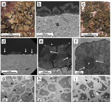
The typical habitus ofV. nigrescensis shown inFig. 2a. Peripheral lobes of aP. teicholytathallus are found in the left (P). BothP. teicholyta (Fig. 2b) andV. nigrescens(Fig. 2c, d) show very light interactions with the substrate (S),finding hyphae inside the substrate very rarely (ar-rows). Following dual sequential laser irradiation at 1064 and 266 nm, both stereoscopic (Fig. 2e) and SEM images (Fig. 2f) show the elim-ination of most V. nigrescens areolae. The remaining areolae show a greenish coloration due to the ablation of the cortical layer (Fig. 2e). The effect of the treatment onP. teicholytais not so evident (P,Fig. 2e).
The cross-section of the interface betweenV. nigrescensand the ceramic substrate examined by SEM-BSE shows numerous zones where the li-chen has become detached (Fig. 2g). Closer observation (Fig. 2h) shows remaining areolae-completely collapsed (white arrow) and colonization inside the substrate also affected by the treatment showing clear hyphal collapse (black arrow). Since the laser extracted the areolae ofV. ni-grescensas shown by stereoscopic and SEM-BSE, no chemical treatment could be applied due to the absence of thalli and consequently there are not images from TEM of the cytological aspects of the bionts.
3.3.2. Tile sample 2
A stereomicroscope image of a typical untreated thallus ofC. deci-piensis shown inFig. 3a. Interaction of lichen thalli with the substrate (S) is scarce and hyphal penetration is very rarely observed (Fig. 3b). Fig. 3c reveals the effect of dual laser irradiation at 1064 and 266 nm on a thallus ofC. decipiensunder the stereomicroscope. Only few areolae were detached by this treatment (Fig. 3c, d). Some thallus areas (Fig. 3c, arrows) have lost the cortical layer due to the laser action exposing the algal layer (green color). SEM observation of the thallus surface also reveals the loss of the cortical layer in some areas.Fig. 3e shows a SEM-BSE image of a cross-section of a treated thallus showing that although some areolae persist some have lost the cortical layer (white asterisk) and thallus is clearly internally damaged with serious injuries in algal and medulla layers (arrows). In this latterfigure the thallus-substrate interface shows the penetration of the mycobiont into the ceramic substrate (black asterisk). The ultrastructure of the pho-tobiont of the untreated thallus is shown inFig. 3f. It corresponds to the typical structure of the genus Trebouxia: a central chloroplast with pyrenoid (p) and dense lipid globules called pyrenoglobuli (arrows). However, following dual laser irradiation the structure of the chlor-oplast changes dramatically (Fig. 3g). Thylakoids and pyrenoids appear unstructured (white arrows), and pyrenoglobuli (black arrows) only appear in peripheral zones of the pyrenoid. The application of the chemical treatment alone causes a deep alteration of the protoplast, which is not separated yet from the cell wall. In some cells the pyrenoid zone is still recognizable (Fig. 3h).
When dual sequential laser irradiation was combined with chemical treatment the effects onC. decipienswere also assessed by TEM, since this is the only technique able of detecting changes at the cell level. Cell destruction in the photobiont was complete. This could be seen as the destruction of the chloroplast and separation of the cell wall from the plasmalemma (arrows), accompanied by intense plasmolysis and cell death (Fig. 3i, white asterisks).
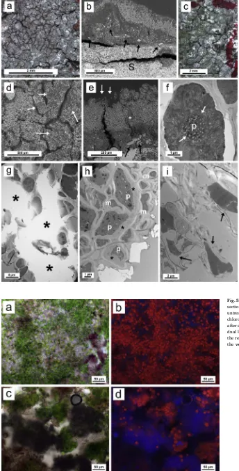
3.3.3. Tile sample 3
Untreated thalli ofP. teicholytaare shown inFig. 4a. A tighter re-lationship with the substrate is observable in this species compared to the previous samples as it is revealed in the SEM-BSE image by the high number of substrate fragments embedded in the lichen thallus (Fig. 4b, Table 1
Resulting data from ceramic roofing tiles characterization.
Sample Matrix Birefringence Type of clay Porosity Inclusions Neoformed
black arrows). Dual laser irradiation at 1064 and 266 nm produced an almost completed removal of upper layers of the thallus, revealing the algal layer (Fig. 4c, green areas), even though no areolae was com-pletely removed using this treatment. SEM examination of the lichen surface also revealed the damage produced by dual laser irradiation (Fig. 4d, white arrows). The effect of this dual laser irradiation on the areolae is shown more clearly inFig. 4e. Algal and cortical layers are ablated from parts of the thallus (white arrows). The damage produced by the lichen thallus to the calcium-rich substrate was evident across several thallus zones and detached layers were as thick as 100–150μm
(black arrows). Moreover, the thallus ofP. teicholytafeatured calcium-bearing (possibly calcium oxalate) mineral, as demonstrated by energy dispersive X-ray (EDS) microanalysis (data not shown). The presence of calcium deposits within lichen thallus has not been detected in lichen thalli growing on tile samples 1 and 2.
The typical appearance of photobiont cells in control thallus is shown in TEM micrographs ofFig. 4f. Partial destruction of both the algal and the medulla layers ofP. teicholytathalli after dual laser irra-diation at 1064 and 266 nm may be observed inFig. 4g. Large parts of the photobiont layer and medulla are ablated (asterisks). The applica-tion of chemical treatment alone causes a deep alteraapplica-tion of the pro-toplast, which is still not separated from the cell wall (Fig. 4h). When subjected to combined dual sequential laser irradiation/chemical treatment, the TEM images showed that the photobiont is significantly damaged, lacking completely its typical ultrastructural features (Fig. 4i, arrows).
Fig. 5shows lichen thalli ofP. teicholytauntreated (a-b) and treated (c-d) with the combined laser and chemical treatment. Either bright
field (Fig. 5a) orfluorescence (Fig. 5b) images of untreated lichen thalli reveal the abundant presence of green algae cells. After laser and che-mical treatment, distinct degradation of algae cells, appeared as dark spots and few green algae, may be appreciated in the brightfield image (Fig. 5c). Fluorescence image obtained from the same area (Fig. 5d) mostly shows blue autofluorescence signal.
3.4. FT-Raman spectroscopy measurements
FT-Raman spectroscopic measurements were undertaken on bare and non-irradiated areas of the ceramic substrate and on those areas where the dual sequential laser irradiation at 1064 and 266 nm induced partial or almost complete elimination of the biodeteriorationfilm. The FT-Raman spectra bands obtained on roofing tile sample 3 colonized by P. teicholytaandC. decipienslichen thalli are listed inTable S2 (Sup-plementary material), together with the corresponding assignments (Long, 1977; Edwards et al., 2003). The spectrum of the bare ceramic substrate acquired on the non biocolonized back side of the roofing tile displays characteristics bands of α-quartz (145, 207, 347, 465, and
1086 cm−1) and calcite (297 and 1086 cm−1). These bands correspond to the main components of this roofing tile (Table S1).
photobiont. Additionally, in the spectra of non-irradiated and irradiated areas of the tile sample, some other bands at 504, 899, 1464, 1490 and 1669 cm−1, could be attributed to calcium oxalate monohydrate (whewellite). The presence of this compound is probably due to the reaction between oxalic acid from the lichen and calcium from the substrate and thus is not present in the bare tile. The bands of the lichen compounds almost disappear after dual laser irradiation of the colo-nized surface, while the bands corresponding to the ceramic substrate weakly emerge in the spectrum, indicating lichen removal.
3.5. Colorimetric measurements
Table 2 displays the colorimetric coordinates of the three areas measured in each roofing tile sample. Tile sample 1 shows a moderate discoloration induced by dual sequential laser irradiation (ΔE* = 16.9).
The main color shift is due to changes inΔa* (−12.3, shift to greener), Δb* (−8.8, shift to bluer) andΔL* (−7.6, shift to a darker hue). Tile sample 2 shows a lower discoloration (ΔE* = 15.3) than tile sample 1.
In this case, the main color shift is due to changes inΔa* (−12.3, shift to greener),Δb* (−7.9, shift to bluer) andΔL* (4.7, shift to a lighter hue). The overall color changes determined on tile sample 3 (ΔE* = 16.1) is ascribed to slight shift to blue color and to the fact that
the laser irradiated area of the tile becomes darker. Finally, after ap-plication of chemical treatment alone it was not possible to carry out colorimetric measurements of the substrate since the chemical treat-ment alone did not destroy the lichen thalli and, consequently, the underlying substrate was not visible.
4. Discussion
Among the three ceramic roofing tiles examined in this research, tile
sample 1, colonized byPyrenodesmia teicholyta and Verrucaria nigrescens showed scarce signs of bioweathering of the ceramic material (Fig. 2b, c). This is in clear contrast with the results of a study which addressed the effects of Nd:YAG laser irradiation on quarry dolostone colonized by V. nigrescens. In this study it was observed that this species did intensely alter the stone substrate (Speranza et al., 2013). The discrepancy could be attributed to the chemical composition of tile sample 1, a non-cal-careous alumina-rich ceramic (Table S1, 4.50 wt % of CaO and 20.06 wt % of Al2O3concentrations) made from an illitic-smectitic clay.C. dec-ipienscolonizing the non-calcareous and also alumina-rich tile sample 2 showed as well little signs of bioweathering (Fig. 3b). All three species grow typically on calciferous substrates, often subjected to nitrogen deposition (Nimis, 2016). Their presence in tile samples 1 and 2 is evidently linked to the abundant nitrogen availability typical of rural environments in which roofing tiles were located.
Fig. 4.Biological colonization (P. teicholyta) of roofing tile sample 3 and its treatment. a) Typical habitus of an un-treated healthy thallus under the stereomicroscope. b) SEM-BSE micrograph showing a cross-section of a thallus. Substrate (S) particles embedded in lichen thallus are pointed out by black arrows. c) Stereomicroscope image of a thallus after dual laser irradiation (1064 and 266 nm). Green areas reveal loss of the cortical layer. d) SEM mi-crograph showing thallus surface after dual laser irradia-tion. White arrows indicate areas with cortical loss. f) SEM-BSE micrographs showing thallus cross-sections after dual laser irradiation treatment. White arrows point out areas where cortical and algal layers were ablated. Black arrows point out mycobiont-substrate interface with substrate fragments embedded in thallus. White asterisk indicates a calcium oxalate deposit. f) TEM micrograph of a healthy algal cell (photobiont) from an untreated thallus, pyrenoid (p). White arrows point out to pyrenoglobuli. g) TEM mi-crograph of thallus section gaps after dual laser irradiation treatment. Gaps in thallus structure are marked with black asterisks. h) Photobionts (p) and mycobionts (m) after chemical treatment with cell alteration and rather good adherence between plasmalem and cell wall. Asterisk in-dicate adhesion between plasmalem and cell wall. i) TEM micrograph of thallus section after dual laser irradiation and chemical treatments. Alteration of photobiont cells showing the lack of ultrastructure features and gaps between plas-malem and cell wall (black arrows). (For interpretation of the references to colour in thisfigure legend, the reader is referred to the web version of this article.)
revealed the accumulation of surprisingly large amounts of calcium in their thalli (Sanz et al., 2017). This suggests that biomobilization of calcium occurs in higher extent in tile sample 3 than in tile sample 1. Such biomobilization processes have been well established (de los Ríos et al., 2004). However, so far it is not possible to establish the relevance of these calcium deposits for the laser treatment.
It is evident that the three ceramic roofing tiles show different patterns in terms of biodeterioration. These patterns seem to be related with chemical composition rather than the state of sintering reached by the ceramic material. Thus, non calcareous and alumina-rich tile sam-ples 1 and 2 barely showed signs of bioweathering in spite of both present medium porosity and even an initial vitrification sintering state in the case of tile sample 1. On the contrary, the best sintered tile sample, the highly calcareous tile sample 3, displayed a thallus pene-tration inside the ceramic substrate although it was fired near to 1000 °C (Table 1). A higher bioweathering has been therefore observed in the roofing tile with the highest content of CaO, whereas bio-weathering of alumina-rich tile samples 1 and 2 seems to be very low. In the present study based on sequential irradiation at 1064 and 266 nm, it has been observed that effectiveness of the treatment de-pends on lichen species. Thus, many areolae were ablated fromV. ni-grescensthalli (Fig. 2e, f), even though the effect onC. decipienswas not so pronounced macroscopically, with only some areolae detached (Fig. 3d). However, the laser treatment caused the total collapse of the lichen thallus, removing most of the cortical layer and exposing the photobiont cells (Fig. 3c). The treatment ofP. teicholytadid not remove thallus areolae (Fig. 4c), although the cortical layer was ablated in most of the surface (Fig. 4c, d). SEM-BSE micrographs (Fig. 4e) show that elimination of upper thallus layers may reach a depth of around 150μm. The collapse of remaining lichen thallus after laser irradiation
is observable in TEM micrograph ofFig. 4g, which reveals large gaps in the thallus structure, while dual sequential laser irradiation plus che-mical treatment produce a complete damage (Fig. 4i) of both photo-biont and mycophoto-biont cells, thereby proving the lack of viability of the remaining lichen structures due to the chemical treatment (Fig. 4h) produces a synergic effect with the laser. Although laser treatment causes the collapse of all lichen species studied, differential effects on laser irradiation was observed. Differences related to lichen species identity have been recently showed, pointing out as potential causes of uneven effectiveness the contrasting thallus structure, the presence of distinct lichen substances and the presence of calcium oxalate (Sanz et al., 2017). On the other hand, the effect of either the laser or the combined treatment (laser + chemical treatment) does not seem to be affected by the chemical composition.
Fluorescence images also reveal the lack of viability after dual laser irradiation and chemical treatment (Fig. 5). Red autofluorescence signal ofP. teicholytathallus (Fig. 5b), which indicates a healthy status of photobiont cells, disappears after dual laser irradiation and chemical treatments, indicating loss of potential viability of the photobiont
(Fig. 5d). This degradation of the algal cells is also evident on the bright field image characterized by the presence of dark brown spots (Fig. 5c). It is largely known that biocides affect lichen viability and that their effect depends on the way they are applied as well as on the target species (Favero-Longo et al., 2017). Moreover, thefluorescence image obtained from the same area (Fig. 5d) mostly shows blue auto-fluorescence coming from rests of dead algae cells. Thus,fluorescence imaging also indicates the destructive action on lichen thalli of the dual laser irradiation plus the chemical treatment. Accordingly, it can be stated that in response to the biocideP. teicholytaundergoes cell col-lapse and that following laser and biocide combined treatment, the structure of the thallus is disrupted and cell death occurs in the sym-bionts.
The naked appearance shown by a portion of the pyrenoid ofC. decipiensphotobiont (Fig. 3h) should be interpreted as a consequence of a dehydrating effect as previously discussed in dehydrated thalli (Brown et al., 1987). In this case, dehydration is due to the effect of the dual laser treatment. Weak staining of parts of its proteinaceous matrix indicates substantial damage to the pyrenoid and thus to the algal cell, since its photosynthetic capacity is disrupted through loss of the Ru-bisco enzyme. Likewise, the photobiont ofP. teicholytacompletely lost its typical cell organization as shown by analyses of its ultrastructure, being impossible to distinguish any eukaryotic cell organelle (Fig. 4i). The measurements by FT-Raman spectroscopy provide with addi-tional evidence of removal of the lichen thalli. In the spectra of the laser irradiated zone no new peaks appear after dual laser irradiation, thereby indicating the absence of chemical changes in the ceramic substrate that could be induced by laser removal of lichens. Light to moderate color changes were determined by colorimetric measure-ments on laser irradiated areas of the tile samples in comparison with the cut edge area which probably was not exactly the color of the ori-ginal clean surface without biodeterioration. This is the most complex issues when discussing the optimum color of the cleaned surface, no matter the cleaning method used to characterize, identify or select the “reference”surface.
5. Conclusions
Bioweathering by lichen thalli of traditional made unglazed ceramic roofing tiles depends mainly on the chemical composition rather than the state of sintering of the ceramic substrate since calcium-rich tiles showed higher bioweathering than alumina-rich ones. A combined treatment based on dual sequential laser irradiation and chemical treatment has resulted to be usefulness for removal of lichen thalli. Laser irradiationfirst damages thallus structure, thereby opening paths to biocide penetration. The biocide seriously alters then the ultra-structure of the lichen symbionts (photobiont and mycobiont). This treatment could be of great help in providing control of biodeteriora-tion processes by lichens in conservabiodeteriora-tion and restorabiodeteriora-tion of unglazed ceramic roofing tiles of historical buildings.
Acknowledgements
This work was supported by research programs Geomateriales 2 (Regional Government of Madrid and EU structural funds, Ref. S2013/ MIT-2914) and IPERION-CH (Integrated Platform for the European Research Infrastructure on Cultural Heritage, Ref. H2020-INFRAIA-2014-2015 nº 654028). Servicios de Microscopia from MNCN-CSIC and CNB-CSIC are gratefully acknowledged. SPO is supported by the grant RYC-2014-16784 and CA and JW by the grant CGL2013-42509P, both from the Spanish Ministry of Economy, Industry and Competitiveness. Finally, the authors are indebted to the TechnoHeritage network on Science and Technology for the Conservation of Cultural Heritage for its professional support.
Table 2
Appendix A. Supplementary data
Supplementary data related to this chapter can be found athttp:// dx.doi.org/10.1016/j.ibiod.2017.10.003.
References
Ascaso, C., Galván, J., Rodríguez-Pascual, C., 1982. The weathering of calcareous rocks by lichens. Pedobiologia 24, 219–229.http://dx.doi.org/10.1007/BF00236519.
Ascaso, C., Sancho, L.G., Rodríguez-Pascual, C., 1990. The weathering action of sax-icolous lichens in maritime Antarctica. Polar Biol. 11, 33–39.http://dx.doi.org/10.
1007/BF00236519.
Brown, D.H., Rapsch, S., Beckett, A., Ascaso, C., 1987. The effect of desiccation on cell shape in the lichenParmelia sulcata. New Phytol. 105, 295–299.http://10.1111/j. 1469-8137.1987.tb00867.x.
Campbell, J.W., Pryce, W., 2003. Brick: a World History. Thames & Hudson, London. Carmona, N., Laiz, L., González, J.M., García-Heras, M., Villegas, M.A., Saiz-Jiménez, C.,
2006. Biodeterioration of historic stained glasses from the Cartuja de Miraflores (Spain). Int. Biodeterior. Biodegr. 58, 155–161.http://10.1016/j.ibiod.2006.06.014. Clauzade, G., Roux, C., 1985. Likenoj de okcidenta europo. Ilustrita determinlibro. Bull.
Soc. Bot. C. Ouest 7 1–893.
Cooper, M., 1998. Laser Cleaning in Conservation: an Introduction. Butterworth Heinemann, Oxford.
Coutinho, M.L., Miller, A.Z., Gutiérrez-Patricio, S., Hernández-Marine, M., Gómez-Bolea, A., Rogerio-Candelera, M.A., Philips, A.J.L., Jurado, V., Saiz-Jiménez, C., Macedo, M.F., 2013. Microbial communities on deteriorated artistic tiles from pena national palace (Sintra, Portugal). Int. Biodeterior. Biodegr. 84, 322–332.http://10.1016/j.
ibiod.2012.05.028.
Coutinho, M.L., Miller, A.Z., Macedo, M.F., 2015. Biological colonization and biodeter-ioration of architectural ceramic materials: an overview. J. Cult. Herit. 16, 759–777.
http://10.1016/j.culher.2015.01.006.
Coutinho, M.L., Miller, A.Z., Martín-Sánchez, P.M., Mirão, J., Gómez-Bolea, A., Machado-Moreira, B., Cerqueira-Alves, L., Jurado, V., Saiz-Jiménez, C., Lima, A., Phillips, A.J.L., Pina, F., Macedo, M.F., 2016. A multi-proxy approach to evaluate biocidal treatments on biodeteriorated majolica glazed tiles. Environ. Microbiol.http://dx. doi.org/10.1111/1462-2920.13380.
Cultrone, G., Rodríguez-Navarro, C., Sebastián, E., Cazalla, O., De la Torre, M.J., 2001. Carbonate and silicate phase reactions during ceramicfiring. Eur. J. Mineral. 13, 621–635.http://10.1127/0935-1221/2001/0013-0621.
de Cruz, A., Wolbarsht, M.L., Andreotti, A., Colombini, M.P., Pinna, D., Culberson, C.F., 2009. Investigation of the Er:YAG laser at 2.94 mm to remove lichens growing on stone. Stud. Conserv. 54, 268–277.http://10.1179/sic.2009.54.4.268.
de los Ríos, A., Ascaso, C., 2002. Preparative techniques for transmission electron mi-croscopy and confocal laser scanning mimi-croscopy of lichens. In: Kranner, I., Beckett, R.P., Varma, A.K. (Eds.), Protocols in Lichenology. Springer, Berlin, pp. 87–151.
de los Ríos, A., Galván, V., Ascaso, C., 2004.In situmicroscopical diagnosis of biodeter-ioration processes occurring in the Convent of Santa Cruz la Real (Segovia, Spain). Int. Biodeterior. Biodegr. 54, 113–120.http://10.1016/j.ibiod.2004.03.020.
de los Ríos, A., Cámara, B., García del Cura, M.A., Rico, V.J., Galván, V., Ascaso, C., 2009. Deteriorating effects of lichen and microbial colonization of carbonate building rocks in the Romanesque churches of Segovia (Spain). Sci. Total Environ. 407, 1123–1134.
http://10.1016/j.scitotenv.2008.09.042.
Edwards, H.G.M., Farwell, D.W., Jenkins, R., Seaward, M.R.D., 1992. Vibrational Raman spectroscopic studies of calcium oxalate monohydrate and dihydrate in lichen en-crustations on Renaissance frescoes. J. Raman Spectrosc. 23, 185–189.http://10.
1002/jrs.1250230310.
Edwards, H.G.M., Newton, E.M., Wynn-Williams, D.D., Coombes, S.R., 2003. Molecular spectroscopic studies of lichen substances 1: parietin and emodin. J. Mol. Struct. 648, 49–59.
Favero-Longo, S.E., Benesperi, R., Bertuzzi, S., Bianchi, E., Buffa, G., Giordani, P., Loppi, S., Malaspina, P., Matteucci, E., Paoli, L., Ravera, S., Roccardi, A., Segimiro, A., Vannini, A., 2017. Species-and site-specific efficacy of commercial biocides and ap-plication solvents against lichens. Int. Biodeterior. Biodegr. 123, 127–137.http://10.
1016/j.ibiod.2017.06.009.
Gazulla, M.F., Sánchez, E., González, J.M., Portillo, M.C., Orduna, M., 2011. Relationship between certain ceramic roofing tile characteristics and biodeterioration. J. Eur. Ceram. Soc. 31, 2753–2761.http://10.1016/j.jeurceramsoc.2011.07.023.
Kiurski, J.S., Ranogajec, J.G., Ujhelji, A.L., Radeka, M.M., Bokorov, M.T., 2005. Evaluation of the effect of lichens on ceramic roofing tiles by scanning electron mi-croscopy and energy-dispersive spectroscopy analyses. Scanning 27, 113–119.http://
10.1002/sca.4950270302.
Laiz, L.L., González, J.M., Saiz-Jiménez, C., Portillo, M.C., Gazulla, M.F., Sánchez, E., 2006. Microbial assessment of the biological colonization on roofing tiles. In: In: Fort, R., Álvarez de Buergo, M., Gómez-Heras, M., Vázquez-Calvo, C. (Eds.), Heritage, Weathering and Conservation, vol. 1. Taylor & Francis, London, pp. 349–353.
Long, D.A., 1977. Raman Spectroscopy. McGraw Hill Intl. Book Company, New York. Maggetti, M., 1982. Phase analysis and its significance for technology and origin. In: Olin,
J.S., Franklin, A.D. (Eds.), Archaeological Ceramics. Smithsonian Institution Press, Washington D.C, pp. 121–133.
Maravelaki-Kalaitzaki, P., Zafiropulos, V., Pouli, P., Anglos, D., Balas, C., Salimbeni, R., Siano, S., Pini, R., 2003. Short free running Nd:YAG laser to clean different en-crustations on Pentelic marble: procedure and evaluation of the effects. J. Cult. Herit. 4, 77s–82s.http://10.1016/S1296-2074(02)01151-2.
Nimis, P.L., 2016. The Lichens of Italy. Edizioni Università di Trieste, Trieste. Nguyen, K.-H., Chollet-Krugler, M., Gouault, N., Tomasi, S., 2013. UV-protectant
meta-bolites from lichens and their symbiotic partners. Nat. Prod. Rep. 13, 1490–1508.
http://10.1039/C3NP70064J.
Nodari, L., Marcuz, E., Maritan, L., Mazzoli, C., Russo, U., 2007. Hematite nucleation and growth in thefiring of carbonate-rich clay for pottery production. J. Eur. Ceram. Soc. 27, 4665–4673.http://10.1016/j.jeurceramsoc.2007.03.031.
Osticioli, I., Mascalchi, M., Pinna, D., Siano, S., 2015. Removal ofVerrucaria nigrescens from Carrara marble artefacts using Nd:YAG lasers: comparison among different pulse durations and wavelengths. Appl. Phys. A 118, 1517–1526.http://10.1007/
s00339-014-8933-y.
Oujja, M., Rebollar, E., Castillejo, M., Domingo, C., Cirujano, C., Guerra-Librero, F., 2005. Laser cleaning of terracotta decorations of the portal of Palos of the cathedral of seville. J. Cult. Herit. 6, 321–327.http://10.1016/j.culher.2005.05.001.
Peters, T., Iberg, R., 1978. Mineralogical changes duringfiring of calcium-rich brick clays. Am. Ceram. Soc. Bull. 57, 503–509.
Sanz, M., Oujja, M., Ascaso, C., de los Ríos, A., Pérez-Ortega, S., Souza-Egipsy, V., Wierzchos, J., Speranza, M., Cañamares, M.V., Castillejo, M., 2015. Infrared and ultraviolet laser removal of crustose lichens on dolomite Heritage stone. Appl. Surf. Sci. 346, 248–255.http://10.1016/j.apsusc.2015.04.013.
Sanz, M., Oujja, M., Ascaso, C., Pérez-Ortega, S., Souza-Egipsy, V., Fort, R., de los Ríos, A., Wierzchos, J., Cañamares, M.V., Castillejo, M., 2017. Influence of wavelength on the laser removal of lichens colonizing heritage stone. Appl. Surf. Sci. 399, 758–768. http://10.1016/j.apsusc.2016.12.032.
Smith, C.W., Aptroot, A., Coppins, B.J., Fletcher, A., Gilbert, O.L., James, P.W., Wolseley, P.A. (Eds.), 2009. The Lichens of Great Britain and Ireland. The British Lichen Society, London.
Speranza, M., Sanz, M., Oujja, M., De los Rios, A., Wierzchos, J., Pérez-Ortega, S., Castillejo, M., Ascaso, C., 2013. ND-YAG laser irradiation damages toVerrucaria ni-grescens. Int. Biodeterior. Biodegr. 84, 281–290.http://10.1016/j.ibiod.2012.02.010.
Tornari, V., Fotakis, C., Georgiou, S., Zafiropulos, V., Anglos, D., 2006. Laser cleaning of
encrustation. In: Fotakis, C., Anglos, D., Georgiou, S., Zafiropulos, V., Tornari, V.
(Eds.), Laser in the Preservation of Cultural Heritage. Principles and Applications. Taylor & Francis, London pp. 336.
Trindade, M.J., Dias, M.I., Coroado, J., Rocha, F., 2009. Mineralogical transformations of calcareous rich clays withfiring: a comparative study between calcite and dolomite rich clays from Algarve. Port. Appl. Clay Sci. 42, 345–355.http://10.1016/j.clay.
2008.02.008.
Wang, Q., Ma, G.Y., He, L.Y., Sheng, X.F., 2011. Characterization of bacterial community inhabiting the surfaces of weathered bricks of Nanjing Ming city walls. Sci. Total Environ. 409, 756–762.http://10.1016/j.scitotenv.2010.11.001.
Warren, J., 1999. Conservation of Brick. Butterworth Heinemann, London. Wierzchos, J., Ascaso, C., 1994. Application of back-scattered electron imaging to the