Corresponding author: [email protected]
The role of virulence factors in
Candida
albicans
pathogenicity
Tri Wibawa
Department of Microbiology, Faculty of Medicine, Universitas Gadjah Mada, Yogyakarta, Indonesia
DOI: http://dx.doi.org/10.19106/JMedSci004801201606
ABSTRACT
Candida albicans is a classical example of causative agent for opportunistic fungal infection. Normally, it colonizes skin, gastrointestinal tract, genital, and mucosal membranes, but in certain condition it may responsible for diseases. This phenomenon was mainly associated with immunological status of the host. However, there were indings that showed the possibility of putative virulence factors work on the transition of commensally to pathogenic role of the yeast. In this review, some virulence factors were discussed. Indeed, there were factors that may be considered as putative virulence factors of C. albicans.
ABSTRAK
Candida albicans adalah contoh klasik jamur penyebab infeksi oportunistik. Jamur ini biasa berkoloni di kulit, traktus gastrointestinal, genital, dan membran mukosa sebagai komensal. Namun demikian, pada kondisi tertentu jamur ini dapat menjadi agen penyebab infeksi. Fenomena ini biasanya dihubungkan dengan status imunitas dari inang. Akan tetapi, terdapat banyak laporan yang mengindikasikan adanya faktor virulensi yang bekerja pada proses transisi dari komensal menjadi patogenik ini. Pada kajian pustaka ini, beberapa kandidat faktor virulensi C. albicans didiskusikan.
Keywords:Candida albicans – opportunistic infection – fungal – pathogenicity - virulence
INTRODUCTION
Candida albicans is a classical example
of causative agent for opportunistic fungal
infection. There are 17 species member of
genus Candida e.g: C. albicans, C. glabrata,
C. parapsilosis and
C. tropicaliswhich are
commonly related with human diseases.
1The
clinical manifestations of C. albicans infection
are related with the host immune response.
The risk of infection is higher in the individual
with risk factors related with immune
response deterioration. It is well known that
C. albicans
is a lora normal in the skin,
gastrointestinal tract, genital, and mucosal
membranes, which is not regularly recognized
as a pathogen in immunocompetence human
being.
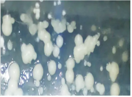
2Candida albicans may be cultured on
several media, such as Sabouraud’s dextrose
agar. It grow very fast, unlike other known
fungus. The yeast colony is white to cream
colored, smooth, glabrous and yeast-like
in appearance. Microscopic morphology
shows spherical to subspherical budding
positive for: Glucose, Maltose, Galactose,
Trehalose, Sucrose (some negative),
D-Xylose, Soluble Starch, D-Mannitol, and
D-Glucitol (Delayed).
3Candida albicans
may be identiied using several methods, i.e.
microscopic, morphological observation on
selective media, germ tube test, assimilation
test, and nucleic acid base identiication
(FIGURE 1).
Candida albicans is the most common
fungal causative agent for urinary tract
infection. In general,
C. albicans is unusual
cause of urinary tract infection in healthy
individual. However, it was reported common
in hospitalized patients, who have risk factors,
such as urinary tract structural problems.
The infection may associate with invasion in
antegrade form blood stream or retrograde
from urethra and bladder.
4A surveillance of nosocomial blood
stream infections (BSI) report in the USA
between April 1995 and June 1996 highlighted
that Candida was the fourth most common
causative agent of nosocomial BSI.
Fifty-two percent of 379 of candidemia cases were
due to C. albicans
,
5and it have reported to be
one of the leading causes of catheter-related
BSI.
6Candidemia affects more than250,000
people worldwide every year and responsible
to more than 50,000 deaths.Incidence rates of
candidemia have been reported to be 2 - 14
casesper 100,000 persons in population-based
studies.
7Epidemiological data showed that Candida
may cause wide spread of vulvovaginal
candidiasis (VVC). VVC is most frequently
caused by C. albicans
, though other species
are emerge.
8This fungal infection affect up
to 75% healthy women, which some of them
develop recurrent infection, known as recurrent
vulvovaginal candidiasis (RVVC). The
RVVC has challenging characteristic which
may responsible for treatment failure.
9Many
factors work to facilitate the transtition of C.
albicans
colonization state to asymptomatic
fungal infection, such as host susceptibility
and host inlammatory responses, as well as
candidal virulence factors.
10,11Candida sp are members of the oral
microlora of humans, they are opportunistic
pathogens that under conditions of host
debilitation can cause an oral infections.
Alteration of oral environment may favor the
overgrowth of Candida, which in turn develop
oral candidiasis. Candida sp is well known
as pathogen responsible for oral diseases.
12In
oral fungal infection,
C. albicans contributes
near 50% of all candidiasis cases.
13Candidiasis (moniliasis) is the third
most common supericial skin fungal
infection after dermatophytosis and pityriasis
versicolor.
14Non-hematogenous primary skin
infections typically occur as intertrigo in skin
folds, which is common in obese and diabetic
patients. Candida is knownresponsible for
neonatal candidiasis syndrome, a widespread
dermatitis due to Candida in neonates. The
syndromes associated with contamination of
amniotic luid, in which limited to the skin for
healthy-term infants.
15There were other various systems and
organs may be involved for C. albicans
infection, such as: liver, spleen, larynx,
lungs, bones, joints, pancreas, peritoneum,
endocardium, eyes, and meninges.
15The C.
albicans clinical manifestation is considered
as transformation from commensal of the fungi
to infection state. There are many factors work
for the transformation, including virulence
factors of C. albicans. This review aims to
discuss several putative virulence factors of
C. albicans
, which isimportant to give more
understanding and insight concerning to
the management of candidiasis which may
involve various organs.
DISCUSSION
There are various putative virulence
factors of C. albicans
documented,
such as: adherence of the fungi to the
host’s surfaces, production of hydrolytic
enzymes, dimorphism, galvanotropism and
thigmotropism, phenotypic switching, bioim
formation, and evasion to the host immune
response.
Adherence
Candida albicans is able to adhere
to various tissues andinanimate surfaces.
For example, buccaland vaginal epithelial
cells, corneocytes, cultured cells (HeLa
and HEp-2) surface, as well as biomaterial
surface.
4Adherence is the irst step of
C.
albicans infection in the oral and other
surfaces. It is an essential stage for the
persistence of the organism in the host, as
shown by the report that number of C. albicans
cells adhered to epithelial cells is signiicantly
higher in the chronic periodontitis group
than in the control group.
16Experimentation
using saliva – coated hydroxiapatite (SHA)
beads showed that C. albicansstrains which
associated with oral candidiasis adhere
better to SHA beads than less pathogenic
strains.
17,18Adhesins are the fungal surface
molecules that mediate binding of C. albicans
to the surface of human or microbes cells, inert
polymers, or proteins.
19There are candidate
genes putatively considered as encoding
adhesins such as: ALA1, ALS1, Hwp1
,
INT1,
MMT1, PMT1, PMT6 and
Als1p.
18-21Other
putative adhesins are mannan, chitin, factor
6 oligomannosaccharide, 66-kDa imbrial
protein, ibronectin binding protein, iC3b
binding protein, fucose binding protein,
GIcNAc or glucosamine, and secreted aspartyl
proteinase (SAP).
18,19Efforts to understand
C. albicans adhesins are hampered by the
limitation of consistency in several studies,
such as: different strain showed different
adherence speciicities and strength, technical
limitation of maintaining adhesins expression
in vitro
, and disagreement of viability yeast
effect to the adherence capability.
18Production of hydrolytic enzymes
Candida albicans is producing many
hydrolytic enzymes that facilitate its
commensally and pathogenic characteristics:
adherence tohost tissue and inert particles,
rupture of host cell membranes, invasion
of mucosal surfaces and blood vessels, and
evasion of the host’s immune response.
There are three major enzymes produced
by
C. albicans
: SAP, phospholipases,and
hemolysins.
4,22Secreted aspartyl proteinases
The Sapproteins of
C. albicans were
SAP9
, and
SAP10.
The Sap1 to Sap10 proteins
are 35- 50 kDa in size and responsible for all
of the extracellular proteolytic activity of C.
albicans.
The characteristic of Sap proteins
are not yet clearly elucidated, though current
evidences showed that the main roles of the C.
albicans
Saps are to provide nutrition for the
yeast cells, to aid penetration and invasion,
and to evade host immune responses.
23Sap
degrades proteins related to structural and
immunologic defenses, such as collagen,
keratin, mucin, antibodies, complement, and
cytokines, during tissue invasion.
4,24The
presence of genes and secretion of aspartic
proteases by C. albicans was demonstrated
to be one of the virulence factors.
25It was
observed that the virulence of C. albicans
associated with the level of Sap activity and
number of SAP genes. C. albicans isolated
from patients who has clinical manifestation,
have higher proteolytic activity compare to
those obtained from healthy individual.
23,26,27Saps expression by
C. albicans is regulated by
several factors, such as nutritional condition,
pH, temperature, and growth phase of the
yeast. Studies on Saps expression revealed
that there are contradiction conclusions on
Sap expression in various
in vivo experiments.
Sap protein speciic types is claimed to be
correlated with organ involve, infection
locataion, and environmental factors in
every study. However, because of different
techniques and experimental model, effort to
formulate conclusive statement on which type
of Sap dominantly active in each candidiasis
cases -- for example which Saps work in oral
candidiasis, vaginal candidiasis, or systemic
candidiasis -- is a very dificult task.
28Phospholipases
Phospholipases hydrolyze
glycerophos-pholipids, which are major components
of mammalian cell membranes. It cleaves
fatty acids from phospholipids which in
turn destabilizing the membranes.
4,29,30There
are seven phospholipase genes have been
identiied i.e.
PLA, PLB1, PLB2, PLC1,
PLC2, PLC3 and PLD1
. However, the role
of the enzymes encoded by these genes
remains unclear.
31Evidence of phospholipase
act as virulence factor was obtained from
comparative study conducted by Ibrahim et
al.
32in which one series of C. albicans obtained
from candidemia patients compared with
isolates obtained from oral cavity of healthy
individuals. It was shown that isolate from
candidemia cases have higher phospholipase
activity, which relected the virulence of these
isolates.
Hemolysins
Hemolytic capacity, which is served by
hemolysins enzyme, is an important putative
virulence factor of the genus Candida to
acquire iron from host tissues, which then is
used by the yeast for metabolism, growth
and invasion during host infection.
33,34In
the human being, iron is found in several
proteins, including hemoglobin located in
the erythrocytes. Candida albicans capable
to bind to erythrocytes, then hemolysis factor
will destroy the erythrocytes. However,
the mechanism of hemolysis caused by C.
albicans
remain unclear.
33,34The hemolytic
activity may be observed readily by culturing
C. albicans on the blood agar media.
It was reported that 98.5% of
C. albicans
isolates have hemolytic activity including
alpha, beta,and gamma hemolysis in aerobic
condition.
35However, the hypothesis of
hemolysins is a virulence factor of C.
albicans
still controversial. Study on
C.
albicans
isolated from HIV patients and
healthy individual showed that the hymolytic
activity were higher in isolates which were
obtained from healthy individual.
36This
inding add another insight that hemolytic
factor without presence of other virulence
factors.
Dimorphism
Candida albicans able to grow in yeast
and mold forms. The transition between yeast
and hyphal forms istermed as dimorphism.
Accordingly it clasiied as dimorphic
fungus.In the yeast form, it may undergo
budding, whereas in mould form, it may
produce new mycelia, or yeast like form. The
transformation between two morphologies can
be induced in vitro with several environmental
conditions, such as pH, temperature, or
chemicals.
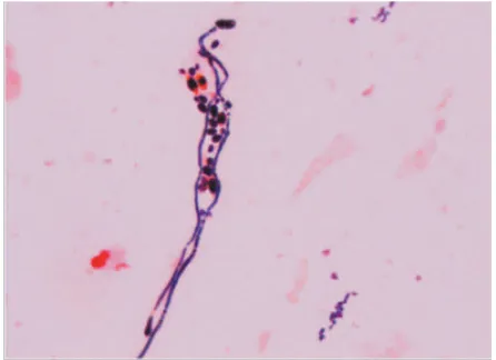
37Dimorphism of C. albicans is a
unique characteristic for pathogenicity of the
yeast (FIGURE 2). Both morphologies have
their own function to support its virulence.
38The hyphal form has been reported to be more
invasive than the yeast form. It was shown
that three C. albicans mutant strains which
compromised in the ability to form hyphae
(
efg1∆/efg1∆, lo8∆/lo8∆,
and
cph1∆/cph1∆
efg1∆/efg1∆
) were signiicantly less virulence
in C. elegans infection model.
39Whereas, the
yeast form is proposed as theform primarily
involved in dissemination of the fungus.
40However, another group found that transition
of C. albicans morphology to yeast form may
not the only factor regulate dissemination
from the gastrointestinal tract to the other
organs in invasive C. albicans infection.
42More than 40 genes were identiied
responsible for dimorphism regulation,
mainly hyphal formation.
42,43Those genes
are working in different stages of infection.
Dimorphism of C. albicansis important
for pathogenicity at both supericial and
systemicinfections. It should be noted that
both yeast and ilamentous form of
C.
albicans were found in infected tissues. The
capability of C. albicans to undergo transition
from yeast to ilamentous form contribute to
numerous nature of its infection stages, such
as adherence to epithelial and endothelial
cells, intercellular invasion, iron acquisition
from intracellular host sources, bioilm
formation, as well as escape from phagocytes
and immune evasion.
44,45FIGURE 2. Microscopic appearance of C. albicans
found in the sputum of patient, after Gram staining. The dimorphism of C. albicans
budding yeast, pseudohyphae, and true hyphae were observed.
Phenotypic switching
Candida albicans has capability to
undergo phenotypic switching that is
commonly called as white-opaque phenotypic
switching.A small proportion of
C. albicans
isolates, which are homozygous at the mating
type locus (
MTL, a/a or α/ α
), able to switch
between two distinct cell morphologies: white
and opaque. It is not known how white-opaque
switching has never been observed in C.
albicans
strains that have heterozygous MTL
genotypes (a/α), though they have all essential
genes for white-opaque switching.
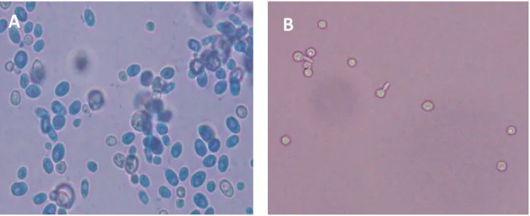
46FIGURE
3 shows different cell morphology between
white and opaque cells. The white cells appear
round and bright under microspcope, while
opaque cells appear darker, polymorphic and
oval. The white-opaqe
cells may be observed
also in their colonies by using simple phloxin
[image:5.595.310.533.185.347.2]white and opaque phenotypes show different
cellular and colony appearances, gene
expression proiles, mating ability and
virulence.
47White and opaque cells differ in their
mating capability as well as expression of
genes that are unrelated with mating process,
such as adhesins and metabolic genes. Opaque
cells are better colonizers of the skin, but they
are less virulent than white cells in a mouse
model of disseminated candidiasis.
48Opaque
cell formed hyphae in very low level in
the suspension cultures, in which white
cell
C. albicans will able to form hyphae.
This inding indicated that opaque cell less
virulent compare to white cells. When opaque
cells able to form hyphae, morphologically
similar with hypae of white cells. However,
genetically still distinc to the hypae formed by
white cells.
49White-opaque switching occurs
at a low frequencyin
C. albicans,
36but certain
environmental conditions can drive the switch
from one phase to the other. Although opaque
cells are less frequently cause systemic
infection than white cells, they have better
optimazion for colonization, such as on the
skin.
50White-opaque switching has been
shown also to affect several virulence factors,
such as susceptibility to antifungal drugs,
proteinase activity, antigenicity, and adhesion
of C. albicans.
4Bioilm formation
Bioilm is a structure made of microbes
consortium supported with extracellular matrix
which attach to the surface of living matter
or inanimate structure. Candida albicans
is well known yeast that able to develop
bioilm. Bioilm of
C. albicans is notorious
because of its deleterious consequences,
such as leads to antifungal resistance, give a
asylum to the yeast because of ability to make
evasion against immune surveillance, and act
as perfect reservoir for source of infection,
as well as several advantages in the fungal’s
perspective: protection from the environment,
resistance of physical and chemical stress,
metabolic cooperation, and a
community-based regulation of gene expression.Indeed,
bioilm formation represents one of the
putative virulence factors contributing to the
pathogenesis of candidiasis.
51-53The bioilm
of
C. albicans may need special attention
since antifungal may not work properly in
FIGURE 3. Microscopic appearance of C. albicans. (A) Opaque cells appear dark, oval and
[image:6.595.121.493.104.256.2]the bioilm setting. Anti-bioilm substance
need to be invented in the near future.
54Bioilm formation is a dynamic process which
is started with adherence of planctonic yeast
cells to the surface, proliferation of the yeast
cells, formation of hyphae, and accumulation
of extracellular matrix. Then the maturation of
the bioilm is complete. Moreover, yeast cells
which construct the bioilm may be detached
and disperse to other part of the body which
may go to the new focal infection.
51,55There
are two types of C. albicans cells involve in
the bioilm formation:small yeast-form cells
(blastospores), and long tubular hyphal cells.
The two cell types have their speciic role in
bioilm formation.
55Study on two series
C. albicans
from HIV infected and healthy
individuals showed that frequency of
C.
albicans
isolates which able to form bioilm
is comparable between two groups.
36The
result showed to us that bioilm as a putative
virulence factor may not work alone. It may
work in a concert with other virulence factors.
Evasion to the host immune responses
Human immune response against C.
albicans
occurs through several mechanisms,
comprising innate and adaptive immune
response. The innate immune response is
nonspeciic and broad. This is the irst line
of host defense against potentially harmful
microbes. Innate immunity comprises of a
group of soluble (complement) and cellular
(neutrophil,
macrophage)
components.
Whereas, the adaptive immune response
recognizes
speciic
antigenicmoieties,
resulting to the development of a targeted
immuneresponse.
56Several mechanisms
have been proposed to explain the mechanism
of
C. albicans evade from the host immune
response, which is considered as virulence
factor of the yeast. Experiment in
TLR2(-/-)
mice showed that C. albicans induce
immunosuppression through TLR2-mediated
IL-10 release, and this leads togeneration of
CD4+CD25+ T-regulatory cells with immuno
suppressive potential.
57Candida albicanswas
shown capable to bind thrombocytesvia
ibrinogen ligands in the blood stream. This
may result to the yeast cells being surrounded
by a group of thrombocytes, which in turn
may camoulage them from the immune
system during dissemination through the
blood stream.
4CONCLUSION
Candida albicans is an opportunistic
pathogen which responsible for various
diseases associated with several organs. It
pathogenicity is not solely because of the
impairment of host immune response. Some
putative virulence factors may work in
concert to facilitate the yeast transition form
commensal to pathogenic. Now, it is important
to further analyze the role of each virulence
factors in every step of the transition, in order
to improve the management of the candidiasis
in the near future.
ACKOWLEDGEMENTS
Author would like to thank Mrs. Mulyani
for preparing the igure of
C. albicans.
REFERENCES
1. The American Thoracic Society. Candida In-fection of the Bloodstream–Candidemia, Am J Respir Crit Care Med 2012; 185:3-4
2. Olsen I. Attenuation of Candida albicans
vir-ulence with focus on disruption of its vacuole
functions. J Oral Microbiol 2014; 6:23898. http://dx.doi.org/10.3402/jom.v6.23898 3. Elis D. Candida albicans, Mycology online,
4. Fisher JF, Kavanagh K, Sobel JD, Kauffman CA, Newman CA. Candida urinary tract in-fection: pathogenesis. Clin Infect Dis 2011; 52 Suppl 6:437-51.
http://dx.doi.org/ 10.1093/cid/cir110
5. Pfaller MA, Jones RN, Messer SA, Edmond MB, Wenzel RP. National surveillance of
nosocomial blood stream infection due to
candida albicans: frequency of occurrence
and antifungal susceptibility in the SCOPE Program. Diag Microbiol Infect Dis 1998; 31(1):327-32. http://dx.doi.org/10.1016/ S0732-8893(97)00240-X
6. Akbari F, Kjellerup BV. Elimination of
bloodstream infections associated with can
-dida albicans bioilm in intravascular cathe-ters. Pathogens 2015; 4(3):457-69.
http://dx.doi.org/10.3390/pathogens4030457 7. Cleveland AA, Harrison LH, Farley MM,
Hollick R, Stein B, Chiller TM, et al. Declin -ing incidence of candidemia and the shift-ing epidemiology of candida resistance in two
US metropolitan areas, 2008–2013: results from population-based surveillance. PLoS ONE 2015; 10(3):e0120452. http://dx.doi. org/10.1371/journal.pone.0120452
8. Vermitsky JP, Self MJ, Chadwick SG, Trama JP, Adelson ME, Mordechai E, et al. Survey of vaginal-lora Candida species isolates
from women of different age groups by use
of species-speciic PCR detection. J Clin Mi-crobiol 2008; 46(4):1501-3.
http://dx.doi.org/10.1128/JCM.02485-07 9. De Bernardis F, Arancia S, Sandini S,
Gra-ziani S, Norelli S. Studies of immune re-sponses in Candida vaginitis. Pathogens 2015; 4(4):697-707
http://dx.doi.org/10.3390/pathogens4040697 10. Sobel JD. Vulvovaginal candidosis. Lancet
2007; 369(9577):1961-71.
h t t p : / / d x . d o i . o r g / 1 0 . 1 0 1 6 / S 0 1 4 0 -6736(07)60917-9
11. Van Schalkwyk J, Yudin MH. Vulvovaginitis:
screening for and management of trichomo
-niasis, vulvovaginal candidiasis, and bacte-rial vaginosis. J Obstet Gynaecol Can 2015;
37(3):266-76. http://dx.doi.org/10.1016/ S1701-2163(15)30316-9
12. Williams D, Lewis M. Pathogenesis and treatment of oral candidosis. J Oral Microbiol 2011; 3:5771. http://dx.doi.org/10.3402/jom. v3i0.5771
13. Zomorodian K, Haghighi NN, Rajaee N, Pakshir K, Tarazooie B, Vojdani M, et al. As-sessment of Candida species colonization and denture-related stomatitis in complete den-ture wearers. Med Mycol 2011; 49(2):208-11. http://dx.doi.org/10.3109/13693786.2010.50 7605
14. Kelly BP. Supericial Fungal Infections. Pe-diatr Rev 2012; 33(4):22-37. http://dx.doi. org/10.1542/pir.33-4-e22
15. Rex JH, Walsh TJ, Sobel JD, Filler SG, Pappas PG, Dismukes WE, et al. Practice
guidelines for the treatment of candidiasis.
Infectious Diseases Society of America. Clin Infect Dis 2000; 30(4):662-78. http://dx.doi. org/10.1086/313749
16. Machado AG, Komiyama EY, Santos SS, Jorge AO, Brighenti FL, Kago-Ito CY. In
vi-tro adherence of Candida albicans isolated
from patients with chronic periodontitis. J Appl Oral Sci 2010; 19(4):384-7. http://dx. doi.org/10.1590/S1678-77572011005000014 17. Schmid J, Hunter PR, White GC, Nand AK,
Cannon RD. Physiological traits associated
with success of Candida albicans strains as
commensal colonizers and pathogens. J Clin Microbiol 1995; 33(11):2920-6.
18. Calderone R, Suzuki S, Cannon R, Cho T, Boyd D, Calera J, et al. Candida albicans: ad
-herence, signaling and virulence. Med Mycol 2000; 38(Suppl 1): 125-37.
h t t p : / / d x . d o i . o rg / 1 0 . 1 0 8 0 / m m y. 3 8 . s1.125.137
20. Tsuchimori N, Sharkey LL, Fonzi WA, French SW, Edwards JE Jr, Filler SG. Reduced viru-lence of HWP1-deicient mutants of Candida
albicans and their interactions with host cells.
Infect Immun 2000; 68(4):1997-2002. http://dx.doi.org/10.1128/IAI.68.4.1997-2002.2000
21. Yang YL. Virulence factors of Candida species. J Microbiol Immunol Ifect 2003; 36(4):223-8.
22. Staniszewska M, Bondaryk M, Siennicka K, Piłat J, Schaller M, Kurzatkowski W. Role of Aspartic Proteinases in Candida albicans Vir-ulence. Part I. Substrate Speciicity of Aspar-tic Proteinases and Candida albicans Patho-genesis. Post Mikrobiol 2012; 51(2):127-35. 23. Naglik JR, Challacombe SJ, Hube B.
Candi-da albicans secreted aspartyl proteinases in virulence and pathogenesis. Microbiol Mol
Biol Rev 2003; 67(3):400-28.
http://dx.doi.org/10.1128/MMBR.67.3.400-428.2003
24. Schaller M, Schackert C, Korting HC, Janus-chke E, Hube B. Invasion of Candida
albi-cans correlates with expression of secreted
aspartic proteinases during experimental in
-fections of human epidermis. J Invest Derma-tol 2000; 114(4):712-7.
h t t p : / / d x . d o i . o r g / 1 0 . 1 0 4 6 / j . 1 5 2 3 -1747.2000.00935.x
25. Mardegan RC, Foglio MA, Gonçalves RB, Höling JF. Candida albicans proteinases. Braz J Oral Sci 2006; 5(16):944-52.
26. Borst A, Fluit AC. High levels of hydrolyt-ic enzymes secreted by Candida albhydrolyt-icans isolates involved in respiratory infections. J Med Microbiol 2003; 52(Pt 11):971-4. http:// dx.doi.org/10.1099/jmm.0.05228-0
27. Monroy-Pérez E, Paniagua-Contreras G, Va-ca-Paniagua F, Negrete-Abascal E, Vaca S. SAP expression in candida albicans strains
isolated from mexican patients with vaginal
candidosis. Int J Clin Med 2013; 4(1):25-31. http://dx.doi.org/10.4236/ijcm.2013.41006
28. Staniszewska M, Bondaryk M, Siennicka K, Piłat J, Schaller M, Kurzatkowski W. Role of Aspartic Proteinases in Candida albicans Virulence. PART II: Expression of SAP 1-10 Aspartic Proteinase during Candida albicans Infection in vivo. Post Mikrobiol 2012; 51 (2): 137–142
29. Ghannoum MA. Potential role of
phospholi-pases in virulence and fungal pathogenesis.
Clin Microbiol Rev 2000; 13(1):122-43. http://dx.doi.org/10.1128/CMR.13.1.122-143.2000
30. Williams DW, Jordan RP, Wei XQ, Alves CT, Wise MP, Wilson MJ, et al. Interactions of
Candida albicans with host epithelial surfac
-es. J Oral Microbiol 2013; 5:22434. http:// dx.doi.org/10.3402/jom.v5i0.22434
31. Samaranayake YH, Dassanayake RS, Cheung BP, Jayatilake JA, Yeung KW, Yau JY, et al. Differential phospholipase gene expression
by Candida albicans in artiicial media and cultured human oral epithelium. APMIS 2006; 114(12):857-66.
http://dx.doi.org/10.1111/j.1600-0463.2006. apm_479.x
32. Ibrahim AS, Mirbod F, Filler SG, Banno Y, Cole GT, Kitajima Y, et al. Evidence
impli-cating phospholipase as a virulence factor
of Candida albicans. Infect Immun 1995; 63(5):1993-8. http://dx.doi.org/10.1111/ j.1567-1364.2009.00570.x
33. Almeida RS, Wilson D, Hube B. Candida al-bicans iron acquisition within the host. FEMS Yeast Res 2009; 9(7):1000-12.
http://dx.doi.org/ 10.1111/j.1567-1364.2009.00570.x
34. Rossoni RD, Barbosa JO, Vilela SF, Jorge AO, Junqueira JC. Comparison of the hemo-lytic activity between C. albicans and non-al-bicans Candida species. Braz Oral Res 2013; 27(6):484-9. http://dx.doi.org/ 10.1590/ S1806-83242013000600007
eval-uation of the effect of anaerobiosis on some
virulence attributes of Candida albicans. J Med Microbiol 2008; 57(Pt 10):1277-81. http://dx.doi.org/10.1099/jmm. 0.2008/ 001107-0
36. Wibawa T, Praseno, Aman AT. Virulence Candida albicans isolated from HIV Infect-ed and non infectInfect-ed individuals. SpringerPlus 2015; 4:408.
http://dx.doi.org/10.1186/s40064-015-1215-0
37. Molero G, Díez-Orejas R, Navarro-García F, Monteoliva L, Pla J, Gil C, et al. Candida
albicans: genetics, dimorphism and pathoge-nicity. Internatl Microbiol 1998; 1(2):95-106. 38. Mayer FL, Wilson D, Hube B. Candida al-bicans pathogenicity mechanisms. Virulence 2013; 4(2):119-28. http://dx.doi.org/10.4161/ viru.22913
39. Pukkila-Worley R, Peleg AY, Tampakakis E, Mylonakis E. Candida albicans hyphal
formation and virulence assessed using a
Caenorhabditis elegans infection model. Eu-karyot Cell 2009; 8(11):1750-8. http://dx.doi. org/10.1128/EC.00163-09
40. Saville SP, Lazzell AL, Monteagudo C, Lo-pez-Ribot JL. Engineered control of cell
morphology in vivo reveals distinct roles for
yeast and ilamentous forms of Candida
al-bicansduring infection. Eukaryot Cell 2003;
2(5):1053-60.
http://dx.doi.org/10.1128/EC.2.5.1053-1060.2003
41. Vautier S, Drummond RA, Chen K, Murray GI, Kadosh D, Brown AJ, et al. Candida albi
-cans colonization and dissemination from the murine gastrointestinal tract: the inluence of
morphology and Th17 immunity. Cell Micro
-biol 2015; 17(4):445-50.
http://dx.doi.org/10.1111/cmi.12388
42. Kumamoto CA, Vinces MD. Contributions of hyphae and hypha-co-regulated genes
to Candida albicans virulence. Cel Mi
-crobiol 2005; 7(11):1546-54 http://dx.doi.
org/10.1111/j.1462-5822.2005.00616.x
43.
44. Han TL, Cannon RD, Villas-Bôas SG. The
metabolic basis of Candida albicans morpho
-genesis and quorum sensing. Fungal Genet Biol 2011; 48(8):747-63.
http://dx.xoi.org/10.1016/j.fgb.2011.04.002 45. Baillie GS, Douglas LJ. Role of dimorphism
in the development of Candida albicans bio
-ilms. J Med Microbiol 1999; 48(7):671-9. http://dx.doi.org/10.1099/00222615-48-7-671
46. Jacobsen ID, Wilson D, Wächtler B, Brunke S, Naglik JR, Hube B. Candida albicans di-morphism as a therapeutic target. Expert Rev Anti Infect Ther 2012; 10(1):85-93.
http://dx.doi.org/10.1586/eri.11.152
47. Xie J, Tao L, Nobile CJ, Tong Y, Guan G, Sun Y, et al. White-opaque switching in natural MTLa/a isolates of Candida albicans:
evolu-tionary implications for roles in host adapta
-tion, pathogenesis, and sex. PLoS Biol 2013; 11(3):e1001525.
h t t p : / / d x . d o i . o rg / 1 0 . 1 3 7 1 / j o u r n a l . pbio.1001525
48. Huang G. Regulation of phenotypic
transi-tions in the fungal pathogen Candida albi
-cans. Virulence 2012; 3(3):251-61. http:// dx.doi.org/10.4161/viru.20010
49. Sasse C, Hasenberg M, Weyler M, Gunzer M, Morschhäusera J. White-opaque switching
of Candida albicans allows immune evasion
in an environment-dependent fashion. Eu-karyot Cell 2013; 12(1):50-8. http://dx.doi. org/10.1128/EC.00266-12
50. Anderson J, Cundiff L, Schnars B, Gao MX, Mackenzie I, Soll DR. Hypha formation in the white-opaque transition of Candida albi-cans. Infect. Immun 1989; 57(2):458-67. 51. Morschhäuser J. Regulation of white-opaque
switching in Candida albicans. Med Micro
-biol Immunol 2010; 199(3):165-72. http:// dx.doi.org/10.1007/s00430-010-0147-0 52. Wibawa T. Candida albicans bioilm:
53. Ramage G, Rajendran R, Sherry L, Wil-liams C. Fungal bioilm resistance. Int J Mi-crobiol 2012; 2012:528521. http://dx.doi. org/10.1155/2012/528521
54. Pierce CG, Chaturvedi AK, Lazzell AL, Pow-ell AT, Saville SP, McHardy SF, et al. A novel
small molecule inhibitor of Candida albicans
bioilm formation, ilamentation and
viru-lence with low potential for the development
of resistance. NPJ Bioilms Microbiomes 2015; 1pii:15012. http://dx.doi.org.10.1038/ npjbioilms
55. Wibawa T, Nurrokhman, Baly I, Daeli PR, Kartasasmita G, Wijayanti N. Cyclosporine a decreases the luconazole minimum
inhibito-ry concentration of Candida albicans clinical
isolates but not bioilm formation and cell growth. Trop Biomed 2015; 32(1): 76-182. 56. Finkel JS, Mitchell AP. Genetic control of
Candida albicans bioilm development. Nat Rev Microbiol 2011; 9(2):109-18. http://dx. doi.org/10.1038/nrmicro2475
57. Naglik JR. Candida Immunity. New J Sci 2014; 2014(ID 390241):1-27.
http://dx.doi.org/10.1155/2014/390241 58. Netea MG, Sutmuller R, Hermann C,Van
der Graaf CA, Van der Meer JW, van Kriek-en JH, et al. Toll-like receptor 2 suppresses
immunity against Candida albicans through
induction of IL-10 and regulatory T cells. J Immunol 2004; 172(6):3712-8.