An efficient method for the production of transgenic plants of
peanut (
Arachis hypogaea
L.) through
Agrobacterium
tumefaciens
-mediated genetic transformation
Kiran K. Sharma *, Vanamala Anjaiah
Genetic Resources and Enhancement Program,Genetic Transformation Laboratory,
International Crops Research Institute for the Semi-Arid Tropics(ICRISAT),Patancheru,Andhra Pradesh502 324,India
Received 20 March 2000; received in revised form 20 May 2000; accepted 22 May 2000
Abstract
Cotyledon explants from mature peanut seeds (Arachis hypogaeaL.) were optimized to obtain adventitious shoot buds with high frequencies (\90%). Efficient transformation of these cotyledons by usingAgrobacterium tumefaciens strainC58 carrying neomycin phosphotransferase II (nptII) and ß-glucuronidase (GUS;uidA), or coat protein gene of the Indian peanut clump virus (IPCVcp) andnptII on binary vectors (pBI121; pROKII:IPCVcp) led to the production of a large percentage (55%) of transgenic plants. Transformed individuals were obtained through selection on medium containing 125 mg l−1kanamycin. A large number
of independently transformed plants (over 75) were successfully transplanted to the glasshouse. Integration of the transgenes and stable genetic transformants in the progeny were assessed by PCR amplification of 700-bp fragment of nptII and 585-bp of IPCVcp genes, and Southern blot hybridizations in the T1 generation of transgenic plants. Analysis of 35 transgenic plants of T1 generation from the progeny of a single transformation event suggested the segregation of a single copy insert in a 3:1 Mendelian ratio. On an average, 120 – 150 days were required between the initiation of explant transformation and transfer of rooted plants to the greenhouse. The cotyledon regeneration system proved to be an excellent vehicle for the production of a large number of independently transformed peanut plants. Shoot formation was rapid and prolific, and a large proportion of these shoots developed into fertile plants. The method reported here provides new opportunities for the crop improvement of peanut via genetic transformation. © 2000 Elsevier Science Ireland Ltd. All rights reserved.
Keywords:Agrobacterium tumefaciens;Arachis hypogaea; Groundnut; Indian peanut clump virus; Shoot regeneration; Transgenic plants www.elsevier.com/locate/plantsci
1. Introduction
Peanut or groundnut (Arachis hypogaea L.) is
an economically important oil and protein rich crop, whose seeds contain about 43% oil and 25% protein that has a significant impact in tropical and sub-tropical regions of Asia, Africa, and North and South America. There are several con-straints to the productivity of the peanut crop that result in great economic losses annually [1].
Al-though some of the wild relatives of A. hypogaea
have been identified as resistance source to several
diseases and pests [2], the success in transferring the desirable traits to cultivated varieties has been limited due to reproductive barriers, and frequent failures in the interspecific crosses. The application of biotechnological methods for the improvement of important crop plants of the semi-arid tropics have been shown to hold great potential [1]. Ge-netic transformation approach allows for
intro-ducing novel genes that are not accessible
normally by conventional cross-breeding, i.e. lim-ited by sexual incompatibility. Although several reports on efficient regeneration from diverse ex-plants of peanut have been published [3 – 6], not much success with genetic transformation of
Arachis species has been achieved. This is due to
* Corresponding author. Tel.:+91-403296161; fax:+91-403241239.
E-mail address:[email protected] (K.K. Sharma).
the lack of efficient protocols to regenerate whole plants through in vitro regeneration of adventi-tious shoot buds from the transformed tissues [7 – 14]. This has prompted some workers to adopt non-tissue culture based approaches that do not depend on the regeneration of adventitious shoot buds for generating transgenic plants of peanut [15,16]. In general, the number of independently transformed plants obtained so far has been very low. Stable engineered resistance requires the pro-duction of numerous independent transformants to allow the selection of those with the appropriate level of gene expression [14]. For successful genetic modification by the production of transgenic plants, effective regeneration and transformation system is imperative.
The transformation protocol reported here was
initially optimized by using marker genes (nptII;
uidA) and later used for transforming with the
coat protein gene of Indian peanut clump virus (IPCVcp) to induce resistance to this virus. The Indian peanut clump virus (IPCV; Pecluvirus) is widespread in India, where it causes clump disease in peanut crop [17]. Despite the screening of more than 10 000 lines of peanut germplasm, no sources of resistance to IPCV have been found. IPCV is
transmitted by Polymyxa gramminis, a soil-borne
protozoan [18] and is difficult to control because of its persistence in soil for several years and the lack of suitable biocides or resistant genotypes. Transformation of plants with DNA encoding the coat protein and replicase genes of viruses is an efficient means to enhance the germplasm for re-sistance to viral diseases [19]. The coat protein gene of IPCV from its RNA-2 has been cloned
and sequenced [20] and its expression inNicotiana
benthamiana studied [21]. At ICRISAT, attempts are underway to induce resistance to IPCV and
other peanut viruses by using biotechnological tools. This study describes a protocol for efficient regeneration of multiple adventitious shoot buds from mature cotyledon explants of peanut and the
production of fertile transgenic plants by A.tume
-faciens-mediated transformation. The transforma-tion system reported here is potentially applicable to a wide range of genotypes.
2. Materials and methods
2.1. Tissue culture system
Mature seeds from different peanut varieties were removed from mature pods and stored at 4°C for further use. Unless mentioned otherwise, vari-ety JL-24 was used in all the experiments to opti-mize adventitious bud formation from cotyledon explants on various media formulations containing
different concentrations of N6-benzyladenine (BA;
2.5, 5.0, 7.5, 10.0, 15.0, 20.0, 25.0 mM) and
2,4-dichlorophenoxyacetic acid (2,4-D; 1.0, 2.5, 5.0,
7.5, 10.0, 15.0, 20.0 mM) in 7×7 combinations.
Five other peanut varieties belonging to two botanical types viz., Spanish and Virginia types (Table 1) were also used to test for shoot bud regeneration response on the optimized medium. Prior to use, the seeds were surface sterilized by rinsing in 70% ethanol for 1 min followed by
treatment with 0.1% (w/v) aqueous mercuric
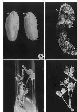
chlo-ride for 10 min and then washed thoroughly four to six times with sterile-distilled water and soaked in sterile water for 2 h before use. After removing the seed coat, the embryo axis was removed surgi-cally and each cotyledon was cut into vertical halves to obtain the cotyledon explants (Fig. 1A). The explants were placed on the shoot induction
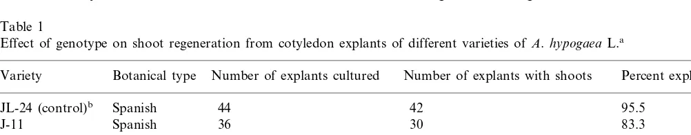
Table 1
Effect of genotype on shoot regeneration from cotyledon explants of different varieties ofA.hypogaea L.a
Botanical type Number of explants cultured
Variety Number of explants with shoots Percent explants
95.5
Virginia 44 38 86.4
ICGS-44
Fig. 1. Regeneration of adventitious shoots from cotyledon explants of A.hypogaea L. (A) Cotyledon explant at the time of culture initiation on shoot induction medium (SIM). Arrows indicate the proximal cut end with high regeneration potential. (B) Induction of adventitious shoot buds from cotyledon explants after 14 days of culture on SIM showing the formation of multiple shoot buds. Arrows indicate the proximal cut end having multiple shoot buds. (C) Production of multiple adventitious roots on elongated shoots after 14 days of culture on root induction medium (RIM). (D) Well-developed roots on an in vitro formed shoot after 28 days of culture on RIM showing normal adventitious roots suitable for transplantation to glasshouse.
medium (SIM) such that the cut edges were em-bedded into the medium. The SIM comprised of modified MS medium (MMS) containing MS inorganic salts [22], organic constituents [23], 3% sucrose at pH 5.8 (adjusted before
autoclav-ing) and solidified with 0.8% (w/v) Difco Bacto
agar in 100×25 mm sterile petri dishes. Modified
MS (MMS) medium was supplemented with
20 mM BA and 10mM 2,4-D prior to autoclaving.
The explants were plated at a density of five cotyledons per petri dish and sealed with
para-film. All the cultures were incubated at 269
1°C under continuous light of 100 mEs−1 m−2
irradiance provided by cool daylight fluorescent lamps.
The explants were transferred to shoot elongation
medium containing MMS with 2mM BA (SEM) for
two to three passages of 28 days each for the development and elongation of adventitious shoot buds. The elongated shoots were transferred to root induction medium (RIM) comprising of MMS
containing 5 mM a-naphthaleneacetic acid (NAA)
for 28 days, and then the plants were transplanted to autoclaved sand-soil (1:1) mixture in plastic pots. The plants were maintained in a growth cabinet at
2591°C with 85% relative humidity for 2 week prior
2.2. Plasmid constructs
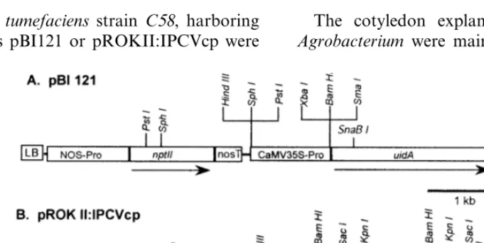
Plasmid pBI121 (13 kb; Fig. 2A) containing the
uidA (GUS) as reporter gene linked to the CaMV
35S promoter and NOS terminater andnptII gene
under the control of a nopaline synthase (NOS) promoter and terminater was used for initial opti-mization [24]. The plasmid pROKII:IPCVcp (Fig. 2B; kindly provided by Dr Mike Mayo, Scottish Crop Research Institute, Dundee, UK), contains the coat protein gene of the Indian peanut clump virus (IPCVcp). The plasmid pROKII:IPCVcp contains coat protein gene having 600 bp
untrans-lated 5% region followed by 650-bp coding region
driven by a CaMV 35S promoter and NOS
termi-nator and chimericnptII gene under the control of
NOS promoter and terminater within the T-DNA borders. Both these plasmids contain kanamycin resistance gene for bacterial selection. Plasmid pRT99gus:IPCVcp was constructed by cloning the
HindIII fragment of plasmid pRTL2:IPCVcp
(car-rying the IPCVcp ORF driven by a double 35S promoter, tobacco etch virus (TEV) leader and
terminated by 35S poly A signal) into the HindIII
site of plasmid pRT99gus [25] that has nptII and
uidA genes. Thus plasmid pRT99gus:IPCVcp
con-tains nptII, uidA and IPCV coat protein gene for
expression in plant tissues and ampicillin resis-tance for bacterial expression.
2.3. Transformation procedure
Disarmed A. tumefaciens strain C58, harboring
binary plasmids pBI121 or pROKII:IPCVcp were
used for transformation. Both plasmids specify the kanamycin resistance in the host bacteria while the
A. tumefaciens strain C58 also has resistance to rifampicin. Thus, the strains were maintained on
LB [26] agar plates containing 50 mg ml−1
kanamycin and 25 mg ml−1 rifampicin. Single
colonies of the individual strain were grown overnight at 28°C in 20 ml of YEB [26] supple-mented with appropriate antibiotics. Bacterial sus-pension (5 ml) was pelleted by centrifugation for 10 min at 5000 rpm and resuspended the cells in
30 ml of 1/2 strength MS-containing 3% sucrose
(1:6 dilution). This suspension was stored at 4°C for 1 – 2 h and used for co-cultivation. The bacte-rial suspension was poured in a sterile petri plate so as to make a thin film (2 – 3 mm) at the base of the petri plate. Freshly excised cotyledon explants from peanut variety JL-24 were taken and their proximal cut ends (Fig. 1A) were immersed into the bacterial suspension for few seconds and im-mediately implanted on SIM with the proximal cut ends embedded in the medium as mentioned above. The cotyledons were co-cultivated with the
Agrobacterium for 72 h and transferred to SIM supplemented with filter-sterilized cefotaxime (250
mg ml−1) and again care was taken to embed their
cut ends into media. Plating density was main-tained at five explants per plate.
2.4. Plant regeneration and selection of stable transformants
The cotyledon explants after treatment with
Agrobacterium were maintained on SIM
contain-Fig. 2. T-DNA regions of the binary plasmids used forA.tumefaciens-mediated transformation. (A) Plasmid pBI121 containing
nptII anduidA genes. (B) Plasmid pROK II:IPCVcp containing nptII and IPCVcp genes, (LB, left border; RB, right border;
ing filter-sterilized 250 mg ml−1 cefotaxime for 2
weeks when multiple shoot buds appeared on at least 70% of the explants while shoot buds con-tinue to form. At this stage, the explants bearing shoot buds were transferred to SIM containing
250 mg ml−1 cefotaxime and 125 mg ml−1
kanamycin to initiate selection and enrichment of transformed cells, organogenic tissues differenti-ated shoot buds for another 2 weeks. Subse-quently, proximal parts of the explants containing multiple adventitious shoot buds were excised and
transferred to SEM containing 125 mg ml−1
kanamycin for two to three subcultures of 4-week duration each. After this stage, the elongated shoots (3 – 4 cm) were cultured on RIM without any antibiotic for rooting of the elongated shoots. All the shoots that were cultured on RIM pro-duced multiple adventitious roots within about 2 weeks.
2.5. Culti6ation of putati6e transgenic plants
The rooted shoots were transferred to pots con-taining autoclaved sand and soil (1:1) mixture and maintained under high humidity (85%) at 25°C in a growth cabinet. After 2 weeks, they were trans-ferred to the glasshouse and allowed to flower and set seed. Upon flowering (2 months after trans-plantation), and pod formation (within 4 months) the mature seeds were collected and analyzed for the presence and expression of the introduced genes. The primary transformants upon transfer to the containment glasshouse were termed as T0 generation while those from subsequent seed
gen-erations were termed as cT1, cT2, cT3, and
so on.
2.6. Histochemical analysis of putati6e
transformants
b-Glucuronidase (GUS) enzyme activity was
de-tected histochemically in unfixed leaves and free-hand sections of petiole and stem sections of
regenerated plants growing in vitro on 125 mg
ml−1 kanamycin-containing medium or in the
containment glasshouse by using X-gluc
(5-bromo-4-chloro-3-indolyl-b-glucuronide) as the substrate
[24]. After the histochemical reaction at 37°C for 6 – 12 h the tissue was cleared in 70% ethanol and examined.
2.7. Molecular analysis
Total genomic DNA was isolated from fresh leaves of in vitro or glasshouse-growing putative transformants (T0 and T1 generations). Leaf tissue (1 g) was ground in liquid nitrogen with a mortar and pestle by following the method based on Dellaporta et al. [27] with some modifications. The ground fine leaf powder was transferred into 30-ml centrifuge tube and 15 ml of extraction buffer was added before thawing of the tissue. The contents were mixed well by inverting the tubes five to six times and 1 ml of 20% SDS was added prior to incubation at 65°C for 10 min. Five mililitres of 5M-potassium acetate was added to the extract
and incubated at −20°C for 20 min. The
superna-tant was collected after centrifugation and the DNA was precipitated with isopropanol and cen-trifuged at 10 000 rpm to collect the DNA pellet. The pellet was washed with ice-cold 70% ethanol, air-dried and dissolved in 10 mM Tris, pH 8.0. After treatment with ribonuclease A (RNase A) the DNA was resuspended in DEAE – cellulose (Whatman DE52) suspension to remove the polysaccharides and proteins followed by purifica-tion [28].
2.7.1. PCR analysis of putati6e transformants
Putative transformants were screened by poly-merase chain reaction (PCR) for the presence of
nptII and IPCVcp genes for routine analysis. The
700-bp region of nptII was amplified by using
22-mer oligonucleotide primers as previously re-ported [29]. A 585-bp coding region of the IPCVcp gene was amplified by using 21-mer
oligonucle-otide primers (IPCVcp primer I:5%-AGT TAC
TCG TGG TGG TGG TCA-3%; and IPCVcp
primer II:5%-GGA GTG GCC GCT GGA TTA
GGG-3%). The amplification reactions were carried
out by using a Techne™ PHC3 thermal cycler under the following conditions — 94°C for 4 min (one cycle), 92°C for 60 s (denaturation), 52°C for 45 s (annealing), 72°C for 90 s (extension) for 28 cycles and final extension at 72°C for 5 min (one
cycle). The PCR was performed by using 200 ng
were transferred to Hybond+nylon membranes (Amersham) by Southern blotting [26] and probed
with nptII or IPCVcp fragments from the
respec-tive plasmids (Fig. 2). The blots were hybridized
with the PstI fragment containing 800-bp coding
sequence of nptII gene or BamHI fragment
con-taining 1200-bp coding sequence of IPCVcp from
pROKII:IPCVcp labeled with non-radioactive
AlkPhos direct system™ (Amersham).
2.7.2. Southern blot analysis
For Southern hybridization analysis, the
ge-nomic DNA (10 – 15mg) from each of the putative
transformants was separately digested with
BamHI that has two restriction sites in the
pROKII:PCVcp orHindIII that has a single
inter-nal site in pBI121 and pROK II:IPCVcp (Fig. 2) to ascertain the integration pattern based on size separation and the number of copies of insert DNA. The digested DNA was run on 0.8% agarose gels to separate the DNA fragments that were transferred onto nylon membranes (Hybond
N+, Amersham) using standard protocols [26].
PCR amplified fragments of respective coding
se-quences (700 bp of nptII and 585 bp of IPCVcp)
were used as probes after labeling with non-ra-dioactive AlkPhos direct system (Amersham). The labeling, hybridization and detection methods were performed according to the manufacturer’s instructions.
3. Results
3.1. Shoot bud differentiation
In preliminary studies, the cotyledon explants (Fig. 1A) from mature seeds of peanut variety JL-24 were cultured on different media formula-tions containing varying concentraformula-tions of BA. Amongst the different media tested MMS
contain-ing 20 mM BA and 10mM 2,4-D (shoot induction
medium; SIM) produced the highest frequency (95.5%) of multiple adventitious shoot buds. The whole cotyledons also underwent regeneration at similar high frequencies (data not shown), but the number of shoots per responding explant was much higher when the cotyledons were split in vertical halves (Fig. 1A). Each half of the split cotyledon responded with similar frequencies while producing greater number of adventitious shoot
buds per explant (Fig. 1B). On SIM, the six peanut varieties produced shoot buds with high frequencies (80.0 – 97.7%) and followed a similar pattern of growth and development (Table 1). Variety JL-24 was used in all the experiments on the optimization of genetic transformation al-though recently, the experiments are also being carried out on variety ICGS-44 with similar results.
On SIM, the explants turned green and under-went considerable enlargement within 3 days of culture initiation. On these explants, multiple shoot buds differentiated at the proximal cut end (Fig. 1B) within 14 days in over 90% of the explants. However, no further elongation of the shoot buds occurred even after 4 weeks on SIM. Hence, the explants bearing shoot buds were cut into two to four pieces and transferred on to MS
medium containing 2 mM BA (shoot elongation
medium; SEM) for at least three passages of 4 week each when elongated shoots were rescued at the end of each passage. Frequently, four to eight shoots were recovered from each explant although over ten shoots could be recovered if the explants were subcultured on SEM for two to three extra subcultures. However, an important consideration for a high frequency of adventitious shoot bud regeneration is that the proximal cut end should be embedded into the medium so that it remains in contact with the medium at least for the first 2 week of culture initiation.
The shoots were micropropagated on SEM through nodal explants for clonal multiplication and eventually rooted on MS medium containing
5 mM NAA. The adventitious roots appeared
within 2 weeks (Fig. 1C) and developed further in 4 weeks (Fig. 1D) when the plants were ready for
transplantation to pots. All the rooted shoots (\
95%) survived after transplantation and appeared phenotypically normal. In the glasshouse, all the transformed plants produced flowers and pods within 4 months and these contained viable seeds. Each plant produced up to 40 pods that provided a total of up to 75 seeds per plant when grown in 12-in. pots.
3.2. Genetic transformation
transfor-mation. The explants were co-cultivated with
Agrobacteriumfor 72 h. Pre-culture of the explants on hormone-free MMS or SIM for 24 h did not have any beneficial effect. After 4 weeks of incuba-tion on the selecincuba-tion medium containing SIM with
250 mg ml−1 cefotaxime and 125 mg ml−1
kanamycin, some of the regenerated shoots (Fig. 1B) underwent bleaching, but it was not consid-ered very reliable for visual selection since some of the non-bleaching shoots were found to be un-transformed. Regenerating explants could survive
very high levels of kanamycin (up to 200 mg
ml−1). Although multiple shoots were formed on
72% of the explants, apparent bleaching was ob-served in less than 50% of the shoots. This obser-vation suggests that in peanut though kanamycin did not result in efficient visual selection of the transformants, it did play a selective role on the suppression of shoot bud induction from the un-transformed cells of the explants. This was also
observed during GUS histochemical and PCR
analysis where over 50% of the randomly selected
shoots tested positive. Other analogs of
kanamycin, i.e. paromomycin and geniticin, were also used for comparisons but none of these provide any distinct advantage over kanamycin (data not shown). The rooted shoots (Fig. 1D) were transplanted first under high relative humid-ity of 80% for 10 days and later to glasshouse where 100% of the shoots survived. Over 100 independently transformed putative transgenic plants were maintained in the glasshouse for fur-ther analysis. Transgenic plants appeared pheno-typically normal and set viable seeds. To date, no apparent undesirable genetic change due to tissue culture or transformation process has been ob-served in the glasshouse grown plants. This proce-dure has been tested with variety ICGS-44 for transformation responses and a large number of
putatively transformed shoots were rescued.
Moleular tests have confirmed their transgenic status (data not shown).
3.3. Characterization of transgenic plants
Each putative independent transformant arising from a treated explant was numbered at the time of isolation and separately maintained for subse-quent DNA analysis and progression of genera-tion. All the seeds collected from an individual transformant were suitably dried and stored at
5 – 10°C until further use. So far in excess of 75 independently transformed plants have success-fully been transplanted to the glasshouse and their T1 generation seed collected. About 40 of these have been advanced to the T2 generation. Trans-formed plants, which were obtained by using the
plasmid pBI121 and selected on 125 mg ml−1
kanamycin, were identified in the T0 generation
(primary transformants) putatively by using GUS
expression in the leaflets and petiole and stem
cross sections. b-Glucuronidase (GUS) expression
in the T1 generation progeny confirmed the
segre-gation of uidA gene in a Mendelian ratio of 3:1
(i.e., 15 out of 21 randomly selected plants from
the progeny of transformant c1R1 tested positive
for GUS expression in histochemical analysis).
Oligonucleotide primers specific to the coding
regions of nptII and IPCVcp genes amplified the
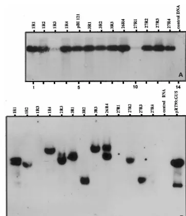
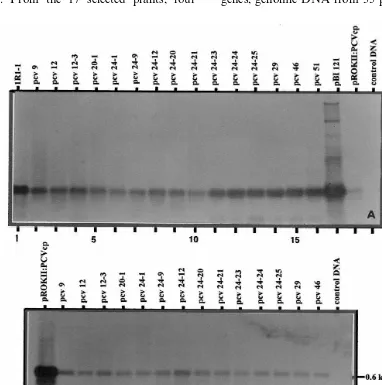
expected size of the respective gene fragments from at least 70% of the analyzed putative trans-formants (Fig. 3A and Fig. 4A, B). PCR analysis was performed on the genomic DNA of the trans-formants obtained with plasmid pBI121. Fig. 3A
shows the amplification ofnptII gene in randomly
selected transformants of T1 generation that origi-nated from four cotyledon explants where the negative plants (lanes 3 and 10) were due to segregation of the negative allele. To determine the authenticity of PCR products, the amplicons were transferred to nylon membrane where they showed
positive Southern hybridization with the npt II
gene probe. Southern hybridization analysis was performed on 14 of these plants to assay the presence and copy number of pBI121 T-DNA.
The results on presence of the npt II gene in PCR
analysis as shown in Fig. 3A were confirmed by Southern hybridization (Fig. 3B). In addition, the results in Fig. 3B also showed that all the selected plants were independent transformants that re-sulted from independent integrations. Eight of the ten positive transformants (80%) possessed a single copy insert whereas two (20%) had two copies. Occasionally, transformants showing multiple in-serts ranging from three to five copies were also observed. An interesting feature of the integration
pattern of nptII gene is that multiple plants
origi-nating from a single explant were due to indepen-dent transformation events (see Fig. 3B, lanes 1 – 4; 6 – 8; 11 – 14) suggesting the high efficiency of the cotyledon system. A similar pattern was also noted
Fig. 3. Molecular analysis ofnptII gene in the genomic DNA of T1 generation of peanut plants transformed with the plasmid pBI121 based inA.tumefaciensstrain C58. (A) PCR amplification of 700-bp fragment ofnptII coding region. The PCR products were resolved on 1.2% agarose gels and probed with non-radio Alkphos-labelednptII gene (0.8-kbPstI fragment of the plasmid pBI121) by Southern hybridization. (B) Southern blot analysis ofnptII gene. The DNA was digested withHindIII to provide a single restriction within the T-DNA. The blot was probed with non-radio Alkphos-labeled 700-bp PCR amplified nptII gene fragment.
nptII and uidA genes co-segregated in the T1
generation progeny.
The binary plasmid pROKII:IPCVcp having
nptII and the coat protein gene of Indian peanut
clump virus (IPCVcp) based in A. tumefaciens
strain C58 was used to obtain transgenic plants
from cotyledon explants. From the T0 and T1 generation, plants growing in the glasshouse puta-tive transformants were randomly selected for
PCR amplification ofnptII and IPCVcp gene
frag-ments from the genomic DNA. To ascertain the fidelity of amplifications in PCR reaction, the
PCR products were transferred to nylon mem-branes for Southern hybridization and probed with non-radio labeled fragments of the respective genes from the plasmids shown in Fig. 2. Trans-genic nature of all the selected plants was
confi-rmed by the expected amplification of the
respective genes (Fig. 4A, B). All the selected
samples of the progeny of plant c PCV 24 (lanes
6 – 13) showed positive amplification for bothnptII
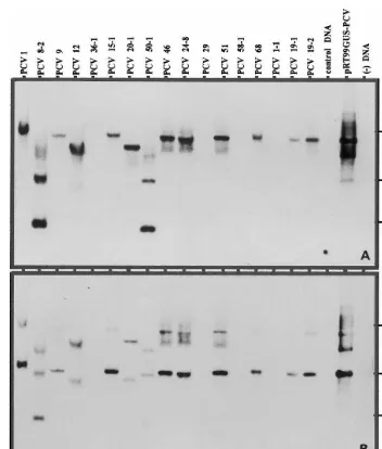
Randomly selected plants from T0 and T1 gen-erations growing in the glasshouse were selected for Southern hybridization to ascertain the
inte-gration and copy number of thenptII and IPCVcp
genes. Genomic DNAs of 17 putative transfor-mants obtained after transformation with the
plas-mid pROKII:IPCVcp were restricted withHindIII
that restricts the T-DNA only once. To maintain uniformity of observations the blot was initially
hybridized with the nptII gene probe and then
rehybridized with the IPCVcp gene probe after
stripping thenptII probe. Apparently the stripping
of nptII probe was not complete that resulted in
some overlap (Fig. 5B). However, a similar pattern
of integration of both nptII and IPCVcp genes
was evident. From the 17 selected plants, four
plants from the segregating generation were nega-tive (lanes 5, 11, 13, 15). The remaining 13 plants
showed positive hybridization for both nptII (Fig.
5A) and IPCVcp (Fig. 5B) genes. Two plants showed the integration of two copies (lanes 2 and 8) while the rest carried single inserts. Rarely plants with more than two and up to five integra-tions were also observed but the frequency of such plants was less than 5% of the total transformants. Interestingly, all the positively transformed plants exhibited distinct patterns of gene integration sug-gesting the origin of plants as a result of
indepen-dent transformation events. The two genes
co-segregated in the T1 progeny plants. To ascer-tain the inheritance pattern of the introduced genes, genomic DNA from 35 plants of T1
Fig. 5. Southern blot analysis ofnptII (A) and IPCVcp (B) genes in the genomic DNA of peanut plants transformed with A.
tumefacienscarrying the binary plasmid pROK II:IPCVcp. The DNA was restricted withHindIII to provide a single restriction within the T-DNA. The blots were probed with non-radio Alkphos-labeled PCR amplified fragment ofnptII (700 bp) and IPCVcp (585 bp) genes, respectively.
nies of one transformant (plant c PCV 24) that
has a single copy integration of IPCVcp gene was processed for Southern hybridization after
restric-tion withHindIII. As expected, a single-copy
inte-gration was observed in about 75% of the plants (data not shown) suggesting the segregation in 3:1 Mendelian ratio that is characteristic of a single locus trait.
4. Discussion
Several methods have earlier been reported to yield transgenic peanut plants that are in general
labor-intensive or often inefficient [8,10 – 16,30]. A comparison of different procedures for
transfor-mation of A.hypogaeaindicates wide variations in
transformation efficiencies as measured by shoot regeneration and yield of transformed shoots. The biolistic-based systems for gene delivery into em-bryogenic calluses and embryo axes is labor inten-sive and requires the bombardment of a large number of explants to obtain a few transformed cell lines (1%) which produce transgenic plants at low frequencies that are often chimeric or result from a few transformation events [8]. To date, the
shoot regeneration from the selected peanut
ex-plants. Although the reported methods using A.
tumefaciens yielded a large percentage of shoot regeneration and numerous transformed calluses, the percentage of confirmed transformed plants
was either not presented or varied between 0.3
and 9.0% [10,11,15,16]. A non-tissue culture based approach that does not rely upon adventitious shoot regeneration by utilizing peanut embryos to generate transgenic plants (in plant transforma-tion) has been adopted to overcome the problems associated with inconsistencies in in vitro shoot regeneration responses [15,16]. However, such methods are of limited use because they require high inputs in terms of the number of treatments to obtain very small number of independent trans-formants. In addition, such approach is labor
intensive requiring 6ir gene induction treatments
such as the use of wounded tobacco leaf extract to
enhance the transformation efficiency of A. tume
-faciens. For example, only three independent transformation events involving 150 embryos re-sulted in 2% transformation frequency [16] that often result in high degree of chimeric shoots where only a sector of T0 transformant may con-tain the T-DNA [15]. As a result, the use of peanut as a model system for gene expression studies has not been widely exploited. Hence, the method reported here is a significant improvement over the previously reported results on peanut transformation where at least 55% of the treated explants resulted in one or more independently transformed shoot.
The value of Agrobacterium-mediated plant
transformation is measured primarily by the num-ber of independently transformed plants carrying the gene of interest per explant used. This can be a function of genotype of the species to be
trans-formed, the Agrobacterium strain (virulence), the
selectable marker, regeneration capacity of the target cells, and the accessibility of the bacterium to the regenerable cells [31]. An additional, less-frequently quantified variable is the amount of labor required to maintain cultures until trans-formed shoots are obtained. The transformation procedure utilizing cotyledon explants reported
here is both highly susceptible to Agrobacterium
-mediated gene transfer and also displays very high
regeneration rates across a wide range ofA. hypo
-gaea varieties, often with numerous independently
transformed shoots per explant. The
manipula-tions used were simple as compared with several previously reported systems. An important aspect of this procedure is that multiple shoots are formed from a single explant. However, we have scored an explant as having a score of ‘1’ whether a single or multiple shoots were produced. These multiple shoots from individual explants are often due to independent transformation events at the surface, which has been confirmed by histological investigations (K.K. Sharma, unpublished data). Considering this observation and data on molecu-lar characterization, in which shoots originating from a single explant showed different integration pattern, the transformation frequency (in terms of number of shoots recovered from a single transfor-mation experiment), in fact, may be much higher than 55%.
The target cells for transformation in our exper-iments were those at the proximal cut surfaces of the cotyledon explants as has previously been
shown in Brassica napus[32] which has resulted in
very high levels of transformation frequencies [31]. The shoot development is very rapid (2 – 3 weeks)
and the cut surface is an ideal target for Agrobac
-terium-mediated transformation as the cells
under-going organogenesis are those most readily
accessed by the Agrobacterium. A key feature of
this procedure is the regenerability of the cells wounded during excision of the cotyledons and these cells are mainly surface cells, readily accessi-ble to the bacterium. This observation was also confirmed by relatively low levels of regeneration and transformation frequencies that we obtained when intact de-embryonated cotyledons were used as explants as compared with the split cotyledon explant.
Cotyledon explants have been shown to be ex-cellent explants for transformation and regenera-tion of fertile plants in several other crop species
including B. napus [31] where kanamycin has been
initial stages of screening. In the present study also, kanamycin selection was beneficial in pro-ducing transgenic plants from cotyledon explants when applied after 2 weeks of cocultivation.
Kanamycin even at even high levels of 200 mg
ml−1 restricted but not always completely
inhib-ited regeneration from control cotyledon explants and did not produce the bleaching effect which is typical of kanamycin on plant tissue cultures. However, kanamycin selection did enrich the growth of transformed tissue in shoot forming cotyledon explants since the proportion of posi-tively transformed plants was very high amongst randomly selected putative transformants.
In conclusion, the cotyledon regeneration sys-tem proved to be an excellent vehicle for the production of a large number of transgenic peanut plants over relatively short periods. In addition,
this explant allowed Agrobacterium-mediated
transformation to be targeted to regeneration competent tissue. Shoot formation was rapid and prolific, and a large proportion of these shoots
developed into phenotypically normal fertile
plants. Some of the transformed plants reported here have been positively tested for gene expres-sion in RT-PCR and Northern hybridization
stud-ies (data not shown). This protocol is
genotype-independent and provides a transforma-tion scheme that allows cost-effective, routine use of peanut transformation as part of basic studies in gene expression. The development of genetically transformed peanut cultivars with resistance to viruses and other biotic constraints should have tremendous impact on crop productivity especially in the resource-poor agricultural systems of the semi-arid tropics.
Acknowledgements
We greatfully acknowledge the technical assis-tance of M. Lavanya, D. Pandary and Jagan Mohan Reddy, and assistance of L. Vidyasagar with photography. Dr R. Ortiz, Dr S. Sivaramakr-ishnan and Dr N. Seetharama provided useful suggestions on the manuscript. We thank Dr Mike Mayo, Scottish Crops Research Institute, for providing plasmid pROKII:IPCVcp, and his con-stant encouragement. Part of the funding for this work was provided by grants from OPEC and Asian Development Bank.
References
[1] K.K. Sharma, R. Ortiz, Program for the application of genetic transformation for crop improvement in the semi-arid tropics, In Vitro Cell. Dev. Biol.-Plant 36 (2000), in press.
[2] H.T. Stalker, J.P. Moss, Speciation, cytogenetics, and utilization of Arachis species, Adv. Agron. 41 (1987) 1 – 40.
[3] M. Cheng, D.C.H. Hsi, G.C. Phillips, In vitro regenera-tion of Valencia-type peanut (Arachis hypogaeaL.) from cultured petiolules, epicotyl sections and other seedling explants, Peanut Sci. 19 (1992) 82 – 87.
[4] Z. Li, R.L. Jarret, R.N. Pittman, J.W. Demski, Shoot organogenesis from cultured seed explants of peanut (Arachis hypogaeaL.) using thidiazuron, In Vitro Cell. Dev. Biol. 30P (1994) 187 – 191.
[5] M. Kanyand, C.M. Peterson, C.S. Prakash, The differ-entiation of emergences into adventitious shoots in peanut, Arachis hypogaea (L.), Plant Sci. 126 (1997) 87 – 95.
[6] P. Venkatachalam, N. Geetha, A. Khandelwal, M.S. Shaila, G. Lakshmi Sita, Induction of direct somatic embryogenesis and plant regeneration from mature cotyledon explants of Arachis hypogaeaL, Curr. Sci. 77 (1999) 269 – 273.
[7] C. Lacorte, E. Mansur, B. Timmerman, A.R. Cordeiro, Gene transfer into peanut (Arachis hypogaea L.) by
Agrobacterium tumefaciens, Plant Cell Rep. 10 (1991) 354 – 357.
[8] P. Ozias-Akins, J.A. Schnall, W.F. Anderson, C. Singsit, T.E. Clemente, M.J. Adang, A.K. Weissinger, Regenera-tion of transgenic peanut plants from stably transformed embryogenic callus, Plant Sci. 93 (1993) 185 – 194. [9] M. Cheng, D.C.H. Hsi, G.C. Phillips, Recovery of
trans-formants of Valencia-type peanut using Agrobacterium tumefaciens, Peanut Sci. 21 (1994) 84 – 88.
[10] M. Cheng, R.L. Jarret, Z. Li, A. Xing, J.W. Demski, Production of fertile transgenic peanut (Arachis hypo
-gaea L.) plants using Agrobacterium tumefaciens, Plant Cell Rep. 15 (1996) 653 – 657.
[11] S. Eapen, L. George, Agrobacterium tumefaciens medi-ated gene transfer in peanut (Arachis hypogaeaL.), Plant Cell Rep. 13 (1994) 582 – 586.
[12] D.M. Livingstone, R.G. Birch, Plant regeneration and microprojectile-mediated gene transfer in embryonic leaflets of peanut (Arachis hypogaea L.), Aust. J. Plant Physiol. 22 (1995) 585 – 591.
[13] C. Singsit, M.J. Adang, R.E. Lynch, W.F. Anderson, A. Wang, G. Cardineau, P. Ozias-Akins, Expression of a
Bacillus thuringiensis cryIA(c) gene in transgenic peanut plants and its efficiency against lesser cornstalk borer, Transgenic Res. 6 (1997) 169 – 176.
[14] H. Yang, C. Singsit, A. Wang, D. Gonsalves, P. Ozias-Akins, Transgenic peanut plants containing a nucleocap-sid protein gene of tomato spotted wilt virus show divergent levels of gene expression, Plant Cell Rep. 17 (1998) 693 – 699.
[15] A.H. McKently, G.A. Moore, H. Doostdar, R.P. Niedz,
(Arachis hypogaeaL.) embryo axes and the development of transgenic plants, Plant Cell Rep. 14 (1995) 699 – 703. [16] V.K. Rohini, K.S. Rao, Transformation of peanut (Arachis hypogaea L.): a non-tissue culture based ap-proach for generating transgenic plants, Plant Sci. 150 (2000) 41 – 49.
[17] D.V.R. Reddy, R. Rajeshwari, N. Iizuka, D.E. Lese-mann, B.L. Nolt, T. Goto, The occurrence of Indian peanut clump, a soil-borne virus disease of peanut (Arachis hypogaea) in India, Ann. Appl. Biol. 102 (1983) 305 – 310.
[18] P. Delfosse, A.S. Reddy, P. Devi, A.K. Murthy, S.V. Wesley, R.A. Naidu, D.V.R. Reddy, A disease of wheat caused by Indian peanut clump virus (IPCV), Plant Dis. 79 (1995) 1074.
[19] D.C. Baulcombe, Mechanisms of pathogen-derived resis-tance to viruses in transgenic plants, Plant Cell 8 (1996) 1833 – 1844.
[20] S.V. Wesley, M.A. Mayo, C.A. Jolly, R.A. Naidu, D.V.R. Reddy, M.K. Jana, V.K. Parnaik, The coat protein of Indian peanut clump virus: relationships with other furoviruses and with barley stripe mosaic virus, Arch. Virol. 134 (1994) 271 – 278.
[21] C. Bragard, G.H. Duncan, S.V. Wesley, R.A. Naidu, M.A. Mayo, Virus-like particles assemble in plants and bacteria expressing the coat protein gene of Indian peanut clump virus, J. Gen. Virol. 81 (2000) 267 – 272. [22] T. Murashige, F. Skoog, A revised medium for rapid
growth and bioassays with tobacco tissue cultures, Phys-iol. Plant 15 (1962) 473 – 497.
[23] O.L. Gamborg, R.A. Miller, K. Ojima, Nutrient require-ments for suspension cultures of soybean root cells, Exp. Cell Res. 50 (1968) 151 – 158.
[24] R.A. Jefferson, Assaying chimeric genes in plants: the GUS gene fusion system, Plant Mol. Biol. Rep. 5 (1987) 387 – 405.
[25] R. Topfer, J. Schell, H.H. Steinbiss, Versatile cloning vectors for transient gene expression and direct gene transfer in plant cells, Nucleic Acids Res. 16 (1988) 8725. [26] J. Sambrook, E.F. Fritsch, T. Maniatis, Molecular Cloning, A Laboratory Manual, second ed., Cold Spring Harbour Laboratory Press, New York, 1989.
[27] S.L. Dellaporta, J. Wood, J.B. Hicks, A plant DNA minipreparation: version II, Plant Mol. Biol. Rep. 1 (1983) 19 – 21.
[28] L. Marechal-Drouard, P. Guillemaut, A powerful but simple technique to prepare polysaccharide-free DNA quickly and without phenol extraction, Plant Mol. Biol. Rep. 13 (1995) 26 – 30.
[29] J.D. Hamill, S. Rounsley, A. Spencer, G. Todd, M.J.C. Rhodes, The use of the polymerase chain reaction in plant transformation studies, Plant Cell Rep. 10 (1991) 221 – 224.
[30] M. Cheng, R.L. Jarret, Z. Li, J.W. Demski, Expression and inheritance of foreign genes in transgenic peanut plants generated by Agrobacterium-mediated transfor-mation, Plant Cell Rep. 16 (1997) 541 – 544.
[31] M.M. Moloney, J.M. Walker, K.K. Sharma, High effi-ciency transformation ofBrassica napususing Agrobac
-terium vectors, Plant Cell Rep. 8 (1989) 238 – 242. [32] J. Hachey, K.K. Sharma, M.M. Moloney, Efficient
re-generation of Brassica campestris using cotyledon ex-plants cultured in vitro, Plant Cell Rep. 9 (1991) 549 – 554.
[33] N.S. Nehra, R.N. Chibbar, K.K. Kartha, R.S.S. Datla, W.L. Crosby, C. Stushnoff, Agrobacterium-mediated transformation of strawberry calli and recovery of trans-genic plants, Plant Cell Rep. 9 (1990) 10 – 13.
[34] H. Mathews, W. Wagoner, J. Kellogg, R. Bestwick, Genetic transformation of strawberry: stable integration of a gene to control biosynthesis of ethylene, In Vitro Cell. Dev. Biol. 31P (1995) 36 – 43.