* corresponding author: [email protected]
α
αα
αα
-Lipoic acid inhibit the decrease of collagen
deposition in ultravioled B-irradiated cultured
normal human skin fibroblasts cell culture
Arum Krismi1*, Satiti Retno Pudjiati2, Yohanes Widodo Wirohadidjojo2 1Faculty of Medicine, Duta Wacana Christian University, Yogyakarta,
2Department of Dermatovenereology Faculty of Medicine, Gadjah Mada University/ Dr. Sardjito Hospital, Yogyakarta, Indonesia
ABSTRACT
Repeated ultraviolet B (UVB) irradiation on human skin has been considered to be responsible in premature aging process because UVB has been proved to inhibit collagen deposition and accelerates collagen degradation. Clinical studies showed that topical usage of 5% α-lipoic acid (ALA) improved the clinical appearance of photoaged skin. However, the effect of ALA on collagen deposition and degradation in UVB-irradiated normal human skin fibroblasts culture has not been reported. The aim of the study was to investigate the effect of ALA on collagen deposition and degradation in UVB-irradiated cultured normal human skin fibroblasts. Culture of normal human skin fibroblasts were treated with 0, 125, 250, 500 µM ALA diluted in complete Dulbecco’s Modified Eagle’s Medium (DMEM) and irradiated with 300 mJ/cm2 UVB. The mean collagen deposition and degradation’s level were measured by Sirius red assay and read with spectrophotometer at λ 550 nm. Mean difference of collagen deposition as expressed by optical density (OD) between normal human skin fibroblasts cell after UVB irradiation and without UVB irradiation was analyzed by Wilcoxon signed-ranks test and Friedman test, while mean difference collagen degradation was analyzed by one way analysis of variance (ANOVA) and pairedt test with 95% confidence level (p<0.05). The results showed that ALA 125 µM inhibited the decrease of collagen deposition significantly (p<0.05), though higher concentrations did not. However, ALA did not inhibit collagen degradation increment (p>0.05). In conclusion, ALA inhibited the decrease of collagen deposition, but did not inhibit collagen degradation in UVB-irradiated normal human skin fibroblasts culture.
Key words:α-lipoic acid - collagen - human skin - fibroblasts – UVB - irradiation
ABSTRAK
Pajanan sinar ultraviolet B (UVB) secara berulang pada kulit manusia dipercaya bertanggungjawab terhadap penuaan dini karena UVB terbukti dapat menurunkan timbunan dan meningkatkan degradasi kolagen. Penelitian klinik membuktikan bahwa penggunaan secara topikal sediaan asam α-lipoat (ALA) 5% dapat memperbaiki tampilan kulit menua dini. Namun, efek ALA pada timbunan dan degradasi kolagen kultur sel fibroblas normal manusia belum pernah dilaporkan. Tujuan penelitian ini adalah mengkaji efek ALA pada timbunan dan degradasi kolagen kultur fibroblas kulit normal manusia yang terpejan sinar UVB. Kultur fibroblas kulit normal manusia yang diberi ALA 0, 125, 250, 500 µM yang dilarutkan dalam DMEM dan dipajani sinar UVB 300 mJ/cm2. Kadar timbunan dan degradasi kolagen diukur menggunakan metode Sirius red dan dibaca dengan spektrofometer pada λ 550 nm. Perbedaan rerata timbunan kolagen sebagaimana ditunjukkan dengan densitas optik (OD) antara sel fibroblas kulit normal manusia setelah pajanan UVB dan tanpa pajanan UVB dianalisis dengan uji Wilcoxon dan uji Fiedman, sedangkan perbedaan degradasi kolagen dianalisi dengan uji t pasangan dan ANOVA satu jalan taraf kepercayaan 95% (p<0.05). Hasil penelitian menunjukkan bahwa ALA 125 µM menghambat penurunan rerata ALA 125 µM menghambat penurunan penimbunan kolagen secara bermakna (p<0.05), tetapi pada kadar lebih tinggi tidak menunjukkan penghambatan yang bermakna. Asam α-lipoat tidak menghambat peningkatan degradasi kolagen secara bermakna (p>0.05). Dapat disimpulkan bahwa ALA dapat menghambat penurunan timbunan tetapi tidak menghambat peningkatan degradasi kolagen kultur fibroblas kulit normal manusia yang terpajan sinar UVB.
INTRODUCTION
Ultraviolet B irradiation on human skin causes the formation of reactive oxygen species (ROS),1 which does not only damage interstitial collagen directly2 but also inactivates tissue inhibitors of metalloproteinase (TIMPs), as well as induces the synthesis and activates matrix metalloproteinases (MMPs),3 and causes an increase in collagen degradation. Repeated UVB irradiation on human skin may accelerate premature aging process since UVB cause a reduction in collagen deposition and an increase in collagen degradation.4
Human skin has a variety of defense mechanism against ROS. One of the mechanisms is non-enzymatic antioxidant system (i.e. vitamin A, vitamin E, vitamin C, polyphenol, and lipoic acid),5 which can be obtained either from endogenous or exogenous sources such as variety of human food consumption.6α-Lipoic acids, a natural cofactor in dehydrogenases complexes, is an endogenous antioxidant and physiologic constituent of mitochondrial membranes.7 In addition to be synthesized de novo in mitochondria by lipoic acid synthase, ALA can also be found in adequate amount in human diet from animal organs with multi-enzyme complexes such as meat, liver, and heart.6,8 Various studies have shown the antioxidant properties of ALA, both in vitro and in vivo.7,9
In regard with its low molecular weight (206.3 Dalton) and its solubility in both aqueous and lipid environment,7 ALA penetrates readily to skin, demonstrated by ALA distribution in dermis and subcutaneous tissue 4 hours after topical application on hairless mice’s skin.10 Clinical trials on human conducted by Perricone11 and Beitner12 have proved that 5% ALA topical cream improved the appearance of premature aging such as facial skin wrinkles and roughness. However, the effect of ALA on inhibition of collagen deposition reduction and collagen degradation increment in UVB-irradiated cultured normal human skin fibroblasts as a basis of premature skin aging appearance has not been reported.
MATERIALS AND METHODS
Samples
Primary cultures of normal human skin fibroblasts were established from 3 volunteers’
normal skin (foreskin) in Dulbecco’s Modified Eagle’s Medium (DMEM; Sigma-Aldrich Corporat-ion, St. Louis, MO, USA) supplemented with 5% bovine serum (BS), 100 µg/mL penicillin-strepto-mycin (Penstrep-Gibco; Invitrogen Corporation, Carlsbad, CA, USA), 100 mg/mL ceftriaxone, and 2.5 µg/mL amphotericine B (Gibco; Invitrogen Corporation, Carlsbad, CA, USA) in a 370C humidi-fied incubator containing 5% CO2. The fibroblasts were cultured to 60% confluence and then subcultur-ed. Cells cultured after 3 passages were used for the experiments. The study has been approved by the Medical and Health Reserach Ehich Committee, Faculty of Medicine, Gadjah Mada University.
α αα
αα-Lipoic acid
α-Lipoic acid was made of Mecola® forte (LAPI Laboratories, Jakarta, Indonesia) caplet containing 600 mg ALA. After the soft capsule was detached from the caplet, the content weight could be determined based on the total capsule weight subtracted by soft capsule weight. We used a mathematical equation to determine the requirement of ALA. The amount of ALA obtained from the calculation were then dissolved in sterile NaCl, diluted in complete DMEM, and used at a final concentration of 125, 250 and 500 ìM.
Treatment
Confluent fibroblasts culture at a density of 2 x 10⁴ cells/µL on a 96-microwell plate (Iwaki; Barloworld Scientific Laboratory, Stone, Stafford-shire, UK) were treated with ALA 0, 125, 250 and 500 ìM. After 24 hours, medium was removed and cells were rinsed twice with sterile phosphate-buffered saline (PBS). Column VII-XII was covered with opaque plaster, then the plate was irradiated with a banks of 6 UVB lamps (Philips UVB TL 40W/12RS; LIPI, Jakarta, Indonesia). The irradiance was 0.8 mW/cm2 at a distance of 25 cm. During irradiation, the culture medium was replaced with PBS to avoid the formation of medium derived toxic photoproducts induced by UV exposure. Subsequently, cells were incubated for 96 hours in complete DMEM.
Collagen measurement
with SpectraMax (Molecular Device Inc., Toronto, Canada) at λ 550 nm against 0.5 N NaOH as blank.
a. Collagen deposition
The medium was removed and cell layers were washed 3 times with PBS. The cell layers were then fixed with Bouin’s solution for 1 hour at room temperature. The solution was removed and plates were washed in running tap water until the yellow stain was removed. The plates were then air-dried in a fume hood overnight. Sirius Red dye solution (1 mg/mL in picric acid) was added to each well for 1 hour and placed under mild shaking. For 96-well plates, 200 µL of dye solution per well was used. After 1 hour, the dye solution was removed and each well was washed 3 times with 200 µL aliquots of 0.1 N HCl to remove unbound dye. The bound dye in each well was eluted with 200 µL of 0.5 N NaOH under mild shaking for 30 min, then the OD was measured.
b. Collagen degradation
Fifty µL aliquots of medium from each control group and sample well were diluted with 50 ìL PBS in eppendorf tubes and were then mixed with 1000 µL of Sirius Red. After centrifugation at 10.000 g for 5 minutes to precipitate the collagen-dye pellet, supernatant was discarded and drained off carefully. The pellet was mixed with 1000 µL of 0.1 N HCl and after centrifugation at 10.000 g for 5 minutes, supernatant was discarded and drained off carefully. Then pellet was mixed with 1000 µL 0.5 N NaOH and 200 µL aliquots of the alkali-dye solutions was transfered from the assay tubes to the 96-microwell plate, then the OD was measured.
Statistical Analysis
Wilcoxon signed-ranks test and Friedman test were used to determine the statistical significance of the mean collagen deposition’s OD differences, while pairedt test and one-way ANOVA were used to determine the statistical significance of the mean collagen degradation’s OD differences with 95% confidence interval (p<0.05).
RESULTS
Irradiation with 300 mJ/cm2 UVB resulted in significant decrease of mean collagen deposition (p<0.05), but did not increase the mean collagen degradation (p>0.05) compared to non-irradiated
cultured fibroblasts (FIGURE 1).
FIGURE 1. The comparison of collagen deposition and degradation OD between non-irradiated and 300 mJ/cm2
UVB-irradiated cultured fibroblast
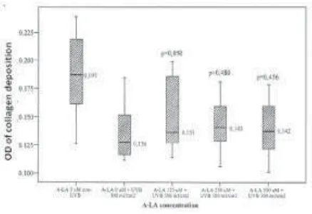
This study also found that treatment with various concentrations of ALA inhibited the decrease of mean collagen deposition on cultured fibroblast irradiated with 300 mJ/cm2 UVB.α-Lipoic acid 125
µM inhibited the decrease of mean collagen deposit-ion significantly (p<0.050) compared to placebo (0 µM ALA). The increase in ALA concentrations did not provide significant inhibitory effects on the mean collagen deposition decrement (FIGURE 2).
FIGURE 2. The comparison of collagen deposition OD in fibroblasts culture treated with various concentration of
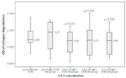
Furthermore, treatment with various concentrat-ions of ALA inhibited the mean collagen degradation increment on cultured fibroblast irradiated with 300 mJ/cm2 UVB.α-Lipoic acid 125 µM provided the
highest inhibitory effect on the mean collagen degra-dation increment despite insignificant (p>0.05) compared to placebo. The increase in ALA concen-trations did not provide significant inhibitory effects on the mean collagen degradation increment (FIGURE 3).
DISCUSSION
Decrease in the mean collagen deposition without an increase in mean collagen degradation (FIGURE 1) showed that irradiation with 300 mJ/ cm2 UVB on fibroblasts cell culture in this study decreased the synthesis of collagen, because the deposition of collagen is the result of a balance between collagen biosynthesis and degradation.13
The decrease of mean collagen synthesis in this study indicated that irradiation procedures have been conducted properly with adequate dose of irradiation and fibroblast culture used in this study did not differ to those used in other studies. It was supported by the decrease of mean collagen synthesis in this study in agreement with those found in the previous studies. Choi et al.14 proved that UVB irradiation increased MMP-1 expression, inhibited TGF-α1 expression, and decreased collagen synthesis. The decrease of mean collagen synthesis in UVB-irradiated fibroblast cell culture might also occur directly due to damage on collagen deposition15 and indirectly due to induction of synthesis and activation
of MMPs (particularly MMP-1) that plays role in collagen degradation.16
The collagen deposition and degradation in this study was possibly influenced by several factors, namely fibroblasts cell, UVB irradiation dose, the time of measurement, as well as the method of measurement. Although UVB irradiation have been proven to induce the synthesis and activation of MMPs, it was predicted that the main factor that affected the decrease of collagen deposition in UVB irradiated fibroblasts cell culture in this study was the decrease of collagen synthesis not the increase of collagen degradation.
The decrease of collagen deposition was inhibited significantly by 125 µM ALA, whereas 250 µM and 500 µM of ALA did not able to provide significant inhibitory effects (FIGURE 2). The inhibition of mean collagen deposition reduction in this study was in agreement with the study by Li et al.17 which proved that the ability of ALA as antioxidants (i.e. ROS scavenging) was greatly depend on its concentration and ALA with a concentration less than 100 µM could not scavenge ROS. Study by Lin et al.18 also proved that 500 µM ALA could not provide photoprotective effect on UV-irradiated skin.
In addition to its function as antioxidants, ALA might also function as pro-oxidants.8,19 Moini et al.19 proved that 250 µM ALA increased the fatty cells’ oxidant level significantly, and the ability of ALA as pro-oxidants increased with its concentration. Ability of 250 µM ALA as pro-oxidants was different from the study by Saliou et al.20 which proved that 250 µM ALA act as antioxidants by partially inhibited the activation of NF-κB on cultured human keratinocyte irradiated with 300 mJ/cm2 UVB. Inability of 250 µM and 500 µM ALA in inhibiting the decrease of mean collagen deposition significantly in this study showed that ALA with a high concentration was possibly more likely to act as pro-oxidants. Thus, this study showed that 125 µM ALA could act as antioxidants by inhibiting the decrease of collagen deposition in UVB-irradiated cultured normal human skin fibroblasts, and could be a potential agent for the prevention of premature skin aging appearance.
On the other hand, inhibition of ALA on the mean collagen degradation increment in this study FIGURE 3. The comparison of mean collagen degradation
(FIGURE 3) could not be concluded since the mean collagen degradation increment in UVB-irradiated cultured fibroblast was not significant compared to those of non-irradiated.
CONCLUSION
α-Lipoic acid inhibited the decrease of collagen deposition, but did not inhibit collagen degradation increment in cultured normal human skin fibroblasts treated with various concentrations of ALA and irradiated with UVB.
ACKNOWLEDGMENT
The authors would like to thank Head of Department of Dermato-Venereology, Faculty of Medicine, Gadjah Mada University/Dr. Sardjito Hospital, Yogyakarta for his permission to perfom this study.
REFERENCES
1. Masaki H, Atsumi T, Sakurai H. Detection of hydrogen peroxide and hydroxyl radicals in murine skin fibroblasts under UVB irradiation. Biochem Biophys Res Commun 1995; 206:474-9.
2. Ma W, Wlaschek M, Tantcheva-Poor I, Schneider A,
Naderi L, Razi-Wolf Z, et al. Chronological ageing and photoageing of the fibroblasts and the dermal connective tissue. Clin Exp Dermatol 2001; 26:592-9.
3. Brenneisen P, Oh J, Wlaschek M, Wenk J, Briviba K,
Hommel C, et al. Ultraviolet B wavelength dependence for the regulation of two major matrix-metalloproteinases and their inhibitor TIMP-1 in human dermal fibroblast. Photochem Photobiol 1996; 64:649-57.
4. Fisher GJ, Wang Z-Q, Datta SC, Varani J, Kang S,
Voorhees JJ. Pathophysiology of premature skin aging induced by ultraviolet light. New Engl J Med 1997; 337: 1419-28.
5. Young AR and Walker SL. Acute and chronic effects of ultraviolet radiation on the skin. In: W Klaus, AG Lowell, IK Stephen, AG Barbara, SP Amy, JL David (eds). Fitzpatrick’s Dermatology in General Medicine, 7th edition,
Vol. I, New York:McGraw-Hill, 2008.
6. Lee J, Koo N, Min DB. Reactive oxygen species, aging, and antioxidative nutraceuticals. Comp Rev Food Sci Saf 2003; 3:21-33.
7. Packer L, Witt EH, Tritschler HJ. α-lipoic acid as a biological antioxidant. Free Radic Biol Med 1995; 19:227-50.
8. Cakatay U. Pro-oxidant actions of α-lipoic acid and dihydrolipoic acid. Med Hypotheses 2006;66:110-7.
9. Biewenga GPh, Haenen GRMM, Bast A. The
pharmaco-logy of the antioxidant lipoic acid. Gen Pharmac 1997; 29:315-31.
10. Podda M, Rallis M, Cuber MG, Packer L, Maibach HI. Kinetic study of cutaneous and subcutaneous distribution following topical application of [7,8-14C]rac-α-lipoic acid onto hairless mice. Biochem Pharmacol 1996; 52:627-33. 11. Perricone NV. Topical 5% α-lipoic acid cream in the treatment of cutaneous rhytids. Aesth Surg J 2000; 20:218-22.
12. Beitner H. Randomized, placebo-controlled, double blind study on the clinical efficacy of a cream containing 5% á-lipoic acid related to photoageing of facial skin. Br J Dermatol 2003; 149:841-9.
13. Uitto J, Chu ML, Gallo R, Eisen AZ. Collagen, elastic fibers, and extracellular matrix of the dermis. In: W Klaus, AG Lowell, IK Stephen, AG Barbara, SP Amy, JL David (eds). Fitzpatrick’s Dermatology in General Medicine, 7th
edition, Vol. II, New York:McGraw-Hill, 2008.
14. Choi CP, Kim YI, Lee JW, Lee MH. The effect of
narrowband ultraviolet B on the expression of matrix metalloproteinase-1, transforming growth factor-α1 and type 1 collagen in human skin fibroblasts. Clin Exp Dermatol 2006; 32:180-5.
15. Menter JM, Patta AM, Sayre RM, Dowdy J, Willis I.
Effect of UV irradiation on type I collagen fibril formation in neutral collagen solutions. Photodermatol Photoimmunol Photomed 2001; 17:114-20.
16. Brennan M, Bhatti H, Nerusu KC, Bhagavathula N, Kang S, Fisher GJ, et al. Matrix metalloproteinase-1 is the major collagenolytic enzyme responsible for collagen damage in UV-irradiated human skin. Photochem Photobiol 2003; 78:43-8.
17. Li Y, Zhao Y, Yu W, Jiang S. Scavenging ability on ROS of α-lipoic acid (ALA). Food Chem 2004;84:563-7. 18. Lin JY, Lin FH, Burch JA, Selim MA, Monteiro-Riviere
NA, Grichnik JM, et al. α-lipoic acid is ineffective as a
topical antioxidant for photoprotection of skin. J Invest Dermatol 2004;123:996-8.
19. Moini H, Packer L, Saris NEL. Antioxidant and prooxidant activities of α-lipoic acid and dihydrolipoic acid. Toxicol Appl Pharmacol 2002; 182:84-90.