Bladder Cancer
Hybrid SPECT-CT: An Additional Technique for Sentinel Node
Detection of Patients with Invasive Bladder Cancer
Amir Sherif
a,*
,1, Ulrike Garske
b,1, Manuel de La Torre
c, Magnus Tho¨rn
daDepartment of Urology, Uppsala University Hospital, Uppsala, Sweden
bDepartment of Medical Sciences/Nuclear Medicine,Uppsala University Hospital, Uppsala, Sweden
cDepartment of Pathology and Cytology, Uppsala University Hospital, Uppsala, Sweden
dDepartment of Surgery, Karolinska Institute, South Stockholm General Hospital, Stockholm, Sweden
a v a i l a b l e a t w w w . s c i e n c e d i r e c t . c o m
j o u r n a l h o m e p a g e : w w w . e u r o p e a n u r o l o g y . c o m
Article info
Article history:
Accepted March 1, 2006 Published online ahead of print on March 20, 2006
Keywords:
Bladder cancer
Computed tomography Lymphoscintigraphy Sentinel node
Abstract
Objectives: To explore the feasibility of performing lymphoscintigraphy com-bined with computed tomography (CT) for preoperative detection of sentinel lymph nodes in patients with invasive bladder cancer.
Materials: Six consecutive patients scheduled for radical cystectomy underwent lymphoscintigraphy after transurethral injection of Albures-technetium 99m in the detrusor muscle peritumourally both with planar imaging and with single-photon emission computed tomography/CT (SPECT/CT). CT for anatomic fusion was performed directly after the SPECT/CT and both investigations were combined to a fused image. Radical cystectomy started with extended lymphadenectomy and intraoperative detection of sentinel nodes with both Geiger probe and dye marker. The conventional planar lymphoscintigraphies and the fused SPECT/CT were compared with each other and with the outcome of intraoperative sentinel node detection and final histopathologic analyses.
Results: The method allowed anatomically detailed preoperative visualisation of 21 sentinel nodes in five of the six patients, whereas planar pictures only visualised two sentinel nodes in two of six patients. Two patients had lymph node metastases and in the other four the nodes were negative. The combined method visualised all metastatic sentinel nodes, whereas planar lymphoscinti-graphy detected only one of six node metastases.
Conclusions: The combination of lymphoscintigraphy with CT enhanced preo-perative anatomic localisation of sentinel nodes in bladder cancer and aided in the identification of sentinel nodes during surgery. The yield of detected sentinel nodes, both metastatic and nonmetastatic, was markedly increased using the combined method compared to conventional planar lymphoscintigraphy.
#2006 European Association of Urology. Published by Elsevier B.V. All rights reserved.
1Both authors contributed equally to this work.
* Corresponding author. Department of Urology, Akademiska University Hospital, SE-751 85 Uppsala, Sweden. Fax: +46 18 50 79 07.
E-mail address:[email protected](A. Sherif).
1. Introduction
Detection of sentinel nodes as a tool for node staging has been explored and implemented with clinical success in the surgical treatment of breast cancer[1]
and malignant melanoma [2]. Recently, we per-formed sentinel node detection in patients with invasive bladder cancer[3]. The dynamic detection of sentinel nodes reveals not only the pathoana-tomic localisation of possible nodal metastases, in contrast to restricted lymphadenectomy, which is based on assumptions of lymph drainage in healthy individuals, but also provides a means to examine early metastatic progression in aspects of clonality and biomarker profiles[4]. The majority of patients undergoing cystectomy and traditional restricted lymphadenectomy have no lymph node metastases according to histopathologic examination [5]. A substantial percentage of these patients still have recurrences and die from disseminated disease. The overall 5-yr survival rate of only 50% in patients undergoing cystectomy is postulated to be caused by undetected metastatic spread to unidentified nodes. It is possible that staging could be improved and result in more accurate assessment of nodal status if sentinel nodes could be identified and examined in a majority of patients with invasive urothelial bladder cancer. The concept of sentinel node detection includes detection of lymph nodes that are sug-gested to be primed in receiving metastatic deposits at one time or another.The individual sentinel node status is thus considered to reflect the actual occurrence of primed sentinel nodes at the moment of excision, with or without metastatic deposits. One technical problem with the interpretation of planar lymphoscintigraphy in the conventional manner, as in our first study, is the difficulty in preoperatively identifying the precise anatomic localisation of the detected node. In most cases the nodes can be identified once intraoperative detection conse-quently is added to the examination, but still the certainty of lymphoscintigraphic localisation of nodes needs to be discussed and reassessed case by case. The problems of limited detection on planar lymphoscintigraphy, with an underestimation of actual number of sentinel nodes, has also been highlighted by other investigators [6]. In our first pilot study, planar lymphoscintigraphy yielded a sentinel node detection rate of 67% and two metastatic sentinel nodes of four, that is, 50%, were also recorded.
The purpose of this trial was to use a technique for better anatomic localisation of sentinel nodes and also to improve detection rates. Therefore we explored a newly described method in which
single-photon emission computed tomography (SPECT) was combined with computed tomography (CT) for anatomic fusion (SPECT/CT). So far this technique has been successful in sentinel node detection of malignant melanoma in two published reports[7,8]
and has been presented as a suitable method for sentinel node detection in prostatic carcinoma[9,10]. In the study of Even-Sapir et al., in 9 of 21 patients with a primary tumour located in the head and neck or trunk region, SPECT/CT fused images detected sentinel nodes that were missed on planar images. Two of these were metastatic sentinel nodes[8]. In a report from Wagner et al., comparing planar lym-phoscintigraphy with SPECT/CT, 30 patients with squamous cell carcinoma of the head and neck region were investigated. Forty-nine sentinel nodes were detected by SPECT/CT and 38 nodes by planar lymphoscintigraphy. The investigators even con-cluded that SPECT/CT is more sensitive to tumor-adjacent sentinel nodes than detection by g-probe,
due to the high activity at the injection site causing obscuring and difficult detection[11].
2. Patients and methods
2.1. Patients
The study included six consecutive patients with urothelial bladder cancer admitted to the University Hospital of Uppsala from October 2002 to January 2003 for cystectomy. The four men and two women were aged 59–75 yr. All six tumours originated from urothelial epithelium and were assessed as muscle invasive, T2a-T2b in five patients and as T1G3 with multiple carcinomas in situ (CIS) in one, according to the TNM classification of 1997 (Union Internationale Contre le Cancer) and histopathologically graded according to criteria of the World Health Organization and the International Pathology Consensus Committee (1988). Preoperative work-up with CT, intravenous pyelogram, and plain chest radiography did not show any local or distant metastases.
None of the patients had received neoadjuvant therapy. The study was approved both by the regional ethical committee and the regional radiation ethical committee. All six patients gave their informed consent to participate.
2.2. Preoperative sentinel node detection
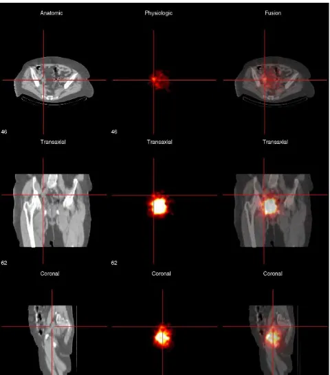
posterior, and lateral views was performed. We defined a sentinel node detected at planar lymphoscintigraphy as a delineated spot with increased radioactivity clearly separated from the sites of injection around the primary tumour. SPECT and subsequent CT scans were performed in one session and both acquisition data were fused to produce a combined picture
for further interpretation (Fig. 1). The CT scan was low-dose CT (2.5 mA compared with 150–250 mA as used for a full diagnostic CT) and no contrast was added. This CT scan did not replace the ordinary CT scan being performed preoperatively for planning and work-up. For SPECT, 60 angles at 30 s each were acquired with a low-energy general (LEG) purpose collimator and a
128128 matrix. The examination was performed with anatomic enhancement on ag-camera Millennium VG5 with
Hawkeye (General Electric); attenuation correction using the CT attenuation map and reconstruction of the raw data were carried out on an Entegra workstation (General Electric). One investigator (U.G.) was the sole interpreter of the lymphoscinti-graphies and the combined pictures of hybrid SPECT/CT.
2.3. Intraoperative sentinel node detection
Prior to major surgery the patient was initially in the lithotomy position and a second cystoscopy was performed. A total of 1 ml Patent blue (Bleu Patente´ V; Guerbet Laboratory, Issy les Moulineaux, France), divided into four equal portions, was injected peritumourally, with the intent to spot the same positions as previously done with radioactive tracer. If>24 h
had elapsed since the latter, renewed injections of Albures-technetium 99m were performed as described above. Intrao-peratively we performed both detections with the help of a handheld g-probe and visual detection of possibly
blue-colored nodes.
2.4. Surgery
One surgeon (A.S.) was the main surgeon in all six laparo-tomies. All six patients underwent cystectomy with lympha-denectomy and received urinary diversions accordingly. Lymphadenectomies were mainly carried out after the cystectomy or cystoprostatectomy, before formation of the urinary outlet. Lymphadenectomy included all sentinel nodes by the different described modalities and nonsentinel lymph nodes in obturator regions and along iliac vessels with the intent to include nodes up to the aortic bifurcation bilaterally.
3. Results
3.1. Lymph node yield
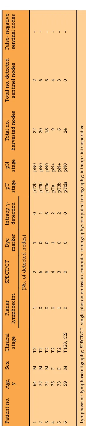
In total, 99 lymph nodes were harvested, with a range of 6–24 nodes. Twenty-one sentinel nodes were detected and excised (Tables 1 and 2). All nodes were subject to routine histopathology, and one of the sentinel nodes was re-examined with immuno-histochemistry (see below).
3.2. Overall sentinel node detection
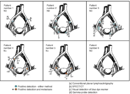
Sentinel nodes (nonmetastatic and metastatic) were detected at various pelvic localisations, and in five of the patients we found bilateral distribution (Fig. 2). Especially in patients 2 and 3 the combined lymphoscintigraphy/CT technique yielded a rela-tively high number of sentinel nodes. The mean number of detected sentinel nodes, calculated on all three methods of detection (SPECT/CT, visual detection of dye marker, and intraoperative g
-detection), was 3.5 and the median was 3.5 (range, Table
0–6). Total detection of sentinel nodes for the hybrid SPECT/CT technique was 21 of 21, 2 of 21 with the conventional planar lymphoscintigraphy, 2 of 21 with visual detection, and 11 of 21 with g-probe
detection. Thus the detailed background anatomy of the SPECT/CT allowed us to surgically retrieve the nodes undetected by intraoperative techniques. We compared SPECT/CT with planar images and it was clear that some of the lymph nodes detected using the new method were not seen when investigating only planar images. This was particularly the case for small-sized sentinel nodes (data not shown).
3.3. Detection of metastatic sentinel nodes
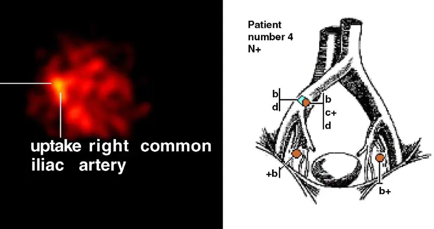
In the two patients with lymph node metastases (patients 4 and 5), the total numbers of metastatic sentinel nodes were three of four and three of three sentinel nodes, respectively. The combined techni-que visualized all metastatic sentinel lymph nodes in both patients (Table 2). In patient number 4, the detected metastatic sentinel node located over the right iliac bifurcation was primarily considered as being devoid of metastatic deposits. The pathologist re-examined the two excised nodes of that area
Table 2 – Results in detection of metastatic sentinel nodes with planar lymphoscintigraphy versus SPECT/CT
Patient no. Total no. metastatic sentinel nodes
No. of metastatic nodes detected by planar lymphoscintigraphy
No. of metastatic nodes detected by SPECT/CT
4 3 0 3
5 3 1 3
SPECT/CT: single-photon emission computer tomography/computed tomography.
using immunohistochemistry with the anticytoker-atin monoclonal antibody AE1/AE3. The result of that examination indicated that one of the nodes had a well-defined micrometastatic deposit (2 mm in size) and thus the patient had three detected metastatic sentinel nodes (Fig. 3). Both of the patients with nodal metastases had bilateral nodal deposits and totally two of metastatic nodes were detected in non-obturator localisations (Fig. 2).
3.4. pN staging
Sentinel node detection and ensuing histopathology adequately staged four patients as pN0, and two patients as pN+. None of the patients could be classified as false negative, considering that no metastases were found in the pathologic examina-tion of the excised nonsentinel nodes.
4. Discussion
Our feasibility study indicates that a combination of lymphoscintigraphy and CT improves preoperative anatomic localisation of sentinel nodes in urinary bladder cancer. We could now identify a higher number of sentinel nodes than with the techniques used in our previous report. The problems with identification of sentinel nodes with planar
lym-phoscintigraphy in urinary bladder cancer have recently been highlighted by another group working with similar concepts. In a series of 75 patients, only 7 of the first 30 patients had detectable sentinel nodes with planar lymphoscintigraphy, and for the remaining 45 patients this mode of investigation was totally abandoned[12].
The identification of bilateral metastatic sentinel lymph nodes was also a new finding in our own findings and adheres to the concept of a mandatory bilateral dissection [13]. The identification of a sentinel node with micrometastatic deposits (patient 4), finally being revealed by immunohisto-chemical examination, encourages us to pursue further refinement of the different techniques included. In that patient we found that the hybrid SPECT/CT technique seemed to be especially valu-able. The sixth patient in our series illustrates one problem of sentinel node detection. This patient reacted with a massive fibrosis in the pelvis minor after the primary transurethral resection, due to perforation of the bladder wall. The fibrosis per-sisted and ultimately constituted a major intrao-perative problem in the course of cystectomy. Our hypothesis is that the fibrosis might have obstructed any detectable lymphatic drainage, thus rendering us no detectable sentinel nodes at all. We find it of specific value to include this patient in our series, as an example of technical problems related to sentinel
Fig. 3 – In patient number 4, hybrid SPECT/CT detected a total of four sentinel nodes. Three (orange circles with a + sign) had metastatic deposits on histopathologic evaluation. On the left side is a close-up of the original SPECT over the right common iliac area, and on the right side a conclusive drawing over the distribution of sentinel nodes. [b] indicates detection
node detection patients with in urinary bladder cancer.
The original concept of sentinel node detection was based on the principle of one tumour-one primary sentinel node. However, the finding of more than one sentinel node is often reported, as in a large breast cancer multicentre study from 1998 (n= 443) where the mean number of sentinel nodes was 2.62.2 [14]. The detection of second- and even third-echelon metastatic sentinel nodes adds more considerations to the questions of defining the actual sentinel node[15]. At the time of presentation and detection, the presence of multiple detectable nodes, and also the presence of a number of detected metastatic sentinel nodes, is thus also being displayed in this present study. The explana-tion might be found both in the individual biologic timetable of the actual tumour/patient, the timing of detection, and the possibility of larger tumours having different sections being drained into anato-mically different lymphatic routes. For example, in a biologically early setting, a patient might have only one single detectable sentinel node, with or without metastatic deposits. Another patient, in a more advanced biologic setting, might have a number of detectable sentinel nodes, with a wide variation of metastatic spread or absence thereof. Simultaneous sentinel nodes might be considered to originate from different parts of a tumour with individual and separate drainage routes being established within the same time frame. That could also serve as an explanation for both crossover phenomena and bilateral distribution of nodal metastatic deposits. We consider all detected nodes as being so called ‘‘hot nodes,’’ thus being detectable nodes by the described methods. It should be pointed out that the term ‘‘hot nodes’’ should be considered as including both first-echelon sentinel nodes (i.e., ‘‘true’’ senti-nel nodes) and second- and third-echelon draining nodes. Until we have morphologic or biochemical markers to differentiate between the position of the nodes in the draining system, we have made the choice to designate all ‘‘hot nodes’’ as sentinel nodes. This is especially the case with the above-mentioned hypothetical argument of having differ-ent sections of the tumour being drained into different subsets of draining areas. We would consider it being suboptimal to just search for a single draining node, especially because we know that usually more than one affected metastatic node can be found in many patients with nodal spread and that distribution of metastatic nodes can be found in various localisations. In a recently pub-lished prospective trial from the Mansourah Center, bilateral nodal involvement was observed in 39% of
the patients with positive nodes [16]. Crossover phenomena were reported in 7% of the patients in the Swiss prospective investigation of 83 patients with positive nodes undergoing cystectomy. This group also noted that of the 39 patients who had a lateralized bladder tumour, 16 had nodes involved on the contralateral side [17]. The insufficiency of the present TNM classification for describing and stratifying node-positive patients with urinary bladder cancer has recently been highlighted in a review discussing lymph node metastasis in this cancer[18].
metastatic sentinel node loses its ability to harbour tracer of any kind[23]. Our feasibility trial includes a low number of patients, and in future attempts we would mainly aim to both increase the number of investigated patients and also to have two separate investigators doing the preoperative imaging. This would allow one investigator to interpret planar lymphoscintigraphy and independently have another investigator assess the SPECT/CT images. Ultimately the request for a more accurate staging of nodal status is linked to our different attempts to tailor treatment options for individual patients. Another attempt to combine different modalities in the investigation of both nodal and distant metastases in advanced bladder cancer is a recently published prospective trial on correlating whole-body fluorine-18 2-fluoro-2-dixoy-D-glu-cose-positron emission tomography (FDG-PET) and CT[24]. Attempts to further refine diagnostic accuracy by combining these methods with sentinel node detection seems to be the next tempting option.
By refining the techniques that can be used for detection of sentinel nodes, one might endeavour to increase the amount of detected sentinel nodes, thus extending the options of further basic research on the intrinsic factors governing routes of meta-static spread, presence or absence of metameta-static nodes, molecular profiling of primary tumours, nonmetastatic and metastatic sentinel nodes, examination of immunologic interactions occurring in primary tumor and sentinel nodes [22], and, finally, also exploring mechanisms of lymphangio-genesis and metastatic tumor-homing.
5. Conclusions
The combination of lymphoscintigraphy with CT enabled preoperative localisation of sentinel nodes in anatomic detail and facilitated detection during operation. Identification of more sentinel nodes than expected might be due to timing of the scintigraphy; some identified nodes may therefore be second- and third-echelon nodes. This additional technique provides us with more information but should be investigated further before any of the other methods of detection are excluded.
Acknowledgements
Dr Per Marits, MD, Department of Internal Medicine Uppsala University Hospital, for providing us with drawings.
References
[1] Schwartz GF. Clinical practice guidelines for the use of axillary sentinel lymph node biopsy in carcinoma of the breast: current update [review]. Breast J 2004;10:85–8. [2] Reintgen D, Balch CM, Kirkwood J, et al. Recent advances
in the care of the patient with malignant melanoma. Ann Surg 1997;225:1.
[3] Sherif A, De La Torre M, Malmstrom PU, Thorn M. Lym-phatic mapping and detection of sentinel nodes in patients with bladder cancer. J Urol 2001;166:812–5. [4] Malmstrom PU, Ren ZP, Sherif A, de la Torre M, Wester K,
Thorn M. Early metastatic progression of bladder carci-noma: molecular profile of primary tumor and sentinel lymph node. J Urol 2002;168:2240–4.
[5] Bassi P, Ferrante GD, Piazza N, et al. Prognostic factors of outcome after radical cystectomy for bladder cancer: a retrospective study of a homogeneous patient cohort. J Urol 1999;161:1494–7.
[6] Jansen L, Nieweg OE, Kapteijn AE, et al. Reliability of lym-phoscintigraphy in indicating the number of sentinel nodes in melanoma patients. Ann Surg Oncol 2000;7:624–30. [7] Kretschmer L, Altenvoerde G, Meller J, et al. Dynamic
lymphoscintigraphy and image fusion of SPECT and pel-vic CT-scans allow mapping of aberrant pelpel-vic sentinel lymph nodes in malignant melanoma. Eur J Cancer 2003;39:175–83.
[8] Even-Sapir E, Lerman H, Lievshitz G, et al. Lymphoscinti-graphy for sentinel node mapping using a hybrid SPECT/ CT system. J Nucl Med 2003;44:1413–20.
[9] Wurm TM, Eichhorn K, Corvin S, Anastidis AG, Bares R, Stenzl A. Anatomic-functional image fusion allows intraoperative sentinel node detection in prostate cancer patients. American Urological Association, San Francisco. J Urol 2004. p. 171, (abstract no. 854).
[10] Kizu H, Takayama T, Fukuda M, et al. Fusion of SPECT and multidetector CT images for accurate localization of pel-vic sentinel lymph nodes in prostate cancer patients. J Nucl Med Technol 2005;33:78–82.
[11] Wagner A, Schicho K, Glaser C, et al. SPECT-CT for topo-graphic mapping of sentinel lymph nodes prior to gamma probe-guided biopsy in head and neck squamous cell carcinoma. J Craniomaxillofac Surg 2004;32:343–9. [12] Liedberg F, Chebil G, Thomas Davidsson T, Gudjonsson S,
Ma˚nsson W. Intraoperative sentinel node detection improves nodal staging in invasive bladder cancer. J Urol 2006;175:84–9.
[13] Ghoneim MA, Abol-Enein H. Lymphadenectomy with cystectomy: is it necessary and what is its extent? [review] Eur Urol 2004;46:457–61.
[14] Krag D, Weaver D, Ashikaga T, et al. The sentinel node in breast cancer—a multicenter validation study. N Engl J Med 1998;339:941–6.
[15] Nieweg OE, Tanis PJ, Kroon BB. The definition of a sentinel node. Ann Surg Oncol 2001;8:538–41.
[17] Mills RD, Turner WH, Fleischmann A, Markwalder R, Thalmann GN, Studer UE. Pelvic lymph node metastases from bladder cancer: outcome in 83 patients after radical cystectomy and pelvic lymphadenectomy. J Urol 2001; 166:19–23.
[18] Liedberg F, Ma˚nsson W. Lymph node metastasis in blad-der cancer. Eur Urol 2006;49:13–21.
[19] Malmstrom PU, Sherif A, Thorn M. Re: extended radical lymphadenectomy in patients with urothelial bladder cancer: results of a prospective multicenter study. J Urol 2004;172:386, author reply p. 386.
[20] Jain RK, Padera TP. Development. Lymphatics make the break. Science 2003;299:209–10.
[21] Alitalo K, Mohla S, Ruoslahti E. Lymphangiogenesis and cancer: meeting report. Cancer Res 2004;64:9225–9. [22] Marits P, Mona Karlsson M, Sherif A, Garske U, Tho¨rn M,
Winqvist O. Detection of immune responses against urin-ary bladder cancer in sentinel lymph nodes. Eur Urol 2006;49:59–70.
[23] Keshtgar MR, Ell PJ. Clinical role of sentinel-lymph-node biopsy in breast cancer [review]. Lancet Oncol 2002;3:105– 10.