Abstract
Fruit of Mangosteen (Garcinia mangostana L.) is well-known in Indonesia and other Southeast Asian countries. Studies have shown that extract of the pericarp of mangosteen contained mostly of xanthones exhibit many biological aciviies, especially as an anitumor. This study aimed to invesigate the cytotoxic acivity and selecivity of Mangosteen Peel Extract (MPE) and α-mangosin against two leukemia cell lines (HL-60 and K-562) and the normal lymphocyte cells from two diferent donors. The cytotoxic acivity was performed using 3-(4,5-dimethylthiazol-2-yl)-5-(3-carboxyme-thoxyphenyl)-2-(4-sulfophenyl)-2H-tetrazolium (MTS) assay. Imainib and Isotreinoin were used as a posiive control to the K-562 and HL-60 cells, respecively. The MPE and α-mangosin revealed higher mortality toward leukemia cell lines rather than toward lymphocyte cells, with more than 80% of HL-60 and K-562 cells died at 6.25 and 25 µg/ml, respecively. MPE was more toxic and selecive against K-562 with IC50 of 2.79 µg/ml and SI of 8.27, while α-mangosin was more toxic and selecive against HL-60 with IC50 of 1.12 µg/ml and SI of 22.34. MPE and α-mangosin showed potent sensiivity and selecivity to leukemia cells, hence these are considered as promising sources for future leukemia treatment.
Cytotoxic Activity of Mangosteen (
Garcinia
mangostana
L.) Peel Extract and α-Mangostin
toward Leukemia Cell Lines (HL-60 and K-562)
Arina Novilla
1, Dedi S. Djamhuri
1, Nurul Fauziah
2, Maesaroh Maesaroh
2, Balqis Balqis
2and
Wahyu Widowati
3*
1School of Health Sciences Jenderal Achmad Yani, Cimahi, Indonesia 2Biomedical and Biomolecular Research Centre, Aretha Medika Utama, Bandung, Indonesia 3Faculty of Medicine, Maranatha Christian University, Bandung, Indonesia; [email protected]
*Author for correspondence
Email: [email protected]
1. Introduction
Cancer has been the second cause of death ater cardiovascular diseases in the world. here were around 8.2 million of people died because of cancer and about 14.1 million of new cancer cases were detected in 20121.
Particularly in Indonesia, there was about 0.1% of total people sufering from cancer with 74.6% of mortality rate
in male and 63.3% in female2. Lung or bronchus cancer,
prostate cancer in men, breast cancer in the female, and colon or rectum cancer reveal as the most deadly cancer worldwide. However, rare cancer such as leukemia is also a cause of death because of its destructive nature3. According
to the GLOBACAN, the percentage of leukemia was 2.5% of total new cases, contributing to 3.2% of total deaths by cancer in 20121 which was mostly sufered by children4.
Keywords: Mangosteen Peel Extract, α-mangosin, acute myeloid leukemia (AML), chronic myeloid leukemia (CML),
Characterized by high numbers of abnormal white blood cells, leukemia has been grouped into lymphoblastic and myeloid leukemia, namely Chronic Lymphocytic Leukemia (CLL) or Acute Lymphocytic Leukemia (ALL) and Chronic Myelogenous Leukemia (CML) or acute Myelogenous Leukemia (AML). Leukemia can be treated using interferon-α (IFN-α), Stem Cell Transplantation (SCT), radiotherapy and chemotherapy. Imatinib, a chemotherapy agent of tyrosine-kinase inhibitor, has been used as the irst-line treatment for CML. It induces apoptosis and inhibition of proliferation by inhibiting the phosphorylation and binding process of Bcr-Abl oncoprotein5. Another chemotherapy agent for leukemia
is retinoic acid, vitamin A derivative such as All-Trans Retinoic Acid (ATRA) and isotretinoin. hey are used for several cancer treatment and remission including for Acute Promyelocytic Leukemia (APL), a subtype of AML by inducing cell apoptosis6. However, the usage of imatinib
and retinoic acid as well as the other chemotherapy agents can generate side efects by damaging the normal cells and lead to the development of the multidrug resistance cancer7.
herefore, the inding of the safe, selective, and sensitive chemicals that have anticancer property is still needed.
Mangosteen (Garcinia mangostana L.) has been used as a traditional medicine to treat diseases related to immune and gastrointestinal for decades. Its extract contains several active compounds, including terpenes, anthocyanins, tannins, phenols, and xanthones8. Xanthones have been shown to have several pharmacological properties, including as an antitumor9. he most abundant and the most studied
xanthones are α-mangostin, β-mangostin, γ-mangostin, garcinone E, and gartanin, and those xanthones possess great biological activities such as anti-allergy, anti-inlammatory, antioxidant, anti-tumor, antibacterial, antifungal and antiviral activities, neuroprotective, cardioprotective, and immunomodulation10–12. Prior studies revealed that α-mangostin were the most abundance xanthone derivative from the aqueous, acetone and methanol extract of mangosteen9,13. herefore, this study aimed to investigate
the cytotoxic activity and selectivity of Mangosteen Peel Extract (MPE) and α-mangostin against two leukemia cell lines (HL-60 and K-562) and the normal lymphocyte cell from two diferent donors. he 3-(4,5-dimethylthiazol-2- yl)-5-(3-carboxymethoxyphenyl)-2-(4-sulfophenyl)-2H-tetrazolium MTS) assay was used to investigate the cytotoxic efect of the MPE, α-mangostin, imatinib, and isotretinoin. hose data are used to reveal the IC50 and Selectivity Index
(SI) of the MPE and three other compounds to the HL-60 and K-562 cell lines. he in vitro result demonstrates that MPE and α-mangostin have a potential to use against leukemia by showing the higher cytotoxic efect and selective index to leukemia rather than the imatinib and isotretinoin.
2. Materials and Methods
2.1 Mangosteen Peel Extract (MPE)
Preparation
he peels of Garcinia mangostana L. from Cisalak-Subang, West Java, Indonesia were determined by the herbarium staf of Biology Department, School of Life Sciences and Technology, Bandung Institute of Technology, Bandung, West Java, Indonesia. Around 350 g of chopped and dried-mangosteen peels were extracted using steady-state extraction (maceration) method. he chopped and dried peels were immersed in ethanol 70% for 24 hours and iltrated. he iltration was repeated until the iltrate became colorless. he iltrate was evaporated with a rotary evaporator at 40°C and the ethanolic extract of mangosteen peel was collected and stored in a freezer with the temperature of -20°C12,13.
2.2 Leukimia Cell Lines Cultivation
Two diferent leukemia cell lines, human acute promyelocytic (HL-60) and human chronic myelogenous (K-562) were obtained from Stem Cells and Cancer Institute (SCI) Jakarta, Indonesia. Cell lines were cultured in supplemented Iscove’s Modiied Dulbecco’s Medium (IMDM) (Biowest, France), 2% Penicillin-Streptomycin (Biowest, France), 10% Fetal Bovine Serum (FBS) (Biowest, France) and were incubated at 37°C in a humidiied incubator with 5% CO2. Ater 24 hours of incubation, the number of viable cells was counted using a haemocytometer with tryphan blue staining to obtain a suicient cell number for the cytotoxic assay.
2.3 Lymphocyte Isolation and Cultivation
the lymphocyte from other leukocytes histopaque (Sigma-Aldrich, U.S.A.) was added into the whole blood. Whole blood then was centrifuged at 1,800 rpm for 5 minutes to obtain the blood bufy coat. he blood bufy coat was added with 1x Phosphate Bufer Saline (PBS) (Biowest, France) and was centrifuged at 1,800 rpm for 10 minutes in 4oC. hen, the pellet was added with 1x PBS and was
centrifuged at 1,500 rpm for 10 minutes in 4oC. he inal
pellet contained leukocytes was maintained in Roswell Park Memorial Institute Medium (RPMI) (Biowest, France) and supplemented with 1% non-essential amino acids, 1% 1-glutamine, 100 U/ml penicillin, 10mg/mL streptomycin and 10% heat-inactivated fetal calf serum (Biowest, France) then incubated at 37oC in humidiied air containing 5%
CO214.
2.4 Cytotoxic Assay
Approximately 100 µL of lymphocyte, HL-60 and K-562 cell suspension contained 105 cells/ml were seeded in each
well of 96-well plate. Following that, 100 µL of Isotretinoin (Roche, Switzerland) in diferent levels (100, 85, 70, 55, 40, and 25 µM) were added into the wells of lymphocyte and HL-60. Imatinib (Glivec, Novartis) in diferent levels (100, 50, 25, 12.5, 6.25, and 3.125 µM) were added into the wells of lymphocyte and K-562. α-mangostin (Cengdu Biopurify Phytochemical, China) (100, 50, 25, 12.5, 6.25, and 3.125 µM) and Mangosteen Peel Extract (MPE) (100, 50, 25, 12.5, 6.25, and 3.125 µg/mL) were added into the wells of lymphocytes, HL-60, and K-562. Cells then incubated at 37°C with 5% of CO2 for 24 hours. hen, 20 µL of 3-(4,5-dimethylthiazol-2- yl)-5-(3-carboxyme-thoxyphenyl)-2-(4-sulfophenyl)-2H-tetrazolium (MTS) (Promega, U.S.A.) was added into each well and plate was incubated at 5% CO2, 37ºC for 3 hours. Finally, the absorbance used to calculate the percentage of cell mortality (Equation 1) was measured at 490 nm with Multiskan GO (hermo Scientiic, U.S.A). Wells of each cell that not supplemented with the compounds were presented as the negative control.
Atreated cell: Absorbance of cells treated with either MPE, α-mangostin, imatinib, or isotretinoin;
Auntreated cell: Absorbance of untreated cells
2.5 Selectivity Analysis
he Selectivity Index (SI) describes the selectivity of the MPE, α-mangostin, imatinib, and isotretinoin toward the leukemic cell lines. he index was calculated from IC50 ratio of the lymphocytes and the cancerous cell line (HL-60 or K-562) (Equation 2)15. he compound with SI higher than
3 revealed a high selective compound against the HL-60 or K-562, and the opposite was a less selective compound.
IC50 of normal cell: he value of median inhibitory concentration (IC50) of lymphocyte cells;
IC50 of cancer cell: he value of median inhibitory concentration (IC50) of cancer cells
2.6 Statistical Analysis
All treatment in this study was done independently and conducted in triplicate. SPSS Statistic 17.0 for Windows was used to calculate the IC50 of the compounds and the signiicant diferences using probit analysis and one-way ANOVA, respectively. he mortality and viability data were presented as mean ± Standard Deviation (SD).
3. Results
3.1 Cytotoxic Efect of Each Compound
toward HL-60, K-562, and Leukocyte
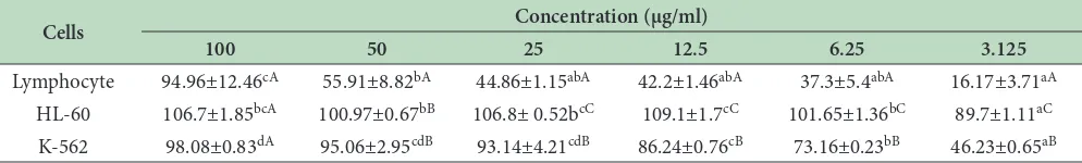
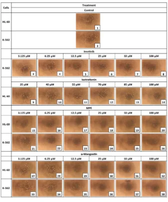
Table 1 to 4 show the cytotoxic efect of imatinib, isotretinoin, α-mangosteen and MPE presented as the percentage mortality of HL-60, K-562, and leukocyte. Isotretinoin and α-mangostin started at the concentration of 25 μM had more than 50% mortality to HL-60 and K-562, respectively. Imatinib and α-mangostin started at 12.5 μM showed more than 50% mortality to K-562 and HL-60, respectively (Table 1-3). hose of MPE started at 3.125 µg/ml and at 6.25 µg/ml revealed more than 50% mortality to HL-60 and K-562, respectively (Table 4). Furthermore, Figure 1 shows HL-60 and K-562 cell lines have lower density in a higher concentration of MPE and α-mangostin treatments. (1)
Cells Concentration (µM)
100 50 25 12.5 6.25 3.125
Lymphocyte 74.67 ± 11.09b 10.71 ± 5.82a 6.09 ± 6.48a 5.67 ± 6.57a -5.81 ± 1.92a -22.88 ± 10.86a
K-562 94.64 ± 0.11f 76.89 ± 1.01e 63.43 ± 0.74d 55.95 ± 0.99c 51.95 ± 0.9b 47.5 ± 0.8a
Table 1: The percentage mortality (%) of Lymphocyte and K-562 treated with Imatinib in six diferent concentrations
Data is presented as mean ± standard deviation, diferent superscript letters in each row (Lymphocyte, K-562) showed a signiicant diference at p<0.05 (Tukey HSD post hoc test) among the concentrations.
Cells Concentration (µM)
100 85 70 55 40 25
Lymphocyte 91.74±12.07b 60.39±17.70b 54.93±34.16b 39.4±14.56ab 15.05±4.31ab -38.14±11.43a
HL-60 99.05±1.11e 95.41±2.19de 83.48±1.02cd 80.53±2.48bc 69.52±4.66b 51.73±2.86a
Table 2: The percentage mortality (%) of Lymphocyte and HL-60 treated with Isotretinoin in six diferent concentrations
Data is presented as mean ± standard deviation, diferent superscript letters in each row (Lymphocyte, HL-60) showed a signiicant diference at p < 0.05 (Tukey HSD post hoc test) among the concentrations.
Cells Concentration (µM)
100 50 25 12.5 6.25 3.125
Lymphocyte 67.53±25.83aA 49.9±1.88aA 33.8±26.82aA 29.32±7.07aA 22.6±37.28aA 19.38±1.11aB HL-60 95.72±0.43dA 94.55±0.07dB 94.51±0.65dA 75.1±0.27cB -29.44±0.5bA -45.36±7.4aA K-562 96.2±0.11dA 94.65±0.3dB 91.29±0.27dA 16.31±0.65cA -21.72±3.08bA -68.47±8.35aA
Table 3: The percentage mortality (%) of Lymphocyte, HL-60, and K-562 treated with α-Mangosteen in six diferent concentrations
Data is presented as mean ± standard deviation, diferent lowercase of superscipt letters in each row (Lymphocyte, K-562, HL-60) showed a signiicant diference at p<0.05 (Tukey HSD post hoc test) among the concentrations, diferent uppercase of superscript letters in each column (100, 50, 25, 12.5, 6.25, 3.125) showed a signiicant diference at p<0.05 (Tukey HSD post hoc test) among the cells.
Cells Concentration (µg/ml)
100 50 25 12.5 6.25 3.125
Lymphocyte 94.96±12.46cA 55.91±8.82bA 44.86±1.15abA 42.2±1.46abA 37.3±5.4abA 16.17±3.71aA HL-60 106.7±1.85bcA 100.97±0.67bB 106.8± 0.52bcC 109.1±1.7cC 101.65±1.36bC 89.7±1.11aC K-562 98.08±0.83dA 95.06±2.95cdB 93.14±4.21cdB 86.24±0.76cB 73.16±0.23bB 46.23±0.65aB
3.2
Sensitivity and Selectivity of Each
Compound toward HL-60, K-562, and
Leukocyte
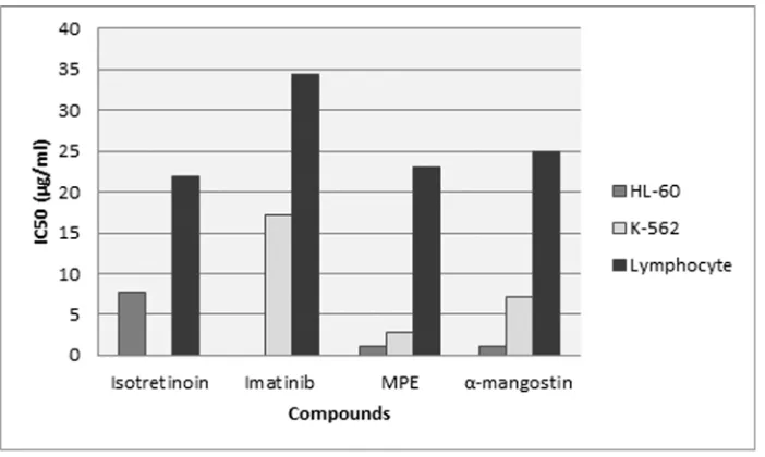
Further analysis of percentage mortality of the compounds using probit regression revealed that the IC50 of imatinib, isotretinoin, MPE, and α-mangostin were high to normal lymphocyte and the IC50 MPE and α-mangostin were comparably low to the leukemia cell lines (Figure 2). MPE
possessed the lowest IC50 (2.79 µg/ml) toward K-562 whereas α-mangostin possessed the lowest IC50 (1.12 µg/ml) to HL-60 (Table 5). he result of selectivity index showed that isotretinoin, a recent chemotherapy agent used to treat leukemia was less selective than MPE and α-mangostin toward HL-60, whilst imatinib was more selective toward K-562 than MPE and α-mangostin. Nevertheless, MPE and α-mangostin showed high selectivity toward both of HL-60 and K-562, with selectivity index was more than 3 (Table 5). Data is presented as mean ± standard deviation, diferent lowercase of superscript letters in each row (Lymphocyte, K-562, HL-60) showed a signiicant diference at p < 0.05 (Tukey HSD post hoc test) among the concentrations, diferent uppercase of superscript letters in each column (100, 50, 25, 12.5, 6.25, 3.125) showed a signiicant diference at p < 0.05 (Tukey HSD post hoc test) among the cells.
Fig. 2. IC50 values of isotretinoin, imatinib, Mangosteen Peel Extract (MPE) and α-mangostin toward leukemia cell lines (HL-60 and K-562) and normal lymphocyte.
4. Discussion
he activities of xanthones and its derivates to inhibit certain molecular target in cancer progression are related to the tricyclic scafold and its position8. As an anti-cancer, xanthones arrest cell cycle, induce apoptosis, and diferentiation but inhibit the tumor cell proliferation, adhesion, invasion, and metastasis16–18. Xanthones were also
reported to prevent initiation stage of cancer by inducing Quinine Reductase (QR) and inhibiting cytochrome P450 (CYP) activity9,19.
In this study, imatinib, isotretinoin, MPE, and α-mangostin showed a cytotoxic efect on the HL-60, K-562, and lymphocyte cells in a concentration-dependent manner. Microscopic images showed the cell density gradually
decreased in the higher concentration of each treatment (Figure 1). his result was supported by the percentage of mortality data that showed the cell mortality increased in higher concentration of each treatment. Based on the Table 5, the IC50 value of MPE and α-mangostin toward HL-60 and K-562 cell lines was lower than the IC50 value toward lymphocyte, with the selectivity index higher than 3. hese indings suggested that MPE and α-mangosteen were safe against the normal lymphocyte, and possessed a high selectivity and sensitivity toward HL-60 and K-5628,15.
hese results were consistent with Matsumoto et al.22 study
that showed the antiproliferative activity of xanthones in mangosteen pericarp against human leukemia20. he
α-mangostin mediates mitochondrial apoptotic pathway in human promyelocytic leukemia (HL-60) by activating
Table 5: The cytotoxicity and selectivity of MPE, α-mangostin, imatinib and isotretinoin against HL-60, K-562, and leukocyte
Compounds
Cell Line
HL-60 K-562 Lymphocyte
IC50 (µg/ml) SIa IC
50 (µg/ml) SIa IC50 (µg/ml) MPEb 1.16 19.83 2.79 8.27 23.08
α-mangostin 1.12 22.34 7.21 3.47 25.02 Isotretinoin 7.66 2.86 - - 21.93
Imatinib - - 2.84 12.07 34.35
the caspase-3 and caspase-921. Together with β-mangostin, γ-mangostin, and methoxy-β-mangostin, these compounds arrest the cell cycle via expression of cyclin proteins in the human colon cancer cells (DLD-1)11. Furthermore, other
anti-tumor activities of mangostins in several cancers have also been reported, including the inhibition of TCF/β-catenin transcription in colon cancer cells and inhibition of cell growth signaling pathways in chondrosarcoma22. hese
indings suggest that xanthones work by various pathways to cancer cells.
In contrast, isotretinoin was either less selective or less sensitive toward the HL-60 cells compared with MPE and α-mangostin. Cancer has been known to be able to develop resistance towards chemotherapy. he resistance of leukemic cell lines to retinoic acid derivate might occur in molecular level by afecting several proteins functions and the mutations of the RARα receptor in APL can block the initiation of diferentiation by retinoic acid23. On the
contrary, Imatinib shows good sensitivity and selectivity toward K-562 human Chronic Myeloid Leukemia (CML) cells. CML is characterized by the presence of a Bcr-Abl fusion gene, which is caused by a reciprocal translocation of chromosomes 9 and 2224. he cytotoxic activity of imatinib
was supported by other study, demonstrated that imatinib was able to inhibit Bcr-Abl kinase activity led to inactivate of survival pathways and induce long-term activation of caspases that responsible for the degradation and inactivation of Bcr-Abl tyrosine kinase as well as apoptosis of the K562 cells25.
5. Conclusion
Take together, our data suggest that MPE and α-mangostin possessed potent sensitivity and selectivity against leukemia. Both of them revealed higher selectivity and sensitivity than isotretinoin toward HL-60 cell line, while MPE also show high sensitivity and selectivity toward K-562 cell line, showing its great potential for pharmaceutical application. We suggest that MPE can be produced as a safe, eicient and low cost of an alternative remedy to ight leukemia. herefore, the further study of mangosteen peel extract in molecular and in vivo study must be conducted.
6. Acknowledgements
We gratefully acknowledge the inancial support from Research Grant of Hibah Bersaing 2016 by Ministry of Research, Technology and Higher Education of the
Republic of Indonesia and Biomedical and Biomolecular Research Centre Aretha Medika Utama, Bandung, Indonesia for research grant, research method and facilities support. We are also thankful to I Dewa Gde Sathya Deva from Biomedical and Biomolecular Research Centre Aretha Medika Utama, Bandung, Indonesia for his valuable assistance.
7. References
1. Ferlay J, Soerjomataram I, Dikshit R, Eser S, Mathers C, Rebelo M, et al. Cancer incident and mortality worldwide: Source, methods and major patterns in GLOBOCAN 2012. Int J Cancer. 2014; 136:349–86.
2. McDonald M, Hertz R, Lowcnthal SW. Pizer facts: he burden of cancer in Asia USA: Pizer Medical Division; 2008.
3. Modak H, Kulkarin SS, Kadakol GS, Hiremath SV, Patil BR, Hallikeri U, et al. Prevalence and risk of leukemia in the multi-ethnic population of North Karnataka. Asian Pac J Cancer Prev. 2011; 12:671–5.
4. Belson M, Kingsley B, Holmes A. Risk factors for accute leukemia in children: A review. Environ Health Persp. 2007; 115(1):138–45.
5. Henkes M, van der Kuip H, Aulitzky WE. herapeutic options for chronic myeloid leukemia: focus on imatinib (Glivec®, GleevecTM). her Clin Risk Manag. 2008; 4(1):163–87.
6. Niles RM. Recent advances in the use of Vitamin A (Retinoids) in the prevention and treatment of cancer. Nutr. 2000; 16:1084–90.
7. Gottesman MM, Fojo T, Bates ES. Multidrug resistance in cancer. Nat Rev Cancer. 2002; 2:48–58.
8. Shan T, Ma Q, Guo K, Liu J, Li W, Wang F, et al. Xanthones from mangosteen extracts as natural chemopreventive agents: potential anticancer drugs. Curr Mol Med. 2011; 11(8):666–77.
9. Foti RS, Pearson JT, Rock DA, Wahlstrom JL, Wienkers LC. In vitro inhibition of multiple cytochrome P40 isoforms by xanthone derivatives from mangosteen extract. Drug Metab Dispos. 2009; 37(9):1848–55.
10. Yang J, Liu RH, Halim L. Antioxidant and antiproliferative activities of common edible nut seeds. Lwt-Food Sci Technol. 2009; 42(1):1–8.
mangostana xanthones extract. BMC Compl Alternative Med. 2012; 12(104):1–10.
12. Darsono L, Hidayat M, Maesaroh M, Fauziah N, Widowati W. Ex vivo study of Garcinia mangostana L. (Mangosteen) peel extract and xanthones as anti-adipogenesis in HepG2 cell model. Int J Med Res Health Sci. 2015; 4(3):566–71. 13. Widowati W, Darsono L, Suherman J, Yelliantty Y,
Maesaroh M. High Performance Liquid Chromatography (HPLC) analysis, antioxidant, antiaggregation of mangosteen peel extract (Garcinia mangostana L.). Int J Biosci Biochem Bioinfo. 2014; 4(6):458–66.
14. Boyum A. Isolation of mononuclear cells and granulocytes from human blood. Isolation of monuclear cells by one centrifugation, and of granulocytes by combining centrifugation and sedimentation at 1 g. Scand J Clin Lab Invest Suppl 1968; 97:77–89.
15. Mahavorasirikul W, Viyanant V, Chaijaroenkul W, Itharat A, Na-Bangchang K. Cytotoxic activity of hai medicinal plants against human cholangiocarcinoma, laryngeal and hepatocarcinoma cells in vitro. BMC Complement Altern Med. 2010; 10(55):1–8.
16. Akao Y, Nakagawa Y, Iinuma M, Nozawa Y. Anti-cancer efects of xanthones from pericarps of mangosteen. Int J Mol Sci. 2008; 9:355–70.
17. Pedraza-Chaverri J, Cardenas-Rodriguez N, Orozco-Ibarra M, Perez-Rojas JM. Medicinal properties of mangosteen (Garcinia mangostana). Food Chem Toxicol. 2008; 46:3227–39.
18. Hung SH, Shen KH, Wu CH, Liu CL, Shih YW. Alpha-mangostin suppresses PC-3 human prostate carcinoma cell metastasis by inhibiting matrix metalloproteinase-2/9
and urokinase-plasminogen expression through the JNK signaling pathway. J Agric Food Chem. 2009; 57(4):1291– 8.
19. Chin YW, Jung HA, Chai H, Keller WJ, Kinghorn AD. Xanthones with quinone reductase-inducing activity from the fruits of Garcinia mangostana (Mangosteen). Phytochemistry. 2008; 69(3):754–8.
20. Matsumoto K, Akao Y, Kobayashi E, Ohguchi K, Ito T, Tanaka T, et al. Induction of apoptosis by xanthones from mangosteen in human leukemia cell lines. J Nat Prod. 2003; 66:1124–7.
21. Matsumoto K, Akao Y, Yi H, Ohguchi K, Ito T, Tanaka T, et al. Preferential target is mitochondria in alpha-mangostin-induced apoptosis in human leukemia HL60 cells. Bioorg Med Chem. 2004; 12(22):5799–806.
22. Krajarng A, Nakamura Y, Suksamram S, Watanapokasin R. Preferential target is mitochondria in alpha-mangostin-induced apoptosis in human leukemia HL60 cells. J Agric Food Chem. 2011; 59:5746–54.
23. Niles RM. Recent advances in the use of vitamin A (Retinoids) in the prevention and treatment of cancer. Nutr. 2000; 16:1084–90.
24. Deininger M, Buchdunger E, Druker BJ. he development of imatinib as a therapeutic agent for chronic myeloid leukemia. Blood. 2005; 105(7):2640–53.