Determination and Genetic Variation Analysis of
Wild-type and Albino’s Mutant of Monascus spp. by
Random Amplification of Polymorphic DNA (RAPD) Method
Tutus Gusdinar1), Marlia Singgih1), Tiana Milanda2), Haryanto Dhanutirto1)
1)
School of Pharmacy ITB-Indonesia
2)
Dept. of Pharmacy Padjadjaran University-Indonesia
ABSTRACT
Determination and genetic variation analysis of wild-type and albino’s mutan of
Monascus spp. isolated from Cikapundung River at Bandung area, were carried out using Random Amplification of Polymorphic DNA (RAPD) method. The albino’s mutant, was obtained by Ethyl Methane Sulphonate (EMS)-treated spores of the wild-type. RAPD result of Monascus spp wild-type was compared with Monascus purpureus CECT 2955T as reference standard, and it was found that both RAPD profiles observed was identical. These result showed that the wild-type is proved to be Monascus purpureus. However, one of the DNA bands of albino’s mutant at 1150 bp was not found on the wild-type and the standard, and it was considered as a genetic variation resulted from the mutation process.
Key words : RAPD, Monascus purpureus, wild-type, albino’s mutant
INTRODUCTION
also used commercially to produce alcohol, organics acids and enzymes (Chen and Tseng, 1989; Martinkova et al., 1995)
M. purpureus was first isolated from Chinese anka in Java, Indonesia (Wong and Bau, 1977). Based on morphological and biochemical reactions, 12 species and two varietas of Monascus were identified from isolates from Asian countries (Lizuka and Lin, 1981). Hawksworth and Pitt (1983) identified Monascus species using microscopic morphological characteristic on different culture media. But variation due to mutations or genetic recombination in reproductive structure can make identification of Monascus
difficult at specific level.
Determination of genetic variation in fungi can be assessed by several biology techniques such as protein profile analysis, isoenzyme analysis, RFLP (Cruz et al., 1996), PCR (Mullis et al., 1986), RAPD (Welsh and and McClelland, 1990) and AFLP (Peleman et al., 1995) have provided rapid, accurate indications of genetic relationships. K. Lakrod et al. (2000) initiated genetic investigation of Monascus spp using RAPD markers and cluster analysis to determine the level of genetic variation within a collection of isolates from food products from Asian countries. S. Campoy et al. (2003) characterized a high-level pigment-producing Monascus IBCC1 as M. purpureus by RAPD method too.
In this work, we determine and analysis of genetic variation of a wild-type and an albino’s mutant of Monascus spp. by RAPD method. Our objective was to identified species of the wild-type and to asses the genetic variation resulted from the mutation process in albino’s mutant. This mutant will be used as cell receptor in a genetic transformation system for Monascus purpureus.
MATERIALS AND METHODS
Strains and culture conditions
All fungal strains were grown in YMP agar (0.3% yeast extract, 0.3% malt extract, 0.6% peptone, 2% glucose, 1% bacto-agar). Spores from one big glasstube of YMP agar were collected using sterile aquadest. The spores were used to inoculate liquid cultures in YMP broth.
Isolation and extraction of fungal DNA
Mycelial masses of each Monascus were grown and increased in 20 mL YMP broth using a spore suspension (105-106 spores/mL) from 7-10 days culture in YMP agar as starter. Liquid cultures were incubated at room temperature on an orbital shaker (150 rpm) for 20 hours. Mycelial masses were harvested by filtration using Whatman No. 1 paper.
DNA’s of each isolate were extracted in CTAB extraction buffer using small
potter for several minutes. Mycelial lysate were incubated at 65°C for 45 minutes, then extracted with chloroform. Nucleic acid were precipiteted using one of isopropanol, then were redissolved in Tris-EDTA pH 8 (100 mM Tris pH 8, 1 mM EDTA). RNA’s were removed by adding DNAse free RNAse. DNA’s were further extracted using phenol-chloroform-isoamylalcohol (25 :24 :1) at least twice, followed by chloroform extraction at least once. DNA’s were precipitated using 0.1 vol 3 M sodium acetate pH 5.2 and 1 vol. isopropanol. DNA pellets were washed with 70% ethanol and redissolved with Tris-EDTA pH 8.
RAPD analysis
Total genomic DNA’s from each Monascus were spectrophotometrically quantified at 260-280 m and were diluted to 50 mg/mL before using as a DNA template in a RAPD analysis. RAPD rections were performed using oligonucleotides CRL9 (5’-CAGCCGCCCC-3’) and CRL12 (5’-CGCCGCCCG-3’), according to Kubelik And Szabo (1995). The PCR reaction were performed in DNA thermocycler (Perkin-Elmer) using 25 µL volume containing 0.5 units of Taq DNA polymerase, Taq polymerase buffer (Sigma Chemical Co.), 90 nmol MgCl2, 8 fmol DNA primer, 5 nmol of each
at 94°C, followed by 44 cycles of 40 s at 94°C, 60 s at 34°C and 120 s at 72°C, with a
final 10 min at 72°C.
RAPD products were electrophoretically separeted in 1.6% agarose in 0.5 TBE. RAPD fragments were examined by ethidium bromide staining and transmitted uv lights. Gels were photographed with a Polaroid camera using Polaroid film.
RESULTS
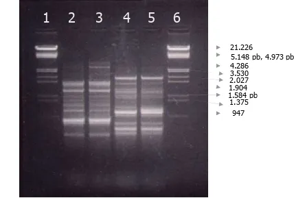
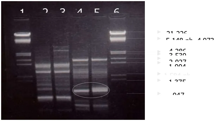
DNAs from Monascus spp. KM1, the albino’s mutant and reference strains (Monascus purpureus CECT 2955T) were compared by RAPD (Fig 1 and Fig.2). Identical bands were observed both of the wild type and the reference. However, one of the DNA bands of albino’s mutant at 1150 bp was not found on the wild-type and the reference.
b
Fig. 1a RAPD amplification of Monascus spp. DNA using oligonucleotides CRL9 and CRL12.
Lane 1 and 6 DNA markers
Lane 2 Monascus purpureus CECT 2955T/CRL9 Lane 3. Monascus spp KM1/CRL9
Lane 5. Monascus spp KM1/CRL12
Fig. 2 RAPD amplification of Monascus spp. DNA using oligonucleotides CRL9 and CRL12.
Lane 1 and 6 DNA markers
Lane 2 Monascus purpureus CECT 2955T/CRL9 Lane 3. Monascus albino’s mutant/CRL9
Lane 4 Monascus purpureus CECT 2955T/CRL12 Lane 5. Monascus albino’s mutant/CRL12
DISCUSSION
The identical of RAPD profiles of Monascus spp wild-type and Monascus purpureus CECT 2955T showed that the wild-type is proved to be Monascus purpureus. One of the DNA bands of albino’s mutant at 1150 bp was not found on the wild-type and the standard, and it was considered as a genetic variation resulted from the mutation process.
ACKNOWLEDGEMENT
REFERENCES
Campoy, S., F. Perez, J.F. Martin, S. Guiterrez and P. Liras (2003) Stable transformants of the azaphilone pigment-producing Monascus purpureus obtained by protoplast transformation and Agrobacterium-mediated DNA transfer. Curr Genet 43, 447-452.
Chen, M. and Y.Y. Tseng (1989) Efficacy of antimicrobial substance from Monascus
metabolites on preservation of meat. Science and Technology2, 483-485.
Endo, A. (1985) Compactin (ML-236B) and related compound s as potential cholesterol-lowering agents that inhibit HMG-CoA reductase. J Med Chem28, 401-405. Fabre, C.E., A.L. Santerre, M.O. Loret, R. Barberian, G. Pareilleux, G. Goma and P.J.
Blanc (1993) Production and food application of the red pigments of Monascus ruber. J Food. Sci 58, 1099- 1110.
Hawksworth, D.L. and J.I. Fitt (1983) A new taxonomy for Monascus species by on cultural and microscopical characters. Australian J Bot. 31, 51-61.
Jacobson, G. and J. Wasileski (1994) Production of food colorants by fermentation In : A. Gabelman (ed) Bioprocess production of flavour, fragrance and color ingredients. Wiley Interscience. New York, 205-237.
Kubelik, A.R., L.J.Szabo (1995) High-GC primers are useful in RAPD analysis of fungi.
Curr Genet28, 384-389.
Lakrod, K., C. Chaisrisook, B. Yongsmith and D.Z. Skinner (2000) RAPD analysis of genetic variation within a collection of Monascus spp. isolated from red rice (ang-kak) and sofu. Res104, 403-408
Lin, C.F. and H. Lizuka (1982) Production of extracellular pigment by a mutant of
Monascus kaoliang sp. nov. Applied and Enviromental Microbiology43, 671-676. Martinkova, L., P. Juzlova and D. Vesely (1995) Biological activity of polyketide
pigments produced by the fungus Monascus. Journal of Applied Bacteriology 79, 609-616.
Mullis, K.B., F.A. Faloona, S. Scharf, R.G. Saiki, G. Horn and H. Erlich (1986). Spesific enzymatic amplification of DNA in vitro : the polymerase chain reactions. Cold Spring Habour Symposium on Qualitative Biology 51, 263-273.
Peleman, J., M. Kuiper and M. .Zaeban (1995) AFLP : a new concept for DNA fingerprinting. Nucleic Acids Research23, 4407-4414.
Vera Cruz, C.M., E.Y. Ardales, D.Z. Skinner, J. Talag, R.J. Nelson, F.J. Louws, H. Leung, T.W. Mew and J.E. Leach (1996) Measurement of halotopic variation in
Xanthomonas oryzae pv. oryzae within a single field by repPCR and RFLP analysis. Phytopathology86.1352-1359.
Welsh, J. and M. McClelland (1990) Fingerprinting genomes using PCR with arbitrary primers. Nucleic Acids Research18, 7213-7218.