I.J. Biotech. Janatunaim et al.
Identi
fi
cation of BSA B1 Bacteria and Its Potency of Puri
fi
ed Cellulase to
Hydrolyze
Chlorella zo
fi
ngiensis
Rifqi Zahroh Janatunaim
1, Radhiyah Mardhiyah Hamid
1, Ghea Putri Christy
1, Yekti
Asih Purwestri
1, Woro Anindito Sri Tunjung
1*1Laboratory of Biochemistry , Faculty of Biology, Universitas Gadjah Mada, Jl. Teknika Selatan, Sekip Utara, Yogyakarta 55281, Indonesia.
Abstract
Cellulase has been widely used as biocatalyst in industries. Production of cellulase from microorganisms has many advantages such as short production time and less expense. Our previous study indicated that one of cellulolytic bacteria from digestive tract of milkfi sh (Chanos chanos), namely BSA B1, showed the highest cellulase activity. The objective of this study was to determine the phylogenetic of BSA B1 strain using 16S rRNA gene sequence. Furthermore, this study also determine the specifi c activity of purifi ed cellulase from BSA B1 strain and its potency to hydrolyze Chlorella zofi ngiensis cellulose. Cellulase was purifi ed using ammonium sulphate precipitation, dialysis, and ion exchange chromatography. The purifi ed cellulase was used to hydrolyze cellulose of C. zofi ngiensis. The result demonstrated that BSA B1 strain was closely related with Bacillus aerius
and Bacillus licheniformis. The specifi c activity of the crude enzyme was 1.543 U mL-1; after dialysis was 4.384 U mL-1; and after chromatography was 7.543 U mL-1. Purifi ed cellulase exhibited activity in hydrolyzed both CMC and C. zofi ngiensis. Compared to commercial cellulase, purifi ed cellulase had lower activity in hydrolyzed CMC but higher activity in hydrolyzed C. zofi ngiensis. Ethanol dehydration could potentially increase the reducing sugar yield in cellulose hydrolysis when used appropriately. Morphologyof C. zofi ngiensis cell has changed after incubation with cellulases and ethanol dehydration indicated degradation of cell wall.
Keywords: cellulase, enzyme purifi cation, cellulose hydrolysis, BSA-B1
*Corresponding author : Woro Anindito Sri Tunjung
Laboratory of Biochemistry, Faculty of Biology Universitas Gadjah Mada, Yogyakarta 55281, Indonesia. Email : [email protected] , Phone: (0274) 580839, Fax.: (0274) 580839
Introduction
Cellulase is the hydrolase enzyme that has been widely used as biocatalyst in various industries.
The uses of cellulase have been reaching 20% of the whole use of enzymes in the world (Mantyla et al. 1998). Cellulase can be used as biocatalyst in the process of composting organic waste and the production of bioethanol or biofuel from materials containing cellulose (Hidayanti, 2011).
Nowadays, single cell protein (SCP) has been developed especially in the
agricultural sector for the provision of quality feed and increased food quality. The SCP and bioethanol can be developed from microalgae, one of them is Chlorella. However the degradation of cellulose into bioethanol and SCP is still diffi cult because
the algae have more complex cellulose cell wall structure. Degradation of cellulose in the cell wall of Chlorella can be done by an enzymatic reaction using pure cellulase like cellulase R-10, Y-23 pektiolase, pectinase, maserosim, and hemicellulase (Sukmadjaja
et al. 2007)
On the other hand, the commercial enzyme is ineffi cient to be used in industry
or agriculture because of its high price. One of them is using cellulase purified from cellulolytic bacteria. Pure cellulase from bacteria is easy to develop because it comes
from cellulolytic bacteria which can be cultivated through cell culture. Therefore the price of bacteria cellulase will be lower than commercial cellulase.
Our previous study has isolated eight cellulolytic bacteria from the digestive tract of milkfi sh (Lathifah et al. 2009). However,
these cellulolytic bacteria has not identifi ed
yet. Hence it is important to reveal that these bacteria are belong to cellulolytic bacteria. Among isolated bacteria, crude cellulase extract of BSA B1 cellulolytic bacteria showed highest enzyme activity at 0.35 U mL-1. The
activity of this cellulase to degrade the CMC was optimum at pH 9 and 50ºC. The highest enzyme activity was reached by purifi cation
with ammonium sulfat at 60% saturation, with the volume of 0.26 U mL-1 (Muhammad
et al. 2012).
Purifi cation of enzymes is an important
stage after the enzyme was isolated (Juhana, 2011). Enzyme purifi cation can be done in
various ways such as by precipitation using organic salts, dialysis, and ion exchange chromatography. In addition, to examine the effi cacy of purifi ed cellulase, it is necessary to
incubate cellulases in cellulose substrate. The objective of this study were to identify the cellulolytic bacteria BSA B1, purify cellulase from BSA B1 isolate and examine ability of this cellulase to hydrolyze cellulose as compared to commercial cellulase. The substrate was C. zofi ngiensis and CMC.
Chlorella is a natural food of fi sh that is used
as a source of microalgae. Hence, the cellulase is expected to degrade complex cellulose on
Chlorella. At this stage, we calculated dry
weight and cells biomass of C. zofi ngiensis
and compared the ability of purifi ed cellulase
and commercial cellulase (Celluclast®) to
hydrolyse cellulose in C. zofi ngiensis.
Materials and Methods
Medium cultivation of C. zofi ngiensis
was fuel 3N + modified vitamin. The substrate used in the degradation is C. zofi ngiensis Dönz (1934). Cellulase that used is cellulase purifi ed from bacteria BSA B1
from milkfi sh (Chanos chanos) digestive organ
and Celluclast® (comercial cellulase). DNA
isolation using SDS buffer lysis, chloroform, proteinase and ethanol. Other materials used in analyzing the enzyme activity and protein levels of cellulase and biomass measurement are material for dinitrosalisilat acid reagent (DNS), phosphate buffer pH 9, Bradford reagent and ethanol 95%. Gel polyacrylamide 10%, aquabidest, running buffer, 10% APS, TEMED, sample buffer, loading buffer, staining and destaining materials.
Amplifi cation of 16S rRNA Gene
The cellulolytic bacteria BSA B1 isolate was identifi ed based on 16S rRNA gene. The
total genomic DNA of cellulolytic bacteria was isolated and purifi ed using the methods
as previously described (Ausubel et al. 1995). Pellet of bacteria was resuspended with mixture of lysis buffer and proteinase then precipitated with chloroform. Lysis buffer consisting of 100 mM Tris- HCl pH 8, 100 mM NaCl, 50 mM EDTA, and 1% SDS . The fi nal stage was washed with 70%
ethanol to obtain DNA from BSA B1. DNA was dissolved in 50 microliters of TE and storage until used. 16S rRNA genes were amplified from bacterial genomic DNA using bacterial universal primers (forward primer 5’-TGGCTCAGAACGAACGCTG GCGGC-3’, reverse primer 3’-TACCTTGT TACGACTTCACCCCAGTC-5’). PCR was performed using thermal cycler (Biorad) with 50 μl of reaction mixture using PrimeStar GXL polymerase reaction (Takara). PCR cycling condition were, denaturation at 98ºC for 1 min, 30 cycles of 98ºC for 10 s, 60ºC for 15 s, and 68ºC for 1 30 s min and the fi nal extension at 68ºC for 5 min.
Amplified product was separated by 1% agarose gel electrophoresis (Nacalai). The DNA product of the correct size (1,500 base pairs) was purifi ed and subjected for
was constructed by MEGA 5.2 (Tamura et
al., 2011), using Neighbor Joining statistical
method and bootstrap consensus trees inferred from 1,000 permutations of the data sets.
S a m p l e P r e p a r a t i o n f o r C e l l u l a s e
Purifi cation
BSA B1 isolates were inoculated on Carboxyl Methyl Cellulose (CMC) media and incubated at room temperature for 24-48 hours. Two loops full of pre-incubated bacteria were inoculated into 100 mL of 1% CMC liquid medium and incubated in a shaking incubator for 24 hours. Starter inocula was poured into 1% CMC liquid media and incubated in a shaking incubator for 24 hour. Enzyme was precipitated by centrifugation at a speed of 4,000 rpm for 30 minutes.
Ammonium Sulphate Precipitation
Ammonium sulphate at various levels of saturation of 60% (23.04 g) was added into 50 mL of crude cellulase enzyme while stirring at 4 °C. Then the crude enzyme was incubated at 4 °C overnight and centrifuged at a speed of 4,000 rpm for 30 minutes. The precipitate obtained was suspended in phosphate buffer 0.01 M pH 9.
Dialysis
The enzyme was put into dialysis membrane tubing 3 x 10 cm and closed by the valve cover dialysis tubing. The dialysis tubing containing enzyme was inserted into 0.01 M buffer phosphate pH 9 and stirred it for 3 hours at a temperature of 4°C. Then, phosphate buffer was replaced and the membrane tubing containing enzyme was dialyzed overnight in the refrigerator without stirring. Again, phosphate buffer was replaced and dialyzed for 3 hours at 4 °C and stirred.
Ion Exchange Chromatography
The cellulase was further purifi ed by
DEAE Sepharose Anion Chromatography.
The column was washed with 5 mL aquabidest and 5 mL substrate buffer (0.025 M Tris buffer pH 9) 2 times. Sample was loaded into the column and then the column was washed with buffer (0.025 M Tris buffer pH 9) two times. Enzyme samples was diluted using elution buffer (0.025 M Tris buffer pH 9 plus 1 M NaCl in the ratio 1: 1) about 5 mL.
Examination of Cellulase Enzyme Activity and Protein Concentration
At each step of purifi cation, cellulase
activity was calculated by using the method of Miller (1959). The protein concentration was measured using the Bradford method. The specifi c activity was calculated from the
enzyme cellulase activity value divided by the protein concentration.
Calculation of Cellulase Molecular Weight
Purified enzyme was analyzed by SDS-PAGE. Then 15 mL of purifi ed enzyme
was added with 5 mL of sample buffer (62.5 mM Tris HCl pH 6.8, 10% glycerol, 5% -mercaptoethanol, SDS, and Brom phenol blue). The mixture was heated at 95 oC for 5
minutes. Samples and marker protein was then applied to 12% polyacrylamide gel, and then electrophoresed using a running buffer of 0.75 M Tris HCl for 2.5 hours at 75 volts. Then the results of SDS-PAGE was stained with silver staining.
Preparation of C. zofi ngiensis
In C. zofi ngiensis preparation was used suspension substrate 1% C. zofi ngiensis (dry weight) in phosphate buffer pH 9. The cell density was calculated to determine the number of cells per mL of dry weight in exponential phase. The number of cells calculated using Haemocytometer Nebauer 1 mm. The cell culture taken 1 mL suspension then dried it in an oven temperature of 50 ºC until constant weight was reached. Degradation assay used 1% C. zofi ngiensis
suspension in phosphate buffer pH 9. Cellulose degradation in C. zofi ngiensis
suspended and dehydrated ethanol. Suspended cells treatment used fresh C.
zofingiensis culture, then centrifuged at
3,300 rpm for 10 minutes and washed with phosphate buffer pH 9 twice. Pellet was resuspended with phosphate buffer pH 9 to obtain a suspension substrate 1% (dry weight of 1 mL C. zofi ngiensis 0.015 mg) and stored in a freezer as a stock.
In ethanol dehydration treatment,
C. zofingiensis cell culture was taken then
centrifuged at 3,300 rpm for 10 minutes and washed with 95% ethanol twice. Pellet was transferred into a porcelain cup and dried by cooling and crushed to a powder. Then,
C. zongifi ensis powder stored in a freezer as a stock.
Degradation of C. zofi ngiensis
Degradation of C. zofi ngiensis was done by purifi ed cellulase from bacteria milkfi sh
(Chanos chanos) BSA B1 (without dilution) and
1% Celluclast® (0.01 mL in 1 mL of phosphate
buffer pH 9) used as a positive control enzyme. Fresh suspended cells were taken each 1.5 mL suspensions of C. zofi ngiensis
and added 1.5 mL of cellulase. Treatment of ethanol dehydration was done by taken each 0.015 grams of powdered C. zofi ngiensis
and suspended in 1.5 mL of phosphate buffer. Then added 1.5 mL cellulase in each suspension and incubated for 24 hours at a temperature of 50ºC with the measurement of enzyme activity at 0, 4, 12, 20, and 24 hours. In these tests, also conducted testing of CMC 1% as positive controls cellulotic substrates using both cellulases. After a 24 hour incubation, C.
zofi ngiensis degradation was observed under
a light microscope at 100x magnifi cation to
see the condition of the cells that have been degraded.
Results and Discussion
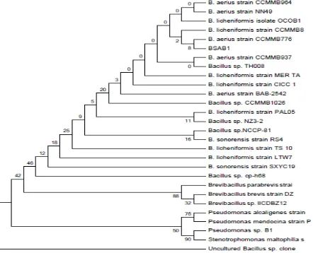
Previous study indicated that one strain of the cellulolytic bacteria isolated from tractus digestivus of milkfi sh, namely BSA B1
showed a highest potency to degrade cellulose compare to other strain. We identifi ed this
isolate based on 16S rRNA gene sequence. BSA B1 included in the same cluster with a strain of B. licheniformis strain CCMMB8 accesion number HG80002.1 with a similarity of 98.24% and B. aerius strain CCMMB776 accesion number KF879292.1 with similarity of 96.54% (Figure 1). It showed that BSA B1 isolate had a higher level of concordance with
B. licheniformis compared to B. aerius. Almost of B. licheniformis was isolated from soil and it can be isolated from various places because of its high ability to produce endospores. Milkfish have a habit to eat detritus that may contain microorganisms that assist for detritus digestion (Aslamsyah, 2012).
Cellulase activity cannot be found in
milkfi sh Chanos chanos (Chiu and Benitez,
1981), but other studies found the microbial cellulase activity in the digestive tract of herbivorous fi sh, especially milkfi sh (Bairagi
et al. 2002). This data was supported by
Das et al. (2014) that detect the presence
of cellulolytic microbes in the digestive tract of four brackish water fish species. Three of them, including Etroplus suratensis,
Scatophagus argus and Terapon jarbua are
kind of omnivorous fish that eat algae as well as milkfish. B. parabrevis and B.
licheniformis were identified from Mystus
guliofi sh. In addition, Prayitno et al. (2014)
identifi ed Shewanella upenei, Bacillus sp, Vibrio
fl uvialis, Shewanella algae, Shewanella sp., and
Photobacterium ganghwense in the digestive
tract of sea bass.
Furthermore, we examined the specifi c
activity of purifi ed enzyme of BSA B1. BSA
B1 bacteria that used for this study was subcultured from previous study. Our previous study have successfuly isolated some strains from tractus digestivus of milkfish. BSA B1 strain have the highest enzyme activity for cellulose degradation compared to other strains.
The crude enzyme has a lowest enzyme activity compared to purifi ed enzyme. This
result probably caused by other biomolecules contained in the crude enzyme which inhibits enzyme to degrade cellulose. Purification started with ammonium sulphate precipitation. Ammonium sulphate
precipitation conducted by using basic principle of protein dehydration that is some water molecules are bound to SO4-2
thus reducing the amount of water available to interact with the enzyme (Dobrev and Zhekova, 2012). Ammonium sulphate precipitation performed at 60% saturation
Table 1.Cellulase specifi c activity in each step of purifi cation
Fraction Enzyme
Volume (mL)
Total Activity (U)
Total Protein (mg)
Specifi c Activity
(U mg-1)
Results (%)
Purity (x)
Crude enzyme 50 9.91 5.70 1.74 100 1.00
Dialisis 5 1.13 0.25 4.61 11.40 2.65
DEAE Sepharose 1 0.30 0.04 7.28 26.43 4.19
*In 30 minutes with molecular weight 1 mol glucose 0.18 mg
which is the best concentration for reaching the isoelectric point of the enzyme.
Cellulase activity can be inhibited by the binding of protein and ammonium sulphate, therefore in this study, enzyme activity was measured after dialysis. Dialysis was based on the principle of osmosis that was the difference of concentration between protein inside dialysis membrane and buffer reagent concentration outside dialysis membrane. Ammonium sulphate will move along gradient concentration through the membrane until it reaches equilibrium. When ammonium sulphate separated from protein, the enzyme purity increased so enzyme activity can be measured.
Next purification step was column chromatography. In this step, enzyme purification was done by the principle of the mobile phase and stationary phase. The mobile phase of this step was purifi ed cellulase
from previous step (dialysis). The purifi ed
cellulase will be absorbed by stationary phase (DEAE Sepharose) while soluble components which were less absorbed will move faster. During the separation process, the solutes undergo adsorption and partition repeatedly. Each solute has a different decreasing rate and it depends on solute partition coeffi cient.
Hence, several layers are formed (Bakare, 2005)
Table 1 showed that each purification step resulted in purer enzyme. The enzyme concentration decreased while enzyme activity increased. Many contaminant proteins probably found in the crude enzyme which increases protein concentration. However, with the high concentration of protein, enzyme activity became less specifi c to the substrate.
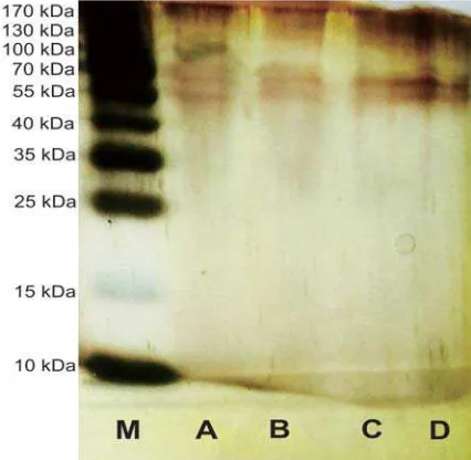
Molecular weight measurement of purifi ed cellulase showed in Figure 2. Thin
protein bands appear because protein content in cellulase decreasing after purifi cation.
The crude enzyme formed three bands with molecular weights of each of them about 59.06 kDa, 54.34 kDa, and 40.6 kDa.
Cellulase after ammonium sulfate precipitation, dialysis, and chromatography
produced two bands with molecular weight about 59.06 kDa and 54.34 kDa. The type of cellulase can be determined by comparing molecular weight with some references about cellulase from microorganisms. These prediction can be seen in Table 2.
According to table 2, our purified cellulase was possibly contained of endoglucanase, cellobiohydrolase, and
-glucosidase.
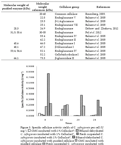
Figure 3 showed specific cellulase activity yields againts C. zofingiensis. Celluclast®, a commercial cellulase showed
lower specific enzyme activity than our purifi ed cellulase. Specifi c enzyme activity
on the incubated substrate at 1% Celluclast®
was 0.0478 U mg-1 in CMC, whereas our
purifi ed cellulase showed specifi c enzyme
activity was 0.7706 U mg-1 in C. zofi ngiensis
dehydrated ethanol. Low specifi c cellulase
activity at 1% Celluclast® possibly because
of its high protein concetration. At 1% Celluclast®, protein concentration was higher
than purifi ed cellulase which was 1.035 mg
mL-1, and 0.041 mg mL-1 respectively. Thus
specifi c activity of 1% Celluclast® was much
lower than purifi ed cellulase.
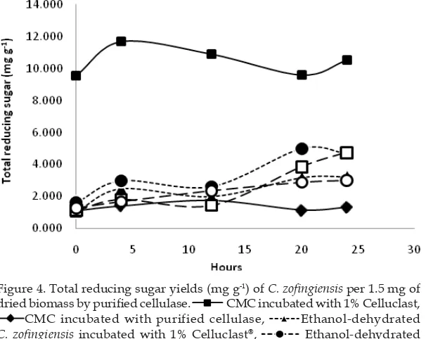
Figure 4 showed that the amount of reducing sugar produced by CMC after being incubated with 1% Celluclast® showed
the highest curve whereas CMC incubated with our purifi ed cellulase had the lowest
curve among other treatments. In the
treatment of 1% C. zofi ngiensis incubated with 1% Celluclast® showed a curve that
was lower than the CMC curve. However, the pattern of the curves were not differ between treatment groups. The optimum time of CMC incubated with 1% Celluclast®
to produce reducing sugar detected after 4 hours while the CMC incubated with
Tabel 2. The molecular weight of purifi ed cellulase.
Molecular weight of purifi ed enzyme (kDa)
Molecular weight references (kDa)
Cellulase group References
20-60 Common cellulase Rosenberg, 2005.
22.8 Endoglucanase V Bakare et al. 2005
23.5 -1,4-glucanase Bakare et al. 2005
25.1 Endoglucanase VII Bakare et al. 2005
28,3 26,9 Endoglucanase Dobrev and Zhekova, 2012
31,5; 53,4 30-55 Endoglucanase Pol et al. 2012
33.4 Endoglucanase IV Bakare et al. 2005
42.2 Endoglucanase II Bakare et al. 2005
45,6 46.0 Endoglucanase I Bakare et al. 2005
48,1 47.2 -Glucosidase I Bakare et al. 2005
53,4; 54,4 52.1 Endoglucanase IV Bakare et al. 2005
52.2 Cellobiohydrolase I Bakare et al. 2005
66,1 75.3 -glucosidase II Bakare et al. 2005
Figure 3. Specifi c cellulase activity yields of C. zofi ngiensis per cell (U
mg-1). CMC incubated with 1% Celluclast®, Ethanol-dehydrated
C. zofi ngiensis incubated with 1% Celluclast®, Fresh suspended C.
zofi ngiensis incubated with 1% Celluclast®, Ethanol-dehydrated C.
zofi ngiensis incubated with purifi ed cellulase CMC incubated with
purifi ed cellulase Fresh suspended C. zofi ngiensis incubated with
According to enzyme activity and protein concentration measured at each stage of purifi cation showed that in every
step our cellulase getting purer, the enzyme concentration decreased, and enzyme activity increased. Furthermore purified cellulase have a molecular weight ranging from 20 to 60 kDa. Less band detected in chromatographic purification stage compare to crude enzyme showed that cellulase getting purer. In this study we used Celluclast®, a commercial cellulase,
as a reference. The activity of commercial cellulase was too high so it cannot be read by spectrophotometer (unpublished data). Thus, we used 1% Celluclast® which predicted had
similar activity with our purifi ed cellulase.
According to SDS PAGE, Celluclast® possibly
mainly contains endoglucanase (CMCase) also exoglucanase and -glucosidase in lower concentration.
Purifi ed cellulase had cellulolytic activity
on CMC. CMC was pure cellulose modifi ed
by the addition of carboxyl groups and has been known as a substrate to test celulase purified cellulase detected in 12 hours.
Furthermore in C. zofingiensis all the optimum treatment detected in 24 hours, except ethanol dehydrated C. zofi ngiensis
incubated in purifi ed cellulase which was
detected after 20 hours of treatment.
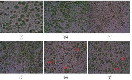
Figure 5 showed that the morphology of the cells changed after enzyme incubation and ethanol dehydration treatment. The addition of ethanol possibly caused C.
zofingiensis cells were dehydrated and
destructed before incubation with cellulase. Incubation with cellulase caused cells were destroyed so we found cell debris while intact cells has not detected. On the other hand incubation of cellulase without ethanol dehydration caused C. zofingiensis lose their cell wall. However a cell membrane still detected whereas the cytoplasm were swollen and formed as morphology similar to protoplasts (pointed with arrow). This condition could be affected by cellulase degrades the cell wall of C. zofingiensis, hence the cells become rigid and form structures similar to protoplasts.
Figure 4. Total reducing sugar yields (mg g-1) of C. zofi ngiensis per 1.5 mg of
dried biomass by purifi ed cellulase. CMC incubated with 1% Celluclast,
CMC incubated with purified cellulase, Ethanol-dehydrated
C. zofi ngiensis incubated with 1% Celluclast®, Ethanol-dehydrated
C. zofingiensis incubated with purified cellulase, Fresh suspended
C. zofingiensis incubated with 1% Celluclast®, Fresh suspended C.
activity. In various studies, CMC is known as substrate-specifi c against endoglucanase, so it
is also called the endoglucanase CMCase. On the other hand, C. zofi ngiensis is a microalgae that developed in many industries. This microalgae is known to have high cellulose content and high potential to be degraded by cellulase from the group -glucosidase (Rodrigues et al., 2015) using C. homosphaera
which has a composition similar to C. zofi ngiensis). Results showed that our purifi ed
cellulase had lower activity on CMC but higher activity in C. zofi ngiensis compare to 1% Celluclast. These data revealed that our purifi ed cellulase contains different type of
cellulase with Celluclast®. The content of
endoglucanase in 1% Celluclast® possibly
suitable for hydrolyzing CMC so that its activity was higher while the content of -glucosidase is possibly dominant in our purifi ed cellulase which more appropriate
to degrade the component of C. zofi ngiensis
cell wall.
Fresh cells was used as model to determined ability of enzyme to produce single cell protein whether ethanol dehydrated cells was used as a model of bioethanol production. Ethanol was used because it can destabilize outer cell walls of algae. This condition would facilitate the cellulase to hidrolyze ultrastructure cellulose of the cell. Hydrolyzed cellulose was expected to raise the amount of resulted reducing sugars. However, our results showed that ethanol was only able to raise the amount of reducing sugar if it was incubated with purified cellulase, whereas in 1% Celluclast®, ethanol
lowering the amount of reducing sugar. This indicated that ethanol can increase the amount of reducing sugar when used appropriate cellulase.
C. zofi ngiensis cell walls contain cellulose that can be hydrolyzed by purifi ed cellulase
and 1% Celluclast®. Fresh cell incubated with
cellulases showed that the cell wall of C.
zofi ngiensis was successfully hydrolyzed by
Figure 5. Comparison of C. zofi ngiensis cell morphology at several treatments after incubating for 24 hours (a), (b) normal cell; (c) 1% Celluclast® and ethanol treatments; (d) Cellulase treatment from purifi cation and ethanol; (e) 1%
Celluclast® treatment; (f) Purifi ed Cellulase from this study.
(a) (b) (c)
both cellulases with the loss of the cell wall whereas cell membrane still intact to form structures such as protoplasts. However, the ethanol treatment caused cells have been destroyed prior cellulase incubation. Hence, in ethanol treatment cells intact were not detected.
Conclusion
BSA B1 strain was closely related with
B. aerius and B. licheniformis. Cellulase from BSA B1 bacteria was successfully purifi ed.
The crude enzyme formed three bands with molecular weights of each of them about 59.06 kDa, 54.34 kDa, and 40.6 kDa. The results of ammonium sulfate precipitation, dialysis, and chromatography produced two bands with molecular weight about 59.06 kDa and 54.34 kDa. Purifi ed cellulase had an activity
to hydrolize CMC and C. zofi ngiensis Dönz. Morphology of C. zofi ngiensis Dönz cells on ethanol dehydration and cellulase incubation showed that the cells collapsed. Fresh cell incubated with cellulases showed that the cell wall of C. zofi ngiensis was successfully hydrolyzed by both cellulase with the loss of the cell wall whereas cell membrane are still intact. However in ethanol treatment cells intact were not detected.
Acknowledgment
This study was fi nancially supported
by PKM-P 2014 Higher Education grants. Publication was supported by BPP UGM Grant 2015.
References
Aslamsyah, S. 2012. Seleksi mikroflora saluran pencernaan ikan bandeng sebagai kandidat probiotik (Selection of gastrointestinal microflora in milkfish
as probiotic candidate). Department of
Fisheries, Faculty of Marine Science & Fisheries. Hasanuddin University. Makassar. [In Bahasa].
Ausubel, F.M., Brent, R., Kingston, R.E., Moore, D.D., Seidman, J.G., Smith, J.A. and Struhl, K. 1995. Short Protocols in
Molecular Biology. John Wiley & Sons. New York
Bairagi, A., Ghosh, K.S., Sen, S.K., and Ray, A.K.. 2002. Enzyme producing
fl ora isolated from fi sh digestive tracts.
Aquaculture Internationa,l 10, 109—121.
Bakare, M.K., Adewale, I.O., Ajayi, A., and Shonukan, O.O. 2005. Purifi cation and
characterization of cellulase from the wild-type and two improved mutants of
Pseudomonas fl uorescens. African Journal of
Biotechnology, 4(9), 898—904.
Dobrev, G.T. and Zhekova, B.Y. 2012. B i o s y n t h e s i s , p u r i f i c a t i o n a n d characterization of endoglucanase from a xylanase producing strain Aspergillus niger B03. Braz. J. Microbiol., 43(1), 70–77. Das, P., Mandal, S., Khan, A., Manna, S.K.,
and Ghosh, K. 2014. Distribution of extracellular enzyme-producing bacteria in the digestive tracts of 4 brackish water
fi sh species. Turk J Zool., 38,79–88.
Hidayanti, A.K. 2011. Isolasi bakteri amilolitik dari Chlorophyta (Amylolytic bacteria isolation from Chlorophyta). Thesis, (S.Si.). Fakultas Biologi UGM. Yogyakarta. [In Bahasa].
Juhana, N. 2011. Karakterisasi enzim amilolitik isolat bakteri amilolitik yang berasosiasi dengan Chlorophyta (Characterization of amylolytic enzyme of isolate amylolytic
bacteria associated with Chlorophyta).
Thesis, (S.Si.). Fakultas Biologi UGM. Yogyakarta. [In Bahasa].
Lathifah, A. N., Hidayanti, A.K., and Ramadingrum, W. A.. 2009. Isolasi bakteri selulolitik dari lambung ikan bandeng (Isolation of cellulolytic bacteria from Milkfi sh
gastrointestinal). Fakultas Biologi UGM.
Yogyakarta. [In Bahasa].
Mäntylä, A., Paloheimo, M., and Suominen, P. 1998. Industrial mutants and recombinant strain of Trichoderma reesei. In: Kubicek CP, Harman GE, Ondik KL, editors.
Trichoderma and Gliocladium, Enzymes,
Muhammad, F. B., Pramana, A.A., and Pertiwi, G.A. 2012. Purifi kasi enzim selulase isolat bakteri selulolitik yang berpotensi sebagai agen biokatalisator pada berbagai bidang industri pengguna selulosa (Purifi cation of sellulolytic enzyme from isolate cellulolytic bacteria with potency as biocatalisator agent in various cellulose-used industries). Research report. Fakultas Biologi UGM. Yogyakarta. [In Bahasa].
P o l , D . , L a x m a n , R . S . a n d R a o , M . 2012. Purification and biochemical characterization of endoglucanase from
Penicilium pinophilum MS 20. Indian
Journal of Biochemistry and Biophysics 49, 189–194.
Prayitno, S.B., Sarwan, and Sarjito. 2014. The diversity of gut bacteria associated with Milkfi sh (Chanos chanos Forssksal) from
northern coast of Central Java, Indonesia.
Procedia Environmental Sciences.23 (2015), 375–384.
Rosenberg, I.M. 2005. Protein analysis and purifi cation. Second edition. Birkhauser. USA: 143.
Sukmadjaja, D.N., Sunarlim, E.G., Lestari, I., Roostika, and Suhartini, T. 2007. Teknik isolasi dan kultur protoplas tanaman padi (Isolation technique and protoplast culture of rice plant. Jurnal Agrobiogen,
3(2), 60–65. [In Bahasa].
Tamura, K., Peterson, D., Peterson, N., Stetcher, G., Nei, M, and Kumar, S. 2011. MEGA5: Molecular evolutionary genetics analysis using maximum likelihood, evolutionary distance, and maximum parsimony methods. Mol. Biol. Evol.,