Korespondensi: Wasilah, email: wasilahyahya@gmail.com, Kedokteran Gigi Institut Ilmu Kesehatan, Jl. K.H. Wahid Hasyim 65, Kediri 64114, Jawa Timur. Telp. (0354) 773299, Faks (0354) 771539
IDJ, Volume 1,No. 1, Tahun 2012 27
The Decrease In Number Of Blood Polymorphonuclear (Pmn) To Periapical Radiographs Dose Of radiation exposure
Amni Adlina1, Wasilah2
1
Fakultas Kedokteran Gigi Universitas Jember, Jember, Indonesia 2Staf Pengajar Kedokteran Gigi Institut Ilmu Kesehatan, Kediri, Indonesia
Abstract
Background: Periapical radiograph is a type of radiographic examinations that is widely used in the field of dentistry. Radiographic X-ray is an ionizing radiation that can cause damage and death to cells, tissues or organs, including the polymorphonuclear (PMN). PMN is cell that serves as the first line of defense against the invasion of organisms. Objective:The purpose of this study is 1) to determine whether there is a decrease in the number of peripheral blood PMN after being given periapical radiograph dose of X-ray radiation exposure, and 2) to determine whether there is any difference in the declining number of peripheral blood PMN after being given a single dose and some different total dose of periapical radiograph of X-ray radiation exposure. Methode: The design of this study is an experimental laboratory research with total sample of 24 strains of
Balb c male mice. The sample is divided into four groups, each consisting 6 mice. Group 1 serves as the control group; Group 2 is given a treatment with a single-dose exposure of periapical radiographs of X-ray radiation; Group 3 is given a treatment of 6 times total doses exposure of periapical radiograph of X-ray radiation; and Group 4 is given a treatment of 14 times total doses exposure of periapical radiograph of X-ray radiation. After 24 hours of radiation exposure, the blood was drown. Conclusion :The calculation is done by multiplying the number of PMN percentage from leukocyte counts with a total leukocyte. The data obtained are statistically tested using One Way Anova and LSD with a significance level of 95%. Result :The result of this study indicates that there is a difference in the peripheral blood PMN counts in male mice after being exposed to radiation doses of periapical radiographs in all groups (P <0.05). There is an increasing number of peripheral blood PMN in male mice after being treatedwith X-ray radiation at a single dose of periapical radiographic exposure, whereas after being exposed with radiation at the dose of 6 and 14,the number of PMN is declining.The highest decreasing number of PMN isfound in the group with exposure of radiation at 14 dose.
28 IDJ, Volume 1,No. 1, Tahun 2012 Abstrak
Radiografi periapikal adalah salah satu pemeriksaan radiografi yang banyak digunakan oleh dokter gigi.radiografi sinar X adalah radiasi ionisasi yang dapat menyebabkan kerusakan dan kematian sel, jaringan atau organ, termasuk sel PMN. PMN berperan sebagai pertahanan pertama terhadap invasi mikroorganisme. Tujuan dari penelitian ini adalah untuk mengetahui penurunan jumlah PMN darah tepi setelah paparan sinar X dosis radiografi periapikal dan mengetahui apakah terdapat perbedaan penurunan jumlah PMN antara dosis tunggal dan dosis total radiasi sinar X dosis radiografi periapikal. Penelitian ini merupakan penelitian jenis eksperimental laboratoris. Sampel yang digunakan sebanyak 24 ekor mencit strain Balb C. jantan yang dibagi menjadi 4 kelompok masing-masing 6 ekor. Kelompok 1 adalah kelompok kontrol. Kelompok 2 adalah kelompok dengan pemberian paparan radiasi sinar X dosis paparan tunggal radiografi periapikal. Kelompok 3 adalah kelompok dengan pemberian paparan radiasi sinar X dosis total 6 kali paparan radiografi periapikal. Kelompok 4 adalah kelompok dengan pemberian paparan radiasi sinar X dosis total 14 kali paparan radiografi periapikal. Darah diambil setelah 24 jam pemberian paparan radiasi. Penghitungan jumlah PMN dilakukan dengan mengalikan persen PMN dari hitung jenis leukosit dengan jumlah leukosit total. Data yang diperoleh dilakukan uji statistik One Way Anova dan LSD dengan tingkat kemaknaan 95%.Hasil penelitian menunjukkan bahwa terdapat penurunan jumlah PMN darah tepi mencit jantan setelah paparan radiasi sinar X dosis radiografi periapikal. Hasil dari penelitian ini menunjukkan bahwa terdapat perbedaan jumlah PMN darah tepi pada mencit jantan setelah paparan radiasi dosis periapikal pada semua kelompok (P <0.05). Terdapat peningkatan jumlah PMN darah tepi pada mencit jantan setelah pemberian satu kali paparan radiasi sinar X dosis periapikal.Sedangkan setelah pemberian paparan radiasi sebanyak 6 dan 14 kali jumlah PMN menurun.Penurunan jumlah PMN terbesar terjadi pada kelompok dengan 14 kali paparan radiasi dosis periapikal.
Kata Kunci :Polimorfonuklear (PMN), Radiasi Sinar-X, Radiografi Periapikal
Introduction
Radiographic examination is routi ne examination in the field of dentistry, thus radiography is a necessity that cannot be avoided, and it can be said to be a very important thing for us 1. Periapical radiograph is a type of radiographic examinations
that is widely used in the field of dentistry 2. Periapical radiograph using X-ray, which is one of ionizing radiations 3.
Amni Adlina: The Decrease In Number Of Blood Polymorphonuclear
IDJ, Volume 1,No. 1, Tahun 2012 29
gray (50 rad) dosage of ionizingradiation decreases neutrophil count in blood cells4. The primary
function of neutrophil
Polymorfonuklear (PMN) is to give imune response by performing phagocytosisand also killing or eradicatingincoming
microorganism5.In low dose radiation, few hours after an exposure the neutrophil count increases. However, after 24 hours and more of exposure neutrophil and leukocyte count is decreasing6.
There are some literatures that support the reduction in number of blood cells. Cole said that ionizing radiation can cause leukopenia. While Miller and Weller state that the ionizing radiation can lead to a reduction of all types of leukocytes. WHO also stated that ionizing radiation can cause an early haematological changes, but until
now the decreased
polymorphonuclear (PMN) due to periapical radiographs dose of X-ray exposure has not been studied 7. The purpose of this study isto determine whether there is a decrease in the number of peripheral blood PMN after being given periapical radiograph dose of X-ray radiation exposure, and to determine whether there is any difference in the declining number of peripheral blood PMN after being given a single dose and some different total dose of periapical radiograph of X-ray radiation exposure.
Materials and Methods
This study is a kind of experimental laboratory research. The study
population is a strain of mice Balb c. male mice.
Grouping of samples stage
The control group (group 1), consisted of 6 healthy mice and not treated. Treatment group (group 2), consisted of 6 healthy mice into treatment group given a one-time exposure of dental radiodiagnostic. Treatment group (group 3), consisted of 6 healthy mice into treatment groups that provided in 6 times dental radiodiagnostic exposure with an interval of time between them is 1 minute. Treatment group (group 4), consisted of 6 healthy mice into treatment groups that provided in 14 times dental radiodiagnostic exposure with an interval of time between them is 1 minute.
Mice fixation stage
The used fixation device is made of white plastic paralon. Bottom of the tube was perforated for the head of mice and six holes on the other edge was made by a stick. The mouse’s head inserted through the hole on the bottom tube and the back is fixed with three crossing sticks between the two holes that made before.
Radiation exposure stages
30 IDJ, Volume 1,No. 1, Tahun 2012 Collection and
countin blood sample stage
Animals were sacrificed after 24 hours of exposure to radiation and the blood was taken from the heart using a 1 ml insulin syringe. The number of PMN was counted by multiplying the percentage of PMN in the leukocyte counts in blood smears by the number of leukocytes per mm3 in the counting room Improver Neubeur.
Statistical analysis
The obtained data were analyzed using the Kolmogrof-Smirnof
normality test and the homogeneity test of Levene, followed by One Way ANOVA parametric test to determine whether there are groups of data that have significant mean differences, with 95% degree of significance
(p<0.05). Followed by LSD test (Least Significant Difference Test) to determine which groups were significantly different.
Result
The result of the study shows that the group with single dental radiodiagnostic exposure the absolute neutrophil count increases as much as 291.816,67. Meanwhile, the group which is given 6 times of dental radiodiagnostic exposure treatment shows that there is a decrease of absolute neutrophil count as much as 260.733,33. The number of absolute neutrophil count in the group that has received a treatment of 14 times exposure is the least one which is 260.733,33 and 236.933,33 (Table 1).
Table 1. Mean and standart deviation of the number of PMN in 24 hours observations post exposure of periapical radiographs dose of X-ray radiation
Treated and Control Mean
Control group P1
P2 P3
280,333.33 291,816.67 260,733.33 236,933.33
(cell/mm3)
P1 = treatedgroup with single dose
P2 = treated group with a 6 times of exposure in total dose P3 = treated group with a 14 times of exposure in total dose
Amni Adlina: The Decrease In Number Of Blood Polymorphonuclear
Fig.1. Histogram of the average number of PMN in post 24 hours observation
Before the data was analyzed, it was tested for normality and homogeneity first. The normality test used
Kolmorgof-Smirnof, whereas the homogeneity test used Levene
test.Both use the error rate (a) 0.05. In the used homogeneity test, P = 0.974 values obtained and the normality test, P value obatined for the four groups of data were > 0.05.
Table 2. One Way Anova test results on the control group and treatment
Anova test showed there were some differences in number of Neutrophyle. To find out which
groups that significantly different, the data were analyzed using the LSD test which is shown in Table 3.
Table 3.LSD test result on four group
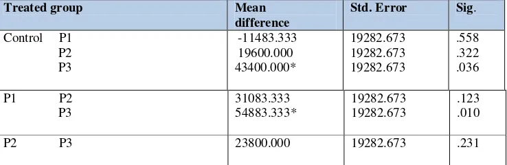
Multiple Comparisons Dependent Variable: The number of neutrophil LSD
Treated group Mean
difference
Std. Error Sig.
Control P1 P2 P3
-11483.333 19600.000 43400.000*
19282.673 19282.673 19282.673
.558 .322 .036
P1 P2 P3
31083.333 54883.333*
19282.673 19282.673
.123 .010
P2 P3 23800.000 19282.673 .231
*.The mean difference is significant at the ,05 level. 0.00
50,000.00 100,000.00 150,000.00 200,000.00 250,000.00 300,000.00 350,000.00
Cont rol group
P1 P2 P3
Sum of Square
df Mean Square F Sig.
Betweengrups Within groups Total
10416571250.0 22309288333.3 32725859583.3
3 20 23
3472190417 1115464417
3.11 3
[image:5.612.126.499.376.454.2] [image:5.612.135.504.555.675.2]32 IDJ, Volume 1,No. 1, Tahun 2012
experimental, shown on Table 4.5 it can be seen that there is a significant difference between the control group and P14 group, between P1 group and P14 group. However, from the sama data set, there is no significant difference between control group and P1 group; control group and P6 group; and P1 and P6 group.
Discussion
X-ray radiation not only providing benefits for all of us but also has a negative effect. Ionizing radiation is one of powerful agent in causing damage and even death of cells, tissues or organs, while the present technology cannot fully protect the body yet from the side effects of ionizing radiation 4.
X-ray radiation from dental radiography units that used in this research is direct through a cone which has 100 cm of surface area. Cone is directed right to the heart of mice as the focus of the organ where blood will be taken, then they fixed so that cannot move and the radiation can certainly lead to cardiac of mice. Blood sampling performed on the heart with blood volume needed considerations for research and focus of the radiation had to be directed to specific organs, so that the heart was chosen as a spot of blood sampling. In this study used mice aged 3 t 4 months, because according to Schalm, mice in that age is not easy to die when irradiated below the lethal dose. Mice lethal dose ranged from 550-640 rad 8.
The result of the research and the statistical test shows that there is a decrease in the number of peripheral
exposure. This is also in line with Underwood’s statatement saying that radiation causes neutrophil reduction in blood circulation (opo circulating blood?)alongside with other blood cells (pancytopenia) 9. Edward also states that radiation has a negative effect to blood cells as it cuts down the number of cells in peripheral circulation4.
The decrease in the number of PMN neutrophil in this research is caused by a condition in which the effect of radiation exposure can be accumulated. This accords Milles’ statement saying that tissues have an ability to repair its damages after being exposed to radiation; however, unrepairable damages can be accumulated10.
Cells damages can be caused both by direct and indirect effect of ionizing radiation. Damages due to direct effect happens when ionizing particles interact (energy transfer to) with biology macromolecule such as
DNA, RNA, protein, or
enzime.Damages due to indirect effect happens when the damage is caused by oxidant agent resulted from ionizing process. 75% of cells is water and this water mollecule is the one that is most ionized by X-rays10.
Amni Adlina: The Decrease In Number Of Blood Polymorphonuclear
IDJ, Volume 1,No. 1, Tahun 2012 33
transported to bone marrow capillaries to move the stored neutrophil into circulation blood4.
This research was conducted on male mice. Whether the results of this study can be equated or generalized to humans, of course, this requires a separate research and discussion, but please note that the lethal dosage (LD) 50/30 of mice is 550-640 rad 11.
. While the LD 50/30 of adult human is 450 rem (~450 rad). LD 50/30 is a whole body radiation dose that is lethal in 50% of the population within 30 days 4.
Conclusions
1. There is a difference of peripheral neutrophil count in male mice Balb cafter given an exposure of dental radiodiagnostic of periapical radiography between control group, single exposure group, 6x exposure group, and 14x exposure group.
2. There is an increase in peripheral neutrophil count in male mice Balb cafter given a single exposure of X-ray radiation of periapical radiography dose; meanwhile after given 6x and 14x of exposure, the number of PMN count is decreasing. The highest number of decrease in peripheral neutrophil count happens to the group with 14x of exposure.
References
1. Yunus, B. 2005: ‘Radiografi Dental sebagai Diagnosa Awal untuk Mencegah Keparahan suatu Penyakit Gigi dan Mulut’,
Majalah Kedokteran Gigi Edisi Khusus Temu Ilmiah Nasional, 4(11) pp: 396-400
2. Suharjo And Endang. 1994: ‘Peranan Teknik dan Interpretasi Radiografi Intraoral Periapikal dalam Perawatan Endodontic’, Jurnal Kedokteran Gigi PDGI, 2(43) pp: 40-43
3. Cotran, R., Robbins, S., Kumar, Abbas and Nelson. 1999: ‘Pathologic Basis of Disease’, (Ed): 7, pp: 67-69
4. Edwards, Cris, Statkiewicz, M.A. and Russell, e. 1990. ‘Radiation Protection for Dental Radiographers’, pp: 43-48
5. Kresno, S.B. 2001: ‘Imunologi:
Diagnosis dan Prosedur
Laboratorium’, (Ed): 7, pp: 30 6. Rosyid, A. 2006. ‘
Temuan-Temuan pada Hasil Pemeriksaan Laboratorium akibat Radiasi‘,
pp: 24
7. Astuti, E.R. 1995: ‘Pengaruh Radiasi Pengion dan Banyaknya Ulangan terhadap Total Leukosit, Hitung Jenis Leukosit, Hb, serta Total Eritrosit Mencit Jantan ‘, pp: 52-60
8. Goaz, P.W. and White, S.C. 1987: ‘Oral Radiology”, (Ed): 2, pp: 104-107
9. Underwood, J. C. E. 2000.
General and Sistematic
Pathology 3rd ed. Edinburgh:
Churchill Livingstone
10. Miles, Van Dis, Jensen, and Ferretti. 1993. Radiographic Imaging for Dental Auxillaries, (Ed): 2, pp: 34