www.elsevier.com/locate/jinsphys
Permeability and disruption of the peritrophic matrix and caecal
membrane from
Aedes aegypti
and
Anopheles gambiae
mosquito
larvae
Marten J. Edwards
a, b, Marcelo Jacobs-Lorena
a,*aDepartment of Genetics, School of Medicine, Case Western Reserve University, 10900 Euclid Avenue, Cleveland, OH 44106-4955, USA bDepartment of Zoology, Ohio Wesleyan University, Delaware, OH 43015, USA
Received 22 October 1999; accepted 11 February 2000
Abstract
In mosquito larvae, the peritrophic matrix (PM) separates the gut contents from the intestinal epithelium. This report describes a new in vivo assay for estimating PM permeability. The assay also allows for assessment of the permeability of the caecal membrane, a structure that separates each caecum from the gut lumen. Permeability was estimated by the appearance of fluorescently-labeled dextrans (size range 4,400 to 2 million Da) within the gastric caecae of mosquito larvae. While the intact peritrophic matrix was impermeable to 2 million Da dextran particles, it was permeable to dextran particles of 148 kDa and smaller. The caecal membrane appears to have considerably smaller pores, being permeable only to dextrans of 19.5 kDa and smaller. The assay was also used to devise a treatment that disrupts the PM sufficiently to allow the passage of virus-sized particles. Dithiothreitol and to a lesser extent, chitinase were effective in disrupting the PM. Cycloheximide had a small effect; Polyoxin D, Pronase and calcofluor did not alter the permeability to 2 million Da dextran particles. Disruption of the PM is discussed in the context of infecting mosquitoes with retroviral transformation vectors.2000 Elsevier Science Ltd. All rights reserved.
1. Introduction
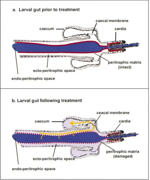
The peritrophic matrix (membrane) (PM) is an acellu-lar chitin-containing sheath that separates the gut con-tents from the secretory/absorptive intestinal epithelium (reviewed by Richards and Richards, 1977; Peters, 1992; Jacobs-Lorena and Oo, 1996; Lehane, 1997; Tellam et al., 1999). In mosquito larvae, the PM is a thin, trans-parent and tubular film composed of proteins, glycosam-inoglycans and chitin. The PM lines the entire digestive tract and is continuously synthesized by a ring of cells that are contained within the cardia. The cardia is located at the junction of the foregut and the midgut. The food bolus is contained within the PM tube in a compartment called the endoperitrophic space. The space between the two tubes — the PM and the midgut epithelium — is called the ectoperitrophic space (Fig. 1).
Fluids in the ectoperitrophic space are separated from
* Corresponding author. Tel.:+1-216-368-2791; fax:+ 1-216-368-3432.
E-mail address:[email protected] (M. Jacobs-Lorena).
0022-1910/00/$ - see front matter2000 Elsevier Science Ltd. All rights reserved. PII: S 0 0 2 2 - 1 9 1 0 ( 0 0 ) 0 0 0 5 3 - 6
the gastric caeca by a semi-permeable caecal membrane. The composition of the caecal membrane appears to be similar to the PM (Volkmann and Peters, 1989a). Volk-mann and Peters (1989b) used FITC (MW=389.3) as a fluorescent marker for fluid flow in larval midguts. The dye accumulated in the anterior region of the gastric caeca, apparently impeded by the caecal membrane.
Digestive enzymes, which are secreted by the midgut epithelium, must cross the PM in order to digest food in the endoperitrophic space (Detra and Romoser, 1979). Likewise, the products of digestion must traverse the PM in the opposite direction before they can be absorbed by midgut epithelial cells. Hence, macromolecular traffic across the larval PM plays a central role in digestion.
Fig. 1. Diagrammatic representation of the larval mosquito gut. (a) Dye is contained within the endoperitrophic space by the peritrophic matrix. (b) Peritrophic matrix and caecal membrane are disrupted. Dye can pass into the ectoperitrophic space and the gastric caeca.
instance, reagents that interfere with chitin synthesis, such as Polyoxin D, and chitinase were effective in dis-rupting the adult PM. However, the adult mosquito PM is a completely different structure from the larval PM (Jacobs-Lorena and Oo, 1996) and results of experi-mental manipulations of the adult mosquito PM may not be applicable to the larval PM. Here we describe a new in vivo assay to estimate mosquito larval PM per-meability and use this assay to devise means of rendering the PM permeable to virus-sized particles.
2. Materials and methods
2.1. Mosquitoes
Anopheles gambiae (PEST strain) and Aedes aegypti (white strain) were obtained from F. Collins (University of Notre Dame) and reared as previously described (Lemos et al., 1996).
2.2. Reagents
FITC-labeled dextrans, blue dextran, chitinase (Serratia marcescens E.C.3.2.1.14), calcofluor white 2RS, latex beads (0.46 pm) and cycloheximide were from Sigma. Dithiothreitol was from GIBCO-BRL,
Sephadex G-10 was from Pharmacia. Pronase and tunicamycin (from Streptomyces lysosuperficus) were from Calbiochem.
2.3. Chemical treatment of larvae
Groups of approximately 100 larvae swam overnight in 5 ml of an aqueous solution of the reagent to be tested. Agar (0.03%) was included as a feeding stimulant as described by Dadd (1975a). For injection experiments,
|0.1 µl of an aqueous solution of polyoxin D (100
µg/ml) were injected free-hand (without using a micromanipulator) into the thorax of cold-immobilized larvae using a thin-walled glass injection needle. Follow-ing treatment, larvae were rinsed thoroughly and trans-ferred to 3 ml of either 0.5 mg/ml FITC dextran (MW 2 million Da) or 2% w/v blue dextran (MW 2 million Da). The FITC dextran solutions were prepared immedi-ately before use, and passed over a 5-ml Sephadex G-10 (Pharmacia) column to remove any unbound FITC and small molecular weight molecules.
these physiological actions can result in an underesti-mate of the number of gastric caeca that would be filled with dye. Thus, filling of the proximal compartment of the caeca was more difficult to detect in larvae that were completely immobilized by chilling on ice. For this rea-son, this in vivo assay must be performed with active larvae, although for ease of observation, their movement should be slowed by moderate chilling. A Leica MZ12 dissecting microscope with a fluorescence module was used for experiments with FITC dextrans.
3. Results
3.1. In vivo assay for the measurement of PM permeability
The gastric caeca are divided into distal and proximal regions by a very thin, chitin-containing caecal mem-brane (Volkmann and Peters, 1989a). This acellular structure is located near the opening of the caeca into the midgut (Fig. 1). Labeled dextrans of various sizes were used to assess permeability of the PM and caecal membrane. Because mosquito larvae are transparent, the movement of the dye-labeled, inert dextran particles within the different compartments of the gut and caeca can be observed in living organisms without invasive procedures. Appearance of dextran blue- or FITC-lab-eled dextrans in the distal region of the gastric caeca was interpreted as the result of the large particles crossing the disrupted PM into the ectoperitrophic space and entering the caeca through the disrupted caecal mem-brane (Fig. 1). Appearance of dextran particles in the proximal, but not the distal region of the caeca was inter-preted as the result of the particles crossing the PM, but not the caecal membrane.
3.2. Effect of the in vitro treatment of the larval PM with chitinase
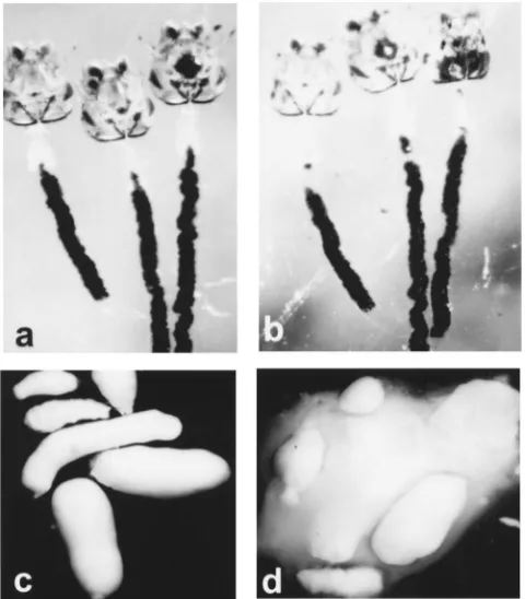
Because chitin is believed to have a crucial role in maintenance of PM structure (Peters, 1992), we first attempted to disrupt the PM by treatment with chitinase. Surprisingly, incubation of dissected larval PM in 1 U/ml chitinase did not appear to have any effect on the structure when observed by light microscopy (Fig. 2a, b). As a control we tested the susceptibility of the adult PM. It was readily disrupted by incubation in a chitinase solution equivalent to that used for the larval PM experi-ments (Fig. 2c, d). We conclude that the larval PM is largely resistant to chitinase disruption and that the adult and larval PMs differ considerably in their susceptibility to chitinase.
Fig. 2. Effect of chitinase on the larval and adult PM. (a) Larval PMs photographed immediately after dissection in PBS. The PM surrounds and contains a black mass of charcoal that was fed to the larvae prior to dissection. (b) The same larval PMs as in panel (a) following 2 h incubation with 1 U/ml chitinase. The charcoal particles remain con-tained, indicating intactness of the PM. The tensile strength of the PM, as estimated by teasing with a forceps, remains unchanged by the chitinase treatment. (c) Adult PMs, dissected 24 h following a meal of latex beads, in phosphate buffered saline (PBS, pH 7.0) and incu-bated for 2 h in PBS. The PM surrounds and contains the white mass of ingested latex beads. (d) The same PMs as shown in panel (c) were photographed following incubation for 2 h with 1 U/ml chitinase. Loss of PM integrity caused the latex beads to disperse.
3.3. Chemical disruption of the PM in vivo
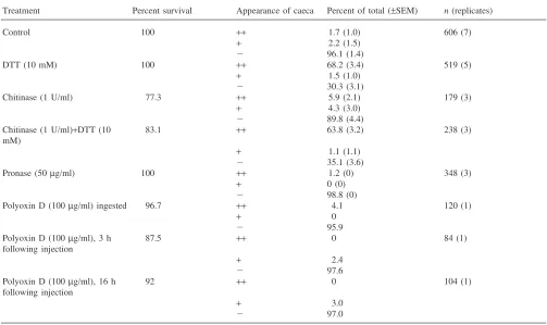
Several reagents were assayed for their ability to dis-rupt the larval PM and the caecal membrane following ingestion of the reagent. Disruption was assessed by the appearance of 2 million Da dextran particles in the ecto-peritrophic space and the gastric caeca. The dextran par-ticles were tagged either with a visible dye (dextran blue: Table 1) or with a fluorescent dye (FITC; Table 2). In addition, the effect of injected polyoxin D was tested to address the possibility that PM secreting cells of the car-dia were not accessible to this inhibitor of chitin syn-thesis when the drug was provided orally.
sig-Table 1
Effect of in vivo chemical treatments on PM permeability using dextran blue (2 million Da) as a markera
Treatment Percent survival Appearance of caeca Percent of total (±SEM) n(replicates)
Control 98.0 + 1.2 (0.7) 598 (4)
2 98.8 (0.7)
DTT (10 mM) 94.0 + 41.0 (0.8) 895 (6)
2 59.0 (0.8)
Chitinase (1U/ml) 94.8 + 24.3 (6.0) 507 (3)
2 75.7 (6.0)
Pronase (50µg/ml) 96.3 + 0.7 (0.2) 460 (3)
2 99.3 (0.2)
Cycloheximide (0.1 mg/ml) 94.7 + 10.3 (0.4) 427 (3)
2 89.7 (0.4)
Calcofluor (1 mg/ml) 99.1 + 1.0 (0.7) 515 (3)
2 99.0 (0.7)
Tunicamycin (10µg/ml) 93.9 + 1.0 (0.3) 521 (3)
2 99.0 (0.3)
aAn. gambiaelarvae were incubated overnight in the indicated chemical (or water for controls) and for 1 h in dextran blue, prior to microscopic
observation. Appearance of caeca: (+) blue dye in the caeca (proximal region or entire caeca filled); (2) caeca completely clear. The data in each category are presented as the percent of total surviving larvae in each replicate ±standard error of the mean.n is the total number of larvae examined followed by the number of replicate experiments in parentheses.
Table 2
Effect of in vivo chemical treatments on PM permeability using FITC-labeled (2 million Da) dextran as a markera
Treatment Percent survival Appearance of caeca Percent of total (±SEM) n(replicates)
Control 100 ++ 1.7 (1.0) 606 (7)
+ 2.2 (1.5)
2 96.1 (1.4)
DTT (10 mM) 100 ++ 68.2 (3.4) 519 (5)
+ 1.5 (1.0)
2 30.3 (3.1)
Chitinase (1 U/ml) 77.3 ++ 5.9 (2.1) 179 (3)
+ 4.3 (3.0)
2 89.8 (4.4)
Chitinase (1 U/ml)+DTT (10 83.1 ++ 63.8 (3.2) 238 (3)
mM)
+ 1.1 (1.1)
2 35.1 (3.6)
Pronase (50µg/ml) 100 ++ 1.2 (0) 348 (3)
+ 0 (0)
2 98.8 (0)
Polyoxin D (100µg/ml) ingested 96.7 ++ 4.1 120 (1)
+ 0
2 95.9
Polyoxin D (100µg/ml), 3 h 87.5 ++ 0 84 (1)
following injection
+ 2.4
2 97.6
Polyoxin D (100µg/ml), 16 h 92 ++ 0 104 (1)
following injection
+ 3.0
2 97.0
aAn. gambiaelarvae were incubated overnight in the indicated chemical (or water for controls) and for 1 h in FITC-labeled dextran, prior to
nificantly more toxic than treatment with DTT (Tables 1 and 2). Treatment with chitinase was sometimes effec-tive in disrupting the PM but less so than with DTT (Tables 1 and 2). Moreover, in some experiments chitin-ase was more toxic than DTT (Tables 1 and 2). Other treatments, including injected polyoxin D, did not appear to disrupt the PM. None of these treatments, including DTT, affected the appearance of dissected PMs when examined by light microscopy.
3.4. PM permeability of untreated larvae
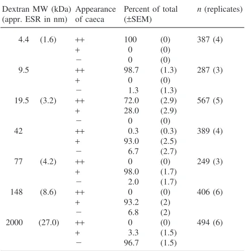
To estimate the permeability of the larval PM and the caecal membrane, FITC-labeled dextran particles of dif-ferent sizes were fed to Ae. aegypti and An. gambiae larvae. For both mosquitoes, the PM was impermeable to 2 million Da dextran particles, partially permeable to dextran particles of 148 kDa and permeable to particles of 77 kDa and less (Tables 3 and 4). The caecal mem-branes of both species were largely permeable to dextran particles of 19.5 kDa and smaller (Tables 3 and 4 and Fig. 3).
Table 3
FITC dextran accumulation in the gastric caeca ofAn. gambiaea
Dextran MW (kDa) Appearance Percent of total n(replicates) (appr. ESR in nm) of caeca (±SEM)
aESR: Einstein–Stokes radius (Pharmacia). Appearance of caeca:
(++) caeca filled with dye; (+) only the proximal region of the caeca filled; (2) caeca are completely clear. The data in each category are presented as the percent of total surviving larvae in each replicate ± standard error of the mean.nis the total number of larvae examined followed by the number of replicate experiments in parentheses.
Table 4
FITC dextran accumulation in the gastric caeca ofAe. aegypti. Further details are given in the footnote to Table 3
Dextran MW (kDa) Appearance Percent of total n(replicates) (appr. ESR in nm) of caeca (±SEM)
The midgut is the first site of interaction between mos-quitoes and the pathogens they transmit. A possible strat-egy to control disease transmission is to express in the adult gut cell genes whose secreted products are noxious to the pathogen. Retroviral vectors have been success-fully employed as a method of introducing genes into the mosquito genome (Jordan et al., 1998), thus circum-venting the laborious procedure of germ line transform-ation. However, retrovirus integration into the host cell genome requires cell division and gut cells of adult mos-quitoes divide only rarely, if at all. This limitation may be circumvented by oral infection of larval gut imaginal cells (the precursor of adult gut cells). A significant obstacle to the infection of the larval imaginal cells is the peritrophic matrix, which is impermeable to virus-sized particles (approximately 100–110 nm; Sharma et al., 1997). Another obstacle to retroviral infection may be the larval epithelium itself, as imaginal cells that give rise to the adult posterior midgut were identified at the bases of the larval midgut cells (O’Brien, 1966). These experiments were initiated in order to develop a non-lethal means of disrupting in vivo the first barrier, i.e. the larval PM.
4.1. In vivo assay of larval PM disruption
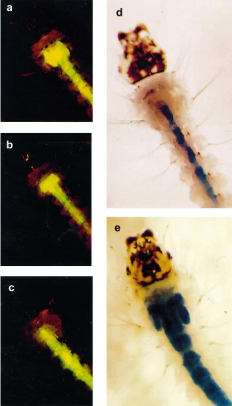
Fig. 3. Appearance of An. gambiae larval gastric caeca following ingestion of FITC-labeled dextran particles. (a) A larva following ingestion of 4.4 kDa FITC-dextran particles. Proximal and distal regions of the caeca are filled with the fluorescent dye. (b) Larva fol-lowing ingestion of 148 kDa FITC-dextran particles. Only the proximal region of the caeca are filled, indicating that the dye did not pass through the caecal membrane. (c) Larva following ingestion of 2 million Da FITC-dextran. No dye is observed in the gastric caeca. (d) Control (untreated) larva following ingestion of dextran blue (MW 2 million Da). Caeca are clear of the dye. (e) Larva following overnight treatment with 10 mM DTT. The caeca fill with dextran blue following ingestion of the dye.
caeca (Volkmann and Peters, 1989a). Consistent with this fluid-transporting role, the gene encoding the V-ATPase B subunit is expressed very strongly in the caeca of Culex quinquefasciatus (Fillippova et al., 1998). Moreover, it appears that contractions of the gut also force fluids from the ectoperitrophic space into the gas-tric caeca (Volkmann and Peters, 1989b). In our assay, filling of the caecal lumen with the dye-tagged dextrans (Fig. 1) is assumed to be driven by these forces. Dextran particles of 2 million Da have an Einstein–Stokes radius
of approximately 27 nm, which approaches that of viral particles (100–110 nm). Thus, the level of PM disruption required for the passage of large dextran particles may also be sufficient for viral particles to enter the ectoperi-trophic space where infection of adult gut imaginal cells could occur.
Of the several reagents assayed for their ability to dis-rupt the larval PM and the caecal membrane, the reduc-ing agent, DTT was the most effective (Tables 1 and 2). DTT is likely to act by breaking protein S–S bonds. We propose that loss of PM and caecal membrane structure is a result of unfolding of their structural proteins and the disruption of protein–protein interactions crucial for the maintenance of PM integrity. Moreover, loss of pro-tein tertiary structure could lead to an increased suscepti-bility of the proteins to digestive proteases. In addition to being effective in disrupting the larval PM and caecal membrane, the treatment has the important advantage of being non-toxic to the larvae (Tables 1 and 2). Larvae that were treated with DTT survived to adulthood at the same rate as control larvae (data not shown). Treatment with Pronase had no effect (Tables 1 and 2). This is not unexpected, if one considers that PM and caecal mem-brane proteins must have evolved resistance to the pro-teolytic enzymes secreted by the larval gut.
Although the larval mosquito PM and caecal mem-brane both appear to contain chitin (Volkmann and Pet-ers, 1989a), treatment of larvae with bacterial chitinase had variable effects (24.3–10.2%; Tables 1 and 2) on the PM permeability to large dextran particles. These obser-vations suggest that chitin is either not a major structural component of the larval PM or that chitin is not readily accessible to the enzyme. However, it is possible that the activity of the bacterial chitinase, which has a pH optimum of around 5.0–6.0 (Brurberg et al., 1996), may have been impaired in the mosquito gut lumen, which is alkaline (Clements, 1992; Dadd, 1975b). In their review of the larval PM, Tellam et al. (1999) mention that theL. cuprinalarval PM is also resistant to chitinase and suggest that the existence of chitin in the larval PM should be re-investigated.
PM but left the old PM structurally intact. Alternatively, the drug may increase the permeability of the larval mos-quito PM, but not sufficiently to allow the passage of 2 million Da dextran particles.
The effect of tunicamycin, a nucleotide antibiotic that blocks the formation of N-glycosidic protein carbo-hydrate linkages, was tested because an important class of PM proteins (mucin-like peritrophins) are heavily gly-cosylated with both N- and O-linked oligosaccharides (Tellam et al., 1999). This treatment did not alter the permeability properties of the PM. Similarly, the chitin synthesis inhibitors, calcofluor white 2RS (Zimmerman and Peters, 1987) and Congo Red (Bartnicki-Garcia et al., 1994) had no effect on PM permeability (Table 1 and results not shown). The protein synthesis inhibitor, cycloheximide, had only a modest effect on PM per-meability.
4.2. Permeability of the larval peritrophic matrix
The same assay was used to estimate the permeability of the PM and the caecal membrane using FITC labeled dextrans of a broad range of sizes (4400 to 2 million Da). In vertebrates, a similar approach has been used to estimate the selectivity of glomerular filtration to macro-molecules of varying size (Oliver and Deen, 1994). One caveat in interpreting the results of this assay is that the three dimensional structure of dextran chains may assume a variety of randomly coiled oval or rod-like shapes. Dextran particles do not conform to the more compact, globular structure of many proteins (Oliver and Deen, 1994; Terra, 1996; Peters and Wiese, 1986). How-ever, the assumption that dextran particles are not digested in the mosquito gut (Peters and Wiese, 1986), combined with the convenience of fluorescent microscopy makes this assay a useful analytical tool.
In bothAe. aegyptiandAn. gambiae, we detected 148 kDa FITC dextran particles in the proximal region of the gastric caeca. This indicates that particles of this size and smaller traversed the PM and entered the ectoperitrophic space. These results are in sharp contrast with those of Peters and Wiese (1986), who investigated the per-meability of mosquito PMs in dissected guts using FITC-labeled dextrans. They found PMs of An. stephensi lar-vae to be impermeable to FITC dextran particles larger than 32 kDa and the Ae. aegyptiPM to be impermeable to FITC dextrans of 2.4 kDa. The discrepancy between our data and those of Peters and Wiese (1986) may be explained by the enhanced resolution of our in vivo assay. Since the ectoperitrophic space is very narrow, it is very difficult to detect dye within this space. Even with careful dissection, it also is possible that the FITC dextrans may have leaked from the ectoperitrophic space, thus eluding detection. In our assay, manipulation is limited to a gentle washing of the living larvae and transfer from a dye solution to water. An advantage of
using a dissecting microscope rather than a compound microscope is that the entire larva, including the gastric caecae can be observed without dissection. This signifi-cantly increases the ease and speed of scoring. Our results are consistent with the fact that macromolecules such as digestive enzymes traverse the PM.
4.3. Permeability of the caecal membrane
Our observation that the peritrophic matrix and the caecal membrane differ in permeability is consistent with Volkmann and Peters (1989b). Our assay indicated that the Ae. aegypti caecal membrane is partially per-meable to 19.5 kDa FITC dextran and the An. gambiae caecal membrane is partially permeable to 42 kDa FITC dextran, whereas the PMs of both species were per-meable to larger FITC dextrans (Tables 1 and 2). How-ever, Volkmann and Peters (1989b) observed that the caecal membrane was not permeable to FITC (MW 389.4) or Evans Blue (MW 960.8). We propose that the discrepancy between these results may be due to our assay being conducted in vivo and the enhanced resol-ution provided by the use of a dissecting microscope with a fluorescence attachment. The lower permeability of the caecal membrane may prevent digestive enzymes from being diverted from the food bolus into the cae-cal lumen.
5. Conclusions
This paper reports on a new in vivo assay for measure-ment of PM and caecal membrane permeability and dis-ruption. The assay is simple, sensitive and easy to per-form. While permeability measurements with FITC-labeled dextrans require a microscope with a fluor-escence attachment, measurement of PM and caecal membrane disruption using Blue dextran can be perfor-med with a simple dissecting microscope. The finding that the PM can be disrupted in vivo with a reducing agent without affecting larval survival may be useful to enhance contact of larger molecules and particles (e.g., enzymes, toxins, baculoviruses, recombinant retroviruses) with the midgut epithelium. The relative inefficiency of chitinase in disrupting the PM was sur-prising, suggesting that the role of chitin in PM structure should be re-evaluated. Finally, the finding that the PM is permeable to molecules of up to 148,000 Da is consist-ent with the need of various digestive enzymes to tra-verse this structure to promote digestion.
Acknowledgements
matrix. This work received financial support from the National Institute of Arthritis and Infectious Diseases and from the John D. and Catherine T. MacArthur Foun-dation. M.J.E. was the recipient of a National Research Service Award.
References
Bartnicki-Garcia, S., Persson, J., Chanzy, H., 1994. An electron micro-scope and electron diffraction study of the effect of calcofluor and Congo Red on the biosynthesis of chitin in vitro. Archives of Bio-chemistry and Biophysics 310, 6–15.
Brurberg, M.B., Nes, I.F., Eijsink, V.G.H., 1996. Comparative studies of chitinases A and B fromSerratia marescens. Microbiology 142, 1581–1589.
Clements, A.N., 1992. The Biology of Mosquitoes, vol. 1. Chapman and Hall, University Press, Cambridge.
Dadd, R.H., 1975a. Ingestion of colloid solutions by filter feeding mos-quito larvae: relationship to viscosity. Journal of Experimental Zoo-1ogy 191, 395–406.
Dadd, R.H., 1975b. Alkalinity within the midgut of mosquito larvae with alkaline-active digestive enzymes. Journal of Insect Physi-ology 21, 1843–1847.
Detra, R., Romoser, W.S., 1979. Permeability ofAedes aegyptilarval peritrophic membrane to proteolytic enzyme. Mosquito News 39, 582–585.
Fillippova, M, Ross, L.S., Gill, S.S., 1998. Cloning of the V-ATPase B subunit cDNA from Culex quinquefasciatus and expression of the B and C subunits in mosquitoes. Insect Molecular Biology 7, 223–232.
Jacobs-Lorena, M., Oo, M.M., 1996. The peritrophic matrix of insects. In: Beaty, B.J., Marquardt, W.C. (Eds.), The Biology of Insect Vec-tors. University Press of Colorado, Niwot.
Jordan, T.V., Shike, H., Boulo, V., Cedeno, V., Fang, Q., Davis, B.S., Jacobs-Lorena, M., Higgs, S., Fryxell, K.J., Burns, J.C., 1998. Pan-tropic retroviral vectors mediate somatic cell transformation and expression of foreign genes in dipteran insects. Insect Molecular Biology 7, 215–222.
Lehane, M.J., 1997. Peritrophic matrix structure and function. Annual Review of Entomology 42, 525–550.
Lemos, F.J.A., Cornel, A.J., Jacobs-Lorena, M., 1996. Trypsin and aminopeptidase gene expression is affected by age and food com-position in An. gambiae. Insect Biochemistry and Molecular Biology 26, 651–658.
O’Brien, J.F., 1966. Origin and Structure of the basal cells of the larval midgut in the mosquito, Aedes aegypti Linnaens. Journal of the New York Entomological Society 74, 59–64.
Oliver, J.D., Deen, W.M., 1994. Random-coil model for glomerular sieving of dextran. Bulletin of Mathematical Biology 56, 369–389. Peters, W., 1992. Peritrophic Membranes. Springer Verlag, New York. Peters, W., Wiese, B., 1986. Permeability of the peritrophic mem-branes of some diptera to labeled dextrans. Journal of Insect Physi-ology 32, 43–49.
Richards, A.G., Richards, P.A., 1977. The peritrophic membranes of insects. Annual Review of Entomology 22, 219–240.
Shahabuddin, M., Kaidoh, T., Aikawa, M., Kaslow, D.C., 1995. Plas-modium gallinaceum: mosquito peritrophic matrix and parasite-vector compatibility. Experimental Parasitology 18, 386–393. Shahabuddin, M., Kaslow, D.C., 1993. Chitinase: a novel target for
blocking parasite transmission? Parasitology Today 9, 252–255. Sharma, S., Murai, F., Friedman, T., 1997. Noninfectious virus-like
particles produced by the Moloney murine leukemia virus-based retrovirus packaging cells deficient in viral envelope become infec-tious in the presence of lipofection reagents. Proceedings of the National Academy of Science, USA 94, 10803–10808.
Stoltz, D.B., Summers, M.D., 1971. Pathway of infection of mosquito iridescent virus. I. Preliminary observations on the fate of ingested virus. Journal of Virology 8, 900–909.
Tellam, R.L., Wijffels, G., Willadsen, P., 1999. Peritrophic matrix pro-teins. Insect Biochemistry and Molecular Biology 29, 87–101. Terra, W., 1996. Evolution and function of the insect peritrophic
mem-brane. Frontiers in Brazilian Research 48, 317–324.
Volkmann, A., Peters, W., 1989a. Investigations on the midgut caeca of mosquito larvae — I. Fine structure. Tissue and Cell 21, 243– 251.
Volkman, A., Peters, W., 1989b. Investigations on the midgut caeca of mosquito larvae — II. Functional aspects. Tissue and Cell 21, 253–261.