Practical Immunology,
Fourth Edition
Frank C. Hay
Olwyn M.R. Westwood
f i r s t e d i t i o n
Sarah-Louise and Rebecca
s e c o n d e d i t i o n
Alexander and Thomas
t h i r d e d i t i o n
Elaine and Frances
f o u r t h e d i t i o n
Practical Immunology
Frank C. Hay
PhDProfessor of Immunology St. George’s Hospital Medical School Cranmer Terrace
London
Olwyn M.R. Westwood
PhDSenior Lecturer in Immunology University of Surrey Roehampton and St. George’s Hospital Medical School London
with the assistance of
Paul N. Nelson
Wolverhampton University, Wolverhampton
F O U R T H E D I T I O N
© 1976, 1980, 1989, 2002 by Blackwell Science Ltd a Blackwell Publishing Company
Editorial Offices:
Osney Mead, Oxford ox2 0el, UK
Tel: +44 (0)1865 206206
Blackwell Science, Inc., 350 Main Street, Malden, MA 02148-5018, USA Tel: +1 781 388 8250
Blackwell Science Asia Pty, 54 University Street, Carlton, Victoria 3053, Australia Tel: +61 (0)3 9347 0300
Blackwell Wissenschafts Verlag, Kurfürstendamm 57, 10707 Berlin, Germany Tel: +49 (0)30 32 79 060
The right of the Authors to be identified as the Authors of this Work has been asserted in accordance with the Copyright, Designs and Patents Act 1988.
All rights reserved. No part of this publication may be reproduced, stored in a retrieval system, or transmitted, in any form or by any means, electronic, mechanical, photocopying, recording or otherwise, except as permitted by the UK Copyright, Designs and Patents Act 1988, without the prior permission of the publisher.
First published 1976 Second edition 1980 Third edition 1989 Reprinted 1991 Fourth edition 2002
Library of Congress Cataloging-in-Publication Data Hay, Frank C.
Practical immunology / Frank C. Hay, Olwyn M.R. Westwood; with the assistance of Paul N. Nelson. a4th ed.
p.; cm.
Rev. ed. of: Practical immunology / Leslie Hudson, Frank C. Hay. 3rd ed. 1989. Includes bibliographical references and index.
ISBN 0-86542-961-8 (pbk.)
1. ImmunologyaLaboratory manuals. I. Westwood, Olwyn M.R. II. Nelson, Paul N. III. Hudson, Leslie. Practical immunology. IV. Title. [DNLM: 1. Immunologic TechniquesaLaboratory Manuals. 2. Allergy and ImmunologyaLaboratory Manuals. QW 525 H412p 2001]
QR183 .H39 2001 616.07’9adc21
2001035417
ISBN 0-86542-961-8
A catalogue record for this title is available from the British Library
Set in 81/2/131/4Stone Serif by Graphicaft Limited, Hong Kong
Printed and bound in Great Britain by MPG Books Ltd, Bodmin, Cornwall
C O N T E N T S v
C O N T E N T S
Foreword to the first edition, vi
Acknowledgements, vii
1: Isolation and structure of immunoglobulins, 1
2: Monoclonal antibodies: production, purification and enzymatic fragmentation, 40
3: Antibody interactions with antigens, 71
4: Antibodies as probes, 115
5: Immunoassay, 163
6: Isolation of cells, 179
7: Phagocytosis, complement and antibody-dependent cytotoxicity, 203
8: Lymphocyte structure, 228
9: Lymphocyte function, 261
10: The cytokines, 301
11: Immunological manipulations in vivo, 326
Appendix A: Buffers and media, 346
Appendix B: Basic techniques and useful data, 353
Appendix C: Equipment and manufacturers index, 377
F O R E W O R D T O T H E F I R S T E D I T I O N
Immunology might well claim to be the most popular and the most glamorous of biological sci-ences today. I suspect that there has been a sharper increase in the number of research workers in immunology over the last two decades than in any other scientific discipline.
Applied immunology, plus the intangibles we lump together as the rising standard of living, has virtually rid the world of smallpox, yellow fever, diphtheria and poliomyelitis and has helped in many other fields. Its prestige lingers on as the major tool of preventive medicine but, as one whose first immunological paper was published more than 50 years ago, I have seen a complete switch in the contemporary importance of immunologyabut not a diminution.
Immunology today is a science in its own right. The enthusiasm of younger workers, like the authors of this book, is primarily directed toward understanding; medical applications of the new knowledge will be wholeheartedly welcomed but they are not central. For me, and to some extent all of us in immunology, the excitement is in the lead that our subject is giving toward a real understanding of the form and strategy of living process. Thanks to the recognisabilityof the
significant molecules, antibody, antigen and the like, we have been able to apply the new tech-niques of molecular biology to the elucidation of one of the essential bodily functions. We are leading the field, for nowhere else have genetics, biochemistry and every other basic science that can help, been so effectively applied to living function. It is the first step toward a sophisticated understanding of what we are and how we became so.
This book is basically an introduction to the techniques and ideas on which immunology is based; to one who grew up with the older, predominantly medical approach, the new version can be sensed everywhere in the authors’ approach.
I wish them every success.
f.m. burnet
A C K N O W L E D G E M E N T S vii
A C K N O W L E D G E M E N T S
Immunology has certainly changed since the first edition of Practical Immunology. Then labor-atory workers had to produce virtually all their own antisera and much of the apparatus as well. Now the majority of reagents are bought ready made with appropriate fluorescent or enzyme labels attached. It was our policy from the start that the book should be complete, so that a technique could be performed without reference to numerous other texts. This has become increasingly difficult as each laboratory has its own preferred make of machine with associated reagents.
We have decided to continue with our original aim of assuming only basic equipment—such as might be available for a class practical. There are instructions for making reagents from first principles, to take account that not everyone using this book will have access to either the equipment or the funds to purchase everything to order.
Each previous edition has grown in size but we have been ruthless in cutting the length for this edition while including much new material. It is our firm intention that this should be an easily carried, working guide for undergraduates and research students. There are other reference tomes for the library shelf. With this edition Leslie Hudson has left for pastures new, and Frank Hay is delighted to welcome instead Olwyn Westwood, who has been extremely busy amassing new material. Paul Nelson has been most helpful in going through the completed text with us over several sessions and has supplied us with further useful material.
Immunology has become a very wide field and we have been grateful to other colleagues, particularly members of the immunology web news group, who helped us in the choice of methods to be included. Also, we are grateful to those who took the trouble to look through the draft manuscript, including Neville Punchard and Brian Ellis, who were most helpful in error trapping. Terry Poulton, Andy Soltys and Emma Frears were generous suppliers of help and advice. Special thanks are due to all our friends at Blackwell Science, especially Andrew Robinson, Fiona Goodgame and Karen Moore.
There are more references to the literature in this edition to guide the reader further, together with some key web sites. We have also set up a web site at http://www.sghms.ac.uk/depts/ immunology/frankhay to maintain updates to methods. Please check the site to look for any modifications and do send us your tips and suggestions so that we can make them available for the benefit of other immunologists.
1 . 1 F R A C T I O N A T I O N B Y S O L U B I L I T Y 1
1
Isolation and structure of immunoglobulins
The following characteristics of the immunoglobulin classes can be used for their isolation and fractionation:
• solubility in aqueous solution; • molecular size;
• electrostatic density; • isoelectric point; and
• affinity for other molecules, e.g. lectins.
1.1
Fractionation by solubility
The relative solubility of proteins in pure water, ethanol or various salt solutions may be used as a basic fractionation technique. Serum may be separated into its euglobulin (insoluble) and pseudoglobulin (soluble) fractions by dialysis against distilled water. Although this is often used as the first step in the purification of immunoglobulin M (IgM), the euglobulin fraction is always contaminated with some immunoglobulin G (IgG).
1.1.1
Euglobulin precipitation to prepare IgM
MATERIALS
Sample: eithermonoclonal antibody culture supernatant of known immunoglobulin (IgM) or
30 ml serum derived from a subject who has fasted overnight (around 50 ml of whole blood) Dialysis membrane tubing
Distilled water
Sephacryl S-200 HR in a column (100×2.5 cm) (see Appendix B.1.2) 0.1Mborate buffer, pH 7.4
UV spectrophotometer
1.1 Fractionation by solubility
1.2 Ultracentrifugation
1.3 Ion-exchange chromatography
1.4 Affinity techniques for immunoglobulins and other molecules
1.5 Purification of antibodies
1.6 Reduction of IgG to heavy and light chains
1.7 Cleavage of polyclonal IgG by proteolytic enzymes
1.8 Enzymic digestion of IgA and IgM
Preparation of serum sample from whole blood
METHOD
1 Collect blood by venesection and allow to clot in a glass container without anticoagulant. 2 Once the clot has formed, separate the serum from the clotted cells by centrifugation at
1000gfor 15 min.
3 Transfer the serum (straw-coloured supernatant) to a suitable container, then proceed to isolation of the immunoglobulins.
Preparation of monoclonal antibody culture supernatant
METHOD
1 Centrifuge the sample at 10 000gfor 30 min at 4°C.
2 Save the supernatant and discard the cell debris, then proceed to next section.
Isolation of the immunoglobulins
METHOD
1 Secure one end of the dialysis tubing and decant in the spun supernatant or serum. 2 Dialyse against water to a volume that is 100 times the sample volume.
3 Collect the dialysed supernatant into a suitable test tube and centrifuge at 15 000gfor 60 min at 4°C.
4 Discard the supernatant.
5 Dissolve the pellet in 5 ml of borate buffer.
6 Prepare a column (100×2.5 cm) with Sephacryl S-200 HR and equilibrate with borate buffer.
7 Load the dialysed supernatant, and allow flow into the column.
8 Elute immunoglobulin with borate buffer, collecting 1 ml fractions, detecting the peaks by UV spectroscopy at 280 nm (IgM is eluted as the first peak).
9 Adjust the IgM to between 1 and 5 mg/ml and store at either 4°C or –70°C. See Appendix B.5.1 (methods for estimation of protein concentration).
TECHNICAL NOTES
• Increasing salt concentration of the medium leads to interference with the interaction of water molecules and the charged polar groups on protein molecules, i.e. rendering them less hydrophilic. This allows a greater hydrophobic interaction between protein molecules and they eventually become insoluble.
• The culture supernatant should contain around 1–50 mg/ml, therefore 500–1000 ml is required for a decent yield of IgM.
1 . 1 F R A C T I O N A T I O N B Y S O L U B I L I T Y 3
1.1.2
Ammonium sulphate precipitation
Ammonium sulphate precipitation is a widely used for the preparation of a crude immunoglo-bulin fraction from whole serum. The use of ammonium rather than sodium sulphate as the precipitating salt offers the advantage of a high solubility that is only minimally dependent on temperature:
(NH4)2SO4 ~ 3% variation between 0°C and 25°C; Na2SO4 5×more soluble at 25°C than at 0°C.
Relatively ‘pure’ IgG may be rapidly prepared by precipitation at a 33.3% saturation of ammonium sulphate. A higher yield of IgG at lower purity (i.e. containing other classes of immunoglobulin) is obtained at 50% saturation. However, smaller fragments of the molecule require higher salt concentrations for precipitation.
Preparation of serum immunoglobulin
MATERIALS AND EQUIPMENT
Ammonium sulphate Dilute ammonia solution Serum
UV spectrophotometer
0.14Msodium chloride solution (saline) Phosphate-buffered saline (PBS)
METHOD
1 Dissolve 1000 g ammonium sulphate in 1000 ml distilled water at 50°C, allow to stand overnight at room temperature and adjust to pH 7.2 with dilute ammonia solution.
2 Dilute 1 part serum with 2 parts saline and add an equal volume of saturated ammonium sulphate solution (prepared in step 1) to a final concentration of 45% saturated v/v.
3 Stir at room temperature for 30 min.
4 Centrifuge off precipitate (1000gfor 15 min at 4°C).
5 Wash precipitate with 45% saturated ammonium sulphate and recentrifuge. 6 Redissolve the precipitate in the same volume of PBS as the original serum. 7 Centrifuge to remove any insoluble material.
8 Reprecipitate the immunoglobulin using a final concentration of 40% saturated ammonium sulphate.
9 Centrifuge off the precipitate and wash with 40% saturated ammonium sulphate. 10 After centrifuging the washed precipitate, redissolve in a minimum volume of PBS. 11 Dialyse the immunoglobulins against five changes of PBS at 4°C (typically five changes of
1 litre). Centrifuge to remove any precipitate.
Calculation of protein content
At 280 nm, an absorbance of 1.0 (1-cm cuvette) is equivalent to an immunoglobulin concentra-tion of 0.74 mg/ml.
Example:
if absorbance of sample diluted 1 : 20=0.95
immunoglobulin concentration=0.95×0.74×20
=14.1 mg/ml.
TECHNICAL NOTES
• Use blood from a person who has fasted overnight as this has a low lipid content.
• Calculation of volume of saturated solution required to achieve a required concentration of ammonium sulphate:
where Vr is volume of saturated solution (ml) to be added per 100 ml volume of protein
solution, Sfis final saturation (fraction, not percentage) and Siis initial saturation (fraction, not percentage).
To minimize excessive volumes of solution when working in bulk, add solid ammonium sulphate according to the nomogram on the front inside cover.
• Determination of protein concentration by UV spectrophotometry is accurate down to about 0.05 mg/ml (see Technical note below).
• Use of protein solutions containing residual ammonium sulphate can interfere with some of the chemical reactions described in this book. It is good practice to test for residual ammonium sulphate by adding 1 drop of dialysate to 0.5 ml acidified barium chloride solution (use 1MHCl to acidify a 10 mg/ml solution of barium chloride in water). If a precipitate forms, continue the dialysis of the protein solution.
• The extinction coefficient varies depending on the species. The figures quoted in Appendix B are for human immunoglobulins and provide a reasonable guide for immunoglobulins from other species. However, it is important to know that the UV absorption is dependent on the proportion of aromatic amino acids such as tryptophan. Polyclonal immunoglobulins will have an average content of these amino acids, but monoclonal antibodies are likely to give aberrant results owing to their unique composition.
Purification of mouse monoclonal antibodies
MATERIALS AND EQUIPMENT
Monoclonal antibody supernatant
Saturated ammonium sulphate solution, pH 7.2 (45–50% final saturation) UV spectrophotometer
V S S
S
r f i f
= −
−
( )
100
1 . 1 F R A C T I O N A T I O N B Y S O L U B I L I T Y 5 METHOD
1 Collect the monoclonal antibody supernatant and remove any contaminating cells by centrifuging at 10 000gfor 30 min at either 4°C or room temperature.
2 Precipitate the immunoglobulin with ammonium sulphate as described above in Section 1.1.2, using 40–50% saturated solution depending on purity required.
After dialysis determine the protein content of the solution using the following conversion factor: at 280 nm, absorbance of 1.0 (in a 1-cm cuvette)=0.69 mg/ml immunoglobulin.
Rapid concentration of immunoglobulins
After column chromatography, samples are often recovered in dilute solution in large volumes of buffer. It is important to concentrate these rapidly as denaturation occurs in dilute solution. Ammonium sulphate precipitation is useful, using the solid salt to limit the total working volume of solution (nomogram, front inside cover).
The method described below is suitable for: • light chains (see Section 1.6);
• Fab regions (see Section 1.7);
• preparing Bence-Jones proteins from the urine of patients with multiple myeloma.
MATERIALS
Material for concentration, for example:
Fab or light chains from column chromatography; urine from patient with multiple myeloma;
urine from a mouse with a transplanted mineral oil-induced plasmacytoma or hybridoma Solid ammonium sulphate
Phosphate-buffered saline (PBS)
METHOD
Steps 1–2 are for urine samples only; otherwise start at step 3.
1 Dialyse the urine against cold, running tap water for 24 h to remove inorganic salts and urea.
2 Centrifuge at 1000gfor 15 min to remove any insoluble material.
3 Adjust to pH 5.5 (salt precipitation is most effective at the isoelectric point of the protein required).
4 Add solid ammonium sulphate to 75% saturation. At 25°C, 575 g solid ammonium sulphate is required for 1000 ml of solution (see also nomogram, front inside cover). Add the salt slowly with stirring, otherwise it will form lumps bound up with protein that are very difficult to dissolve.
5 When all the salt has been added, stir for 1 h at room temperature to equilibrate. 6 Centrifuge at 1000gfor 15 min and discard the supernatant. (Take care to wash any salt
TECHNICAL NOTES
• Ammonium sulphate precipitation is often used to prepare crude γ-globulin fractions from whole serum. For many applications this may provide protein of sufficient purity, but even if highly purified material is required, salt precipitation may provide a useful first step in the isolation procedure.
• The redissolved precipitates still containing residual ammonium sulphate can be stored at 4°C or dialysed against the appropriate buffer system before use.
Combined ammonium sulphate and polyethylene glycol precipitation of IgM
Euglobulin precipitation can produce pure IgM but tends to give low yields, but it can be of use when a source rich in IgM is available, e.g. Waldenström’s macroglobulinaemia serum.
When using polyethylene glycol precipitation of serum proteins it is necessary to remove lipid, e.g. by adsorption to silicon dioxide.
Tatum (1993) developed a method involving:
• low strength ammonium sulphate precipitation to remove lipids and fibrinogen (e.g. useful for plasma samples);
• high strength ammonium sulphate to isolate immunoglobulins; • subsequent separation of IgM with polyethylene glycol.
MATERIALS AND EQUIPMENT
Plasma or serum
Saturated ammonium sulphate (SAS)
Polyethylene glycol 6000 (PEG-6000), 24% w/v in distilled water Phosphate-buffered saline (PBS)
Distilled water Centrifuge Conductivity meter pH meter
Dialysis tubing 1 Mphosphoric acid
METHOD
1Add 42 ml of SAS to 100 ml plasma with gentle stirring over 5 min. 2Continue stirring at room temperature for 30 min.
3Centrifuge for 20 min at 4000g. 4Discard pellet.
5Add a further 50 ml SAS to the supernatant with stirring over 5 min. 6Continue stirring at room temperature for 30 min.
7Centrifuge for 30 min at 4000g. 8Remove as much supernatant as possible.
9Resuspend the precipitate in 100 ml 50% SAS and stir gently for 5 min at room temperature. 10 Centrifuge for 20 min at 4000g.
11 Remove as much supernatant as possible and redissolve the precipitate by the slow addition of a minimal volume of distilled water.
12 Adjust the conductivity to 80 mÙ–1/cm3.
1 . 3 I O N - E X C H A N G E C H R O M A T O G R A P H Y 7 13 Adjust the pH to 6.5–7.0 with 1Mphosphoric acid.
14 Add 1 volume 24% PEG-6000 for every 3 volumes of protein solution over 5 min with gentle stirring.
15 Continue stirring for 30 min. 16 Centrifuge at 4000g.
17 Remove as much supernatant as possible and redissolve pellet in 5–10 ml PBS (or other desired buffer).
18 Dialyse to remove residual PEG and ammonium sulphate.
TECHNICAL NOTES
• The yield can be expected to be about 60–80% with a purity of over 90%, the major contam-inants being IgG and IgA. These may be removed by gel filtration (Fig. 1.1; see Appendix B.1.2). • Increasing the final PEG concentration from 6 to 7.5% will increase the yield of IgM at the
expense of greater IgG contamination.
• Individual monoclonal antibodies, e.g. from patients with Waldenström’s macroglobulinaemia, may need optimization of the method to get maximum yield. Lowering the temperature of PEG precipitation to 4°C can be helpful.
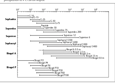
102 103 104 105 106 107 108
Fig. 1.1Effective fractionation ranges for gel filtration media.
Sephadex G-10, G-50 and G-75: available in a range of bead sizes. The finest beads give better resolution but at the expense of slower flow rates.
Superdex Peptide and 30–200: prepacked columns. Superose 6 and 12:
Sephacryl S100–S400:
1.2
Ultracentrifugation
Analytical and preparative ultracentrifugation have been widely applied in immunochemistry, for both molecular weight determinations and isolation procedures.
Preparative ultracentrifugation in sucrose density gradients is useful for the isolation of chicken IgM. Chicken IgM cannot be easily isolated by gel filtration as the IgG readily forms soluble aggregates, so appears within the excluded fraction of Sephacryl S-200 as a major con-taminant of the IgM. However, the difference in size between the IgG dimers and the IgM is still sufficiently great to allow good resolution in the ultracentrifuge.
A detailed treatment of the basic techniques available, for example rate separation and isopycnic separation, both with and without a density gradient, is beyond the range of this book (see Lechner 1994).
1.3
Ion-exchange chromatography
Ion-exchange chromatography is an extremely useful method for the separation of proteins and the isolation of immunoglobulins. Proteins are bound electrostatically onto an ion-exchange matrix bearing an opposite charge. The degree to which a protein binds depends upon its charge density. Proteins are then eluted differentially by:
(a) increasing the ionic strength of the medium. As the concentration of buffer ions is increased they compete with the proteins for the charged groups upon the ion exchanger;
(b) alteration of the pH. As the pH of the buffer approaches the isoelectric point of each protein, the net charge becomes zero and so the protein no longer binds to the ion exchanger. Both cation (e.g. carboxymethyl (CM)–cellulose) and anion exchangers (e.g. diethy-laminoethyl (DEAE)–cellulose) are available. DEAE is used more widely for the fractionation of serum proteins. Cellulose remains the favoured support for the diethylaminoethyl group. Various forms are available to suit particular applications, and high-pressure liquid chromatography columns are available for analytical work.
1.3.1
Batch preparation of rabbit IgG with DEAE–cellulose
DEAE–cellulose and other ion exchangers can be used in columns or in batches. The batch tech-nique is useful when large volumes of serum must be processed under standardized conditions. DEAE–cellulose is equilibrated under conditions of pH and ionic strength which allow all the serum proteins to bind except IgG. The serum must be pre-equilibrated to the same pH and ionic strength as the DEAE–cellulose, then simply stirred with the cellulose prior to recovering the supernatant containing IgG.
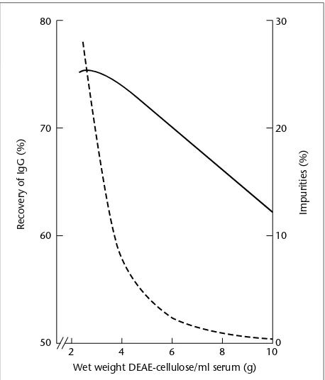
1 . 3 I O N - E X C H A N G E C H R O M A T O G R A P H Y 9 Fig. 1.2Relationship of purity
(dashed line) and yield (solid line) of IgG to the amount of ion exchanger used per ml of serum.This illustrates a universal rule of all protein purifications that the higher one tries to make the yield the lower will be the purity.
80
70
60
50
30
20
10
0
2 4 6 8 10
Wet weight DEAE-cellulose/ml serum (g)
Recovery of IgG (%)
Impurities (%)
Preparation of DE52 cellulose (DEAE)
MATERIALS
Diethylaminoethyl (DEAE)–cellulose DE52 (Whatman) 0.01Mphosphate buffer, pH 8.0
1.0MHCl
METHOD
1 Place 100 g DE52 in a 1-l flask and add 550 ml 0.01Mphosphate buffer, pH 8.0. 2 Titrate the mixture back to pH 8.0 by adding 1.0MHCl.
3 Leave the slurry to settle for 30 min, then remove the supernatant with any fines it may contain. Resuspend the cellulose in enough phosphate buffer to fill the flask.
4 Repeat this cycle of settling, decantation and resuspension twice.
5 Pour the slurry into a Buchner funnel containing two layers of Whatman no. 1 filter paper. Suck the cellulose ‘dry’ for 30 s to leave a damp cake of cellulose.
Preparation of IgG
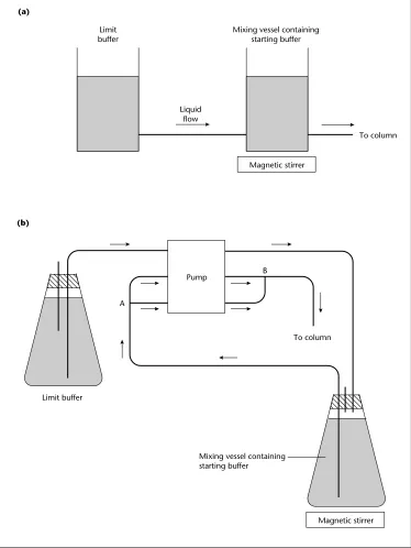
Fig. 1.3 Simple apparatus for the production of an exponential gradient.When the buffer concentration in the limit vessel is greater than the initial concentration in the mixing vessel a convex gradient is produced. Sophisticated pumps are now supplied for fast performance liquid chromatography (FPLC) and high performance liquid chromatography (HPLC) systems, with microprocessor controllers, which allow the programming of an almost limitless range of gradients. Arrows indicate the direction of flow.
To column Pump
Magnetic stirrer
Mixing vessel containing starting buffer
Limit buffer
METHOD
1 Weigh the cellulose into a beaker; for every 10 ml serum use 50 g wet weight of cellulose. Mix 10 ml serum with 30 ml distilled water, to lower its ionic strength, and add to the cellulose at 4°C.
2 To equilibrate stir thoroughly every 10 min for 1 h at 4°C.
3 Pour the slurry onto a Buchner funnel and suck through the supernatant; this contains the required IgG. Rinse the cellulose with 3 volumes of 20 ml 0.01Mphosphate buffer, pH 8.0. 4 Collect and combine all the filtrates.
Examination of IgG preparation
1 If a determination of yield is required, then the IgG content in the original serum and the filtrate may be measured immunologically; e.g. using either rate nephelometry (see Section 3.6) or radial immunodiffusion (see Section 3.4).
2 The purity of the preparation may be determined by comparing the IgG content (measured above) of the filtrate with its total protein content (determined by UV spectrometry). 3 Use SDS-PAGE or immunoelectrophoresis against anti-whole rabbit serum to identify the
main contaminants of the IgG (see also Appendix B.2.1 and Sections 3.2–3.9).
1.3.2
Preparation of IgG with an ionic strength gradient
1 . 3 I O N - E X C H A N G E C H R O M A T O G R A P H Y 11
MATERIALS AND EQUIPMENT
Serum sample
Diethylaminoethyl (DEAE)–cellulose DE52 (Whatman)
Column and fraction collection apparatus (a short wide column is preferable; e.g. 25×3.3 cm; see Appendix B, Fig. B.1)
Gradient device (commercially available, or constructed as in Fig. 1.3) Conductivity meter
Phosphate buffers, pH 8.0, 0.005Mand 0.3M
METHOD
A Equilibration of ion exchanger
1 Place the ion exchanger in a beakerause 2–5 g (wet weight) DE52 for every 1 ml of serum.
2 Add the basic component to the phosphate buffer (0.5Mdisodium hydrogen phosphate) until the pH reaches 8.0.
3 Add 0.005Mphosphate buffer, pH 8.0. There should be 6 ml buffer for every 1 g of wet ion exchanger.
4 Disperse the cellulose and pour into a measuring cylinder and allow to settle (settling time [min]=2×height of the slurry [cm]). Remove the supernatant that contains cellulose ‘fines’; these may block the column.
5 Add a volume of 0.005Mbuffer equal to half the volume of settled cellulose and resuspend. 6 Pour the slurry into the column with the flow-control valve open.
7 Pack the column by pumping 0.005M, pH 8.0, phosphate buffer through at 45 ml/h for each cm2internal cross-section.
8 Monitor the buffer effluent with a conductivity meter. When the ionic strength of the effluent is the same as that of the original buffer, the ion exchanger is equilibrated. If a meter is not available, pass 2–3 l of buffer through the column.
B Running the column
1 Dialyse the sample against the starting buffer (0.005M, pH 8.0 phosphate buffer). 2 Centrifuge the sample. (Some protein will precipitate at this low ionic strength.)
3 Apply the serum to the column and pump through the starting buffer (about 60–100 ml/h). Monitor the effluent for protein. Most of the proteins should bind to the ion exchanger. 4 Elute the proteins with a gradient of increasing ionic strength (see below). Collect fractions
of approximately 5 ml.
TECHNICAL NOTE
If a high concentration of protein is detected in the column effluent prior to the application of the ionic strength gradient, either (a) the ion exchanger or serum was not fully equilibrated or (b) the absorbing capacity of the cellulose has been exceeded.
Ionic strength gradient
this system a discontinuous gradient can be formed automatically and greatly increases the resolu-tion of ion-exchange chromatography.
A continuous exponential gradient may be produced as shown in Fig. 1.3. The limit buffer enters the mixing vessel at the same rate as the buffer is pumped onto the column. The gradient is established according to the following equation:
Cm=Cl−(Cl−C0)e−v/vm
where Cm is concentration in mixing vessel, Clis concentration in limit vessel, C0 is initial
concentration in mixing vessel, vis volume removed from mixing vessel and vmis volume of the mixing vessel.
For ease of calculation this equation may be rewritten as:
When Cl>C0the gradient is convex; when C1<C0the gradient is concave. (The latter is used
for density gradient formation not ion-exchange chromatography: the highest ionic strength buffer will emerge first and elute everything off the column.)
Linear gradients may be established using an open mixing vessel (Fig. 1.4a) or by means of a multichannel pump as shown in Fig. 1.4(b). In this case the equation for the gradient is:
where v0is initial volume of buffer in the mixing vessel and other symbols are as in the equation
for exponential gradient above.
Distribution of serum proteins
Assuming that the ion exchanger has not been overloaded with protein the first peak should con-tain only IgG. This is the only pure protein that can be isolated under these conditions of pH and buffer molarity; the remaining peaks contain several proteins. Beta-lipoproteins, haptoglobin and α2-macroglobulin will contaminate the IgA and IgM fractions.
Regeneration of the ion exchanger
1 Remove the ion exchanger from the column by washing out with distilled water. 2 Add 0.1mHCl (0.5–1×bed volume of cellulose).
3 Place on Buchner funnel and rinse through with distilled water.
4 Add 0.1mNaOH (volume equivalent to the HCl used) and then rinse through with distilled
water.
5 Wash through with full strength buffer and then re-equilibrate with low ionic strength buffer.
TECHNICAL NOTE
To store cellulose ion exchangers add chlorhexidine to a concentration of 0.002% for anion exchangers and sodium azide to 0.02% for cation exchangers.
Note: Azide is a dangerous chemicalado not discard down the sink.
C C C C v
v
m= 0+ ( 1− )0 0
2
2 303 1
1
.
⋅
log −− =
−
C C
C C
v v
m
1 . 3 I O N - E X C H A N G E C H R O M A T O G R A P H Y 13
To column
Magnetic stirrer Mixing vessel containing
starting buffer Limit buffer
Pump B
A
Magnetic stirrer Limit
buffer Mixing vessel containingstarting buffer
Liquid flow
(a)
(b)
To column
1.3.3
Mass production and mini-column ion-exchange chromatography
Conditions for running ion-exchange columns are less critical than for gel filtration; it is there-fore possible to set up large numbers of mini-columns in cheap apparatus such as disposable syringe barrels. The conditions are sufficiently reproducible that fixed volumes of cellulose, serum and buffer give reproducible preparations of IgG and are technically so simple that up to 20 syringe columns in a rack with gravity flow may be run simultaneously. The following procedure will rapidly give very pure IgG.
MATERIALS AND EQUIPMENT
Serum (human)
Saturated ammonium sulphate
Phosphate buffers 0.02Mand 0.2M, pH 7.2
1Mpotassium chloride in 0.02Mphosphate buffer, pH 7.2
Disposable syringe with a central nozzle (e.g. 10-ml hypodermic syringe) Diethylaminoethyl (DEAE)–cellulose DE52
Glass or nylon wool, or sintered plastic disc
Caution: Wear gloves when handling glass wool.
METHOD
1Add 1 ml saturated ammonium sulphate dropwise to 2 ml human serum to give a 33% saturation. Stir for 30 min. Precipitating the serum with ammonium sulphate eliminates much of the material which would otherwise bind to the ion exchanger and reduce its capacity.
2Spin the precipitate at 1000 gfor 15 min and resuspend the pellet in 40% saturated
ammonium sulphate.
3Stir for 10 min and then spin at 1000 gfor 15 min. 4Resuspend the pellet in 0.02Mphosphate buffer, pH 7.2. 5Dialyse the sample against 0.02Mbuffer overnight.
6Block the outlet of a disposable syringe with a little glass or nylon wool or a sintered plastic disc.
7Place 3 g (wet weight) of DEAE–cellulose in the syringe and wash through with 5 ml 0.02M phosphate buffer containing 1MKCl.
8Wash the column with 20 ml 0.02Mphosphate buffer (without KCl). 9Add the dialysed protein sample to the cellulose.
10 Elute the IgG with 15 ml 0.02Mphosphate buffer and collect 3 ml fractions.
11 Determine the absorbance at 280 nm of the fractions and pool those containing protein. These contain the IgG.
12 Calculate the yield of IgG using the extinction coefficient given in Appendix B.
13 Elute the bound protein from the column with 0.02Mphosphate buffer containing 1MKCl. 14 Regenerate the DE52 column as in step 8 above.
TECHNICAL NOTE
1 . 3 I O N - E X C H A N G E C H R O M A T O G R A P H Y 15 Continued on p. 16 and equilibrate both as above. Filtration of the protein sample through the Sephadex G-25 will allow sample equilibration by buffer exchange prior to interaction with the DE52 cellulose. Using this procedure, many samples of highly purified IgG may be prepared during one working day.
1.3.4
QAE–Sephadex isolation of IgG
Quaternary aminoethyl (QAE)–Sephadex is a strongly basic anion exchanger that is particularly suitable for the column separation of proteins using pH gradient elution as the swelling of QAE– Sephadex is not affected by changes in pH. The advantage is that IgG may be prepared using a volatile buffer and freeze dried without prior salt removal. It is advisable to remove β-lipoproteins from the serum before chromatography, otherwise they may break through and contaminate the IgG.
MATERIALS AND EQUIPMENT
Human serum Aerosil
Diamino ethane–acetic acid buffer, ionic strength 0.1, pH 7.0 Acetic acid–sodium acetate buffer, ionic strength 0.1, pH 4.0 Quaternary aminoethyl (QAE)–Sephadex A-50
Column and fraction collection apparatus 1.0Msodium hydroxide
Polyethylene glycol 8000 (PEG-8000) Dialysis membrane tubing
Centrifuge capable of 12 000g
UV spectrophotometer
METHOD
1 Swell QAE–Sephadex A-50 in the diamino ethane–acetic acid buffer. A bed volume of 20 ml of swollen gel is required per 10 ml serum.
2 Pack the gel into a suitable chromatography column and equilibrate with the diamino ethane–acetic acid buffer.
3 Remove b-lipoprotein from the serum by adding 0.2gAerosil to 10 ml serum and stir at
room temperature for 4 h.
4 Centrifuge the serum at 12 000gfor 30 min and remove the lipid layer.
5 Equilibrate the serum with the diamino ethane–acetic acid buffer by dialysis or column buffer exchange.
6 Dilute the equilibrated serum with an equal volume of diamino ethane–acetic acid buffer. (If column buffer exchange was used the sample will have already been diluted by passing through the column.)
7 Apply the sample to the column at a flow rate of 8 ml/cm2/h and continue the elution with the diamino ethane–acetic acid buffer. IgG will come straight through the column while other proteins will be retained. Assess completion of the elution by monitoring the optical density (OD) of elute using a UV spectrophotometer.
9Regenerate the column by running through two bed volumes of diamino ethane–acetic acid buffer.
10 Concentrate the IgG in the first peak to 1/10 volume as quickly as possible; e.g. using dialysis tubing and PEG-8000.
11 The concentrated sample may now be freeze dried without removing salt as the buffer is volatile.
TECHNICAL NOTES
• It is important to concentrate the sample prior to lyophilization otherwise an insoluble pre-cipitate may form.
• The yield of IgG should be about 70% of the serum IgG.
• Conditions should be optimized when preparing immunoglobulins from other species.
1.4
Affinity techniques for immunoglobulins and other molecules
The series of techniques described in this chapter combine the two most sought-after attributes in any purification procedure: (i) large gains in purity in single-step procedures; and (ii) technical simplicity.
In affinity chromatography the technique (summarized in Fig. 1.5) is (a) achieved by the selection of an affinity ligand that shows strong, selective and reversible binding to the molecule being purified (in operational terms, the ligand’s substrate) and (b) facilitated by the use of an insoluble (and, preferably, chemically inert) affinity matrix thus permitting rapid partitioning of the ligand and its substrate.
Axen, Porath and Ernbach introduced a general technique for affinity chromatography whereby molecules containing primary amino groups could be coupled to insoluble polysaccha-ride matrices activated by cyanogen bromide. This route of derivatization is still the most widely employed today, even though the matrix so formed has the disadvantage of charged isourea groups, leading to a bioselective matrix with ion-exchange properties, and unstable covalent bonds between the matrix and ligand, which are susceptible to nucleophilic attack.
Support matrices such as beaded agarose gels may be used. Commercially prepared agarose beads consist of linear chains of agarobiose units in which the ionic charge of the repeating
1 . 4 A F F I N I T Y T E C H N I Q U E S F O R I M M U N O G L O B U L I N S A N D O T H E R M O L E C U L E S 17
change Desorb purifiedsubstrate while
regenerating the affinity matrix
(the theoretical upper limit of the affinity matrix may be calculated from the amount of ligand bound and the stoichiometry of the ligand–substrate interaction). This can be frequently overcome by the use of a ‘spacer arm’ between the support and ligand (see Further reading at end of chapter).
(b) Capture of the substrate molecules. Practical considerations are very important at this stage: e.g. the mixture containing the substrate should be in complete solution (this can be a particular problem with detergent-solubilized cells); the insolubilized ligand and substrate should have sufficient time to interact (do not run the columns too fast and recycle the column effluent several times); and the final washing of the column should be exhaustive to ensure that no unbound or weakly bound material is trapped in the interstices of the column.
1,3-linked β-d-galactopyranose and 2,4-linked 3,6-anhydro-α-galacto-pyranose moieties is
removed by reduction with sodium borohydride under alkaline conditions.
As there are no natural covalent bonds between the linear polysaccharides, these are intro-duced by treatment with epi-chlorohydrin, improving the mechanical and chemical properties of the gel, thus permitting higher flow rates without compression of the gel bed and leading to improved stability at higher temperatures and in the presence of denaturing or chaotropic agents, etc. (Sepharose CL-4B is a commercially available gel with these physical and chemical properties.)
Matrix derivatization–cyanogen bromide activation
Cyanogen bromide reacts with the vicinal diols of agarose (also dextran and cellulose) to produce an activated matrix which will react with ligands (or spacer arms) containing unprotonated prim-ary amines as summarized in Fig. 1.6. The isourea group is positively charged at physiological pH and can act as an ion-exchange matrix with negatively charged proteins.
O HO O
O
OH CH2OH
O OH
O
O HO O
O
OH CH2OH
O OH
O
O HO O
O
OH CH2OH
O OH
O O 1,3-linked D-galactose
Agarobiose repeating unit
2,4-linked 3,6-anhydro-L-galactose
BrC N Cyanogen bromide
Imido carbonate and cyanate-activated intermediates
Protein–NH2
O
C NH2
H N
Protein
O
Isourea derivative
1 . 4 A F F I N I T Y T E C H N I Q U E S F O R I M M U N O G L O B U L I N S A N D O T H E R M O L E C U L E S 19
1.4.1
Preparation of immunoglobulin isotypes
by affinity chromatography
Affinity chromatography may be used to purify immunoglobulin isotypes as an alternative to the physical chemical methods. The most obvious way to use affinity adsorption: prepare an insol-uble antibody specific for the required isotype. However, this requires that the purified isotype first be available to prepare the antibody for immunosorption. Fortunately, immunoglobulins have affinity for a range of other molecules. For example:
IgG binds strongly to protein A, a cell-wall protein derived from Staphylococcus aureus;
IgM binds to mannan-binding protein;
IgA1binds to the lectin jacalin, and mouse IgD binds to Griffonia simplicifoliaI lectin.
The above examples of affinity methods for isolating immunoglobulins largely (except for protein A and G) depend on the recognition of sugars. While particular sugars are associated with various immunoglobulin isotypes the relationship is not absolute and may be altered in disease. Using these methods always has some danger of contamination and yields will not be 100%.
1.4.2
Preparation of IgG on protein A–agarose
The IgG binding properties of protein A make affinity chromatography with protein A–agarose immunoadsorbents a very simple method for preparing IgG. IgG subclasses show differential binding: e.g. human IgG subclasses 1, 2 and 4 bind to protein A but IgG3does not.
MATERIALS
Human serum
Protein A–agarose, e.g. protein A–Sepharose CL-4B Phosphate-buffered saline (PBS)
0.1Mglycine–HCl, pH 2.8
1Msodium hydroxide or solid tris (hydroxymethyl)-aminomethane (Tris)
METHOD
1 Swell 1.5 g protein A–Sepharose CL-4B in 10 ml PBS for 1 h at room temperature and then pack it into a small chromatography column, e.g. a 10-ml disposable hypodermic syringe. Store and use this column at 4°C.
2 Dilute 10 ml human serum with an equal volume of PBS. 3 Filter the serum through the column at a flow rate of 30 ml/h.
4 Wash through unbound proteins with PBS until no more protein leaves the column (monitor the protein with a UV flow cell).
5 Elute the bound IgG with glycine–HCl buffer, pH 2.8.
6 Titrate the pH of the purified IgG solution to near neutrality with NaOH or solid Tris, and dialyse against PBS.
TECHNICAL NOTES
• The protein A content of the swollen gel is 2 mg/ml and the binding capacity for human IgG is approximately 25 mg/ml of packed gel.
• Small quantities of some types of IgM will bind to protein A. You should be aware of this pos-sibility and monitor the IgG preparations if absolute purity is required. Remove the IgM by gel filtration.
• Protein G is also useful for preparing IgG. It has a slightly different range of subclass specificities and is particularly good for preparing rat IgG. Generally it has a high capacity for binding IgG and similar conditions may be used as for protein A isolation of IgG.
As well as IgG, protein A binds to the VH3 region of other immunoglobulin subclasses.
Protein G shows greater specificity for IgG. Protein L, derived from Peptostreptococcus magnus, binds to immunoglobulins through interaction with klight chains, particularly human kI, kIII and kIV and mouse kI. It therefore binds all immunoglobulin classes, but omits all antibodies with llight chains. Protein L can be bound to agarose gels using cyanogen bromide and used as for protein A. (See De Chateau et al. (1993) which discusses the interaction between protein L and
immuno-globulins of various mammalian species.)
1.4.3
Isolation of IgG subclasses using protein A–agarose
Although in both human and mouse the IgG subclasses differ markedly from each other in their biological properties, they are structurally very similar. This similarity has made it almost im-possible to isolate single subclasses using physical chemical techniques. Fractionation of the IgG subclasses is possible using protein A affinity chromatography and pH gradient elution.
Isolation of mouse subclasses
IgG is common to mammalian species, but further evolution has occurred since subclasses of IgG are present in many animals but there is no clear relationship between subclasses in different species. IgG1, IgG2, IgG3and IgG4are found in humans; IgG1, IgG2a, IgG2band IgG3in mice; and IgG1, IgG2a, IgG2band IgG2cin rats.
Mouse serum may be fractionated on protein A–agarose by: 1 allowing all the IgG to bind to the adsorbent; and then
2 eluting the separate subclasses with a stepped gradient of increasing acidity.
MATERIALS AND EQUIPMENT
Mouse serum
Protein A–Sepharose CL-4B Phosphate-buffered saline (PBS) 0.1Mphosphate buffer, pH 8.0
0.1Mcitrate buffers, pH 6.0, 5.5, 4.5, 3.5
1.0Mtris (hydroxymethyl)-aminomethane(Tris)–HCl buffers, pH 8.5, 9.0 Chromatography column or 10-ml disposable syringe
1 . 4 A F F I N I T Y T E C H N I Q U E S F O R I M M U N O G L O B U L I N S A N D O T H E R M O L E C U L E S 21 METHOD
1 Swell 1.5 g protein A–Sepharose in 10 ml PBS for 1 h at room temperature and then pack it into a small chromatography column. Store and use this column at 4°C.
2 Equilibrate the column with 0.1Mphosphate buffer, pH 8.0.
3 Add 2 ml 0.1Mphosphate buffer, pH 8.0 to 4 ml mouse serum and adjust to pH 8.1 with 1MTris–HCl buffer, pH 9.0.
4 Apply the diluted serum to the column and wash through with 30 ml 0.1Mphosphate buffer, pH 8.0 (flow rate 0.4–0.5 ml/min throughout).
5 Elute the IgG1with 30 ml 0.1Mcitrate buffer, pH 6.0. 6 Wash the column with 25 ml 0.1Mcitrate buffer, pH 5.5.
7 To minimize the denaturation of the IgG2aand IgG2bantibodies, collect the eluates from steps 7 and 8 into tubes containing 1.0MTris–HCl buffer, pH 8.5.
8 Elute the IgG2awith 30 ml 0.1Mcitrate buffer, pH 4.5. 9 Elute the IgG2bwith 25 ml 0.1Mcitrate buffer, pH 3.5. 10 Re-equilibrate the column to pH 8.0.
11 Determine the composition of each fraction with specific antisera, preferably using immunoassay.
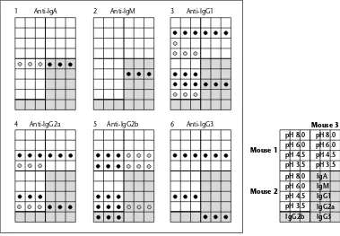
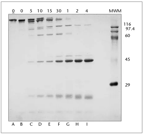
Figure 1.7 shows the purity of fractions obtained from a protein A fractionation of mouse serum. An enzyme-linked immunosorbent assay has been used to examine the protein fractions dot blotted onto nitrocellulose.
TECHNICAL NOTE
IgG3usually elutes with the IgG2afraction. Immunoaffinity chromatography on subclass-specific antibody affinity columns is required to remove this contamination.
Isolation of human IgG subclasses
Protein A may be used to obtain fractions of human IgG which, although not completely pure, are certainly much enriched for individual subclasses. IgG3does not bind to protein A, so if total IgG is filtered through a column of protein A–Sepharose, IgG1, IgG2and IgG4will bind to the adsorbent but IgG3will come straight through. IgG1and IgG2may be differentially eluted from the adsorbent with a pH gradient of increasing acidity. Although IgG4is a slight contaminant in the IgG2fractions, this problem may be reduced by starting with IgG prepared using DEAE– cellulose ion-exchange chromatography (see Section 1.3); this is relatively deficient in IgG4.
MATERIALS AND EQUIPMENT
Protein A–Sepharose CL-4B
Human IgG, DEAE–cellulose purified or human serum 0.15Mcitrate–phosphate buffers, pH 7.0, 5.0, 4.5 0.1Mcitric acid, pH 2.2
Antisera to human IgG subclasses
pH 8.0 pH 6.0 pH 4.5 pH 3.5
pH 8.0 pH 6.0 pH 4.5 pH 3.5 pH 8.0 pH 6.0 pH 4.5 pH 3.5
IgA IgM IgG1 IgG2a IgG3 IgG2b
Mouse 3
Mouse 1
Mouse 2
1 2 3
4 5 6
Anti-IgA Anti-IgM Anti-IgG1
Anti-IgG2a Anti-IgG2b Anti-IgG3
Fig. 1.7 Protein A fractionation of mouse IgG subclasses.Sera from MRL/lpr mice (an inbred mouse strain characterized by lymphoproliferation and high immunoglobulin levels) have been fractionated on protein A–agarose. The material coming straight through the column at pH 8.0 and fractions eluted at pH 6.0, 4.5 and 3.5 have been collected and dot blotted, in triplicate, on to six nitrocellulose membranes. Each membrane has been incubated with a biotinylated specific anticlass or subclass antibody. The blots (1–6) were then incubated with streptavidin labelled with peroxidase. Grid key to each blot shown on right of main figure. Commercially obtained, purified myeloma IgA, IgG1, IgG2a, IgG2b, IgG3and HPLC-purified IgM were included on the blots as standards. The blots show that it is possible to considerably enrich fractions for single subclasses from whole mouse serum. Some slight cross-reactivity is seen with the antibodies reacting with the myeloma proteins. Here there is the problem of determining whether the myeloma proteins or the antisera (all obtained commercially) are impure, or even possibly both. This experiment cautions the reader against always taking manufacturers’ publicity at face value.
Continued METHOD
1 Swell 1 g protein A–Sepharose with 10 ml citrate–phosphate buffer, pH 7.0. 2 Pack the swollen gel into a small chromatography column and equilibrate with
citrate–phosphate buffer, pH 7.0.
3 Load either 5 mg human IgG or 0.5 ml human serum (premixed with 0.5 ml buffer, pH 7.0) onto the column.
4 Wash the column through with pH 7.0 buffer. If purified IgG was used on the column, pure IgG3will come out with the washing buffer. Otherwise, it will come out mixed with all the other non-IgG serum proteins.
1 . 4 A F F I N I T Y T E C H N I Q U E S F O R I M M U N O G L O B U L I N S A N D O T H E R M O L E C U L E S 23 Continued on p. 24 citrate–phosphate buffer, pH 4.5, and the final chamber, which is connected to the column, 6 ml of citrate–phosphate buffer, pH 5.0.
6 Use a flow rate of 12 ml/h and monitor the eluate with a UV flow cell. Two overlapping peaks will be obtained, the first being enriched for IgG2, the second for IgG1.
7 Each peak should be concentrated and recycled on the re-equilibrated protein A column to increase resolution.
8 To re-equilibrate the column, wash sequentially with 6 ml 0.1Mcitric acid and 30 ml citrate–phosphate buffer, pH 7.0.
9 Check the purity of the IgG subclasses with specific antisera, preferably using immunoassay.
1.4.4
Preparation of human IgA
1on jacalin–agarose
IgA is a comparatively difficult immunoglobulin to isolate by physicochemical methods. The lectin jacalin, obtained from the seeds of the jackfruit, Artocarpus integrifolia, binds human IgA1, but not IgA2. Binding is through O-glycosidically linked oligosaccharides containing galactosyl (β-1,3) N-acetylgalactosamine, in the presence or absence of sialic acid. This lectin is available
conjugated to agarose for use in affinity chromatography methods.
An immunoglobulin fraction prepared with ammonium sulphate must be applied to the column, as non-immunoglobulin serum proteins also bind to the lectin. Bound IgA1 is then eluted with melibiose or galactose.
MATERIALS
Jacalin–agarose
Jacalin storage buffer: (4-(2-hydroxyethyl)piperazine-1-ethane sulphonic acid) HEPES 10 mM, pH 7.5, containing 150 mMsodium chloride, 100 mMcalcium chloride, 20 mMgalactose and 0.08% w/v sodium azide
175 mMtris(hydroxymethyl)-aminomethane (Tris)–HCl buffer, pH 7.5
Immunoglobulin preparation, e.g. 45% saturated ammonium sulphate precipitate of human serum Melibiose 0.1Mor galactose 0.8Min 0.175MTris–HCl buffer
Chromatography column or 10-ml disposable syringe
Note: Azide is a dangerous chemicalado not discard down the sink.
METHOD
1 Pour 2 ml of jacalin–agarose gel into a small chromatography column (or 5-ml disposable syringe barrel with the outlet covered by glass or nylon wool).
2 Wash the gel thoroughly with 50 ml of Tris buffer to remove the sugars used to stabilize the lectin during storage.
3 Slowly add 5 ml of human immunoglobulin (10 mg/ml) in Tris–HCl buffer.
4 Wash the column through with 20 ml Tris–HCl buffer (or until the absorbance returns to base line, if you are using a flow-through UV monitor).
5 Elute the IgA with 5 ml 0.1Mmelibiose or 0.8Mgalactose (if using a UV monitor, elute until the protein peak has been collected).
Continued 7 Pool the fractions containing protein and examine for IgA1content and purity by SDS-PAGE
(see Appendix B.2.1) and Western blotting (see Section 4.11) with isotype-specific antisera, or alternatively by immunoelectrophoresis with antisera to IgA and whole human serum. 8 Regenerate the column by washing through with 20 ml storage buffer and store the
jacalin–agarose gel at 4°C.
TECHNICAL NOTES
• The binding capacity of the jacalin–agarose gel will vary between batches but is typically 4.0 mg monomeric IgA1per ml of gel.
• IgA2is lost with other immunoglobulins which do not bind to jacalin. Loomes et al. (1991) provide a method for the purification and characterization of IgA1and IgA2from serum that requires a series of column preparations including gel filtration, DEAE and affinity chromato-graphy on jacalin sepharose.
1.4.5
Preparation of IgM on mannan-binding protein (MBP)
Immobilized mannan-binding protein is commercially available from Pierce (Rockford, Illinois, USA) but can be prepared in-house by immobilizing mannan for isolation of MBP, then coupling the MBP to cyanogen bromide-activated Sepharose 4B (adapted from Nevens et al. 1992).
Preparation of immobilized mannan for isolating MBP
Activation of Sepharose 4B with cyanogen bromide
MATERIALS
Sepharose 4B
Cyanogen bromide (this chemical is very toxic and must be handled in a fume cupboard)
Crushed ice
5Msodium hydroxide
0.1Msodium bicarbonate solution Yeast mannan
1.0Methanolamine, pH 9.0
1.25MNaCl containing 20 mMCaCl2and 10 mMimidazole, pH 7.8
METHOD
1Wash 100 ml Sepharose 4B in 1.6 l of distilled water, then remove the water by suction until it is dried.
2Transfer the Sepharose 4B into a beaker and suspend in 100 ml of distilled water.
3Put beaker containing the Sepharose 4B and water onto a magnetic stirrer and insert a pH meter probe.
1 . 4 A F F I N I T Y T E C H N I Q U E S F O R I M M U N O G L O B U L I N S A N D O T H E R M O L E C U L E S 25 4 Slowly add 20 g of solid cyanogen bromide over a 20-min time period, maintaining the
temperature at around 20°C by adding crushed ice to the stirring slurry.
5 Keep the pH of the slurry between 10.5 and 11.0 by adding a concentrated solution of sodium hydroxide (dropwise).
6 Wash the cyanogen bromide-activated Sepharose using a glass Buchner funnel with around 2 l of ice-cold 0.1Msodium bicarbonate, then dry by suction.
7 Dissolve 1.78 g of yeast mannan in 100 ml 0.1Msodium bicarbonate solution, add to the activated Sepharose beads and stir overnight at room temperature.
8 Wash the activated Sepharose beads with 1 litre of distilled water, then suspend in 160 ml 1.0Methanolamine, pH 9.0, at room temperature for 60 min.
9 Wash with 1 litre of distilled water and pack into a glass columnathe Sepharose is now ready for isolating the MBP.
10 Equilibrate the column with 10 column volumes of 1.25MNaCl containing 20 mMCaCl2 and 10 mMimidazole, pH 7.8.
Coupling of cyanogen bromide-activated Sepharose 4B and MBP
MATERIALS
Sepharose 4B
Solid cyanogen bromide (this chemical is very toxic and must be handled in a fume cupboard)
0.1Msodium bicarbonate buffer, pH 8.5 1.0MNaCl
10 mMtris(hydroxymethyl)-aminomethane (Tris) containing 1.25MNaCl and 2 mMethylene diamine tetra-acetic acid (EDTA), pH 7.4
Mannan-binding protein (MBP) solution Coomassie protein assay reagent
1 Activate the Sepharose 4B with cyanogen bromide by the same procedure as in steps 1–5 (above).
2 Wash the activated Sepharose with 200 ml of ice-cold distilled water followed by 100 ml 0.1Msodium bicarbonate buffer, pH 8.5, then dry by suction.
3 Mix the MBP solution and activated Sepharose and stir overnight at 4°C.
4 Filter the gel suspension and wash with 200 ml 1.0MNaCl, followed by 200 ml water. 5 Block the excess reactive groups on the MBP coupled to cyanogen bromide-activated
Sepharose 4B column with 10 ml 1.0Methanolamine, pH 9.0, and stir at room temperature for 60 min.
6 Wash with 200 ml of distilled water followed by 200 ml 10 mMTris containing 1.25MNaCl and 2 mMEDTA, pH 7.4.
Note: Test the coupling of MBP with the activated Sepharose by mixing 200 µl of
Isolation of IgM using MBP coupled to cyanogen bromide-activated Sepharose 4B
MATERIALS
Sample: serum or monoclonal antibody culture supernatant MBP coupled to cyanogen bromide-activated Sepharose 4B
Hypodermic syringe and glass wool (Caution: Wear gloves when handling glass wool)
10 mMtris(hydroxymethyl)-aminomethane (Tris) containing 1.25MNaCl with 0.02% w/v sodium azide, pH 7.4
10 mMTris containing 1.25MNaCl, 20 mMCaCl2with 0.02% w/v sodium azide, pH 7.4 10 mMTris containing 1.25MNaCl, 2 mMEDTA with 0.02% w/v sodium azide, pH 7.4
Note: Azide is a dangerous chemicalado not discard down the sink.
METHOD
Steps 1–8 are performed at 4°C; therefore all buffers must be ice cold.
1Dialyse the monoclonal antibody supernatant or sample containing IgM overnight against two changes of 1000 ml 10 mMTris containing 1.25MNaCl with 0.02% w/v sodium azide, pH 7.4. (This step is to remove any phosphate ions that could form a precipitate with the calcium ions in subsequent steps.)
2Dilute the sample with an equal volume of 10 mMTris containing 1.25MNaCl, 20 mM CaCl2with 0.02% w/v sodium azide, pH 7.4.
3Load the MBP coupled to cyanogen bromide-activated Sepharose 4B into a 100-ml hypodermic syringe plugged with glass wool.
4Wash the column with 5 column volumes of 10 mMTris containing 1.25MNaCl, 20 mM CaCl2with 0.02% w/v sodium azide, pH 7.4.
5Apply the diluted sample, allow to flow completely into the column, collect the eluate and re-apply to the column.
6Repeat step 5 around five times for the best yield of IgM antibodies.
7Allow the sample to incubate on the column for about 30 min by clamping the eluate tubing, and keep the top of the gel from drying by adding 200 µl of 10 mMTris containing 1.25MNaCl, 20 mMCaCl2with 0.02% w/v sodium azide, pH 7.4.
8After the incubation, wash the column with 10 column volumes of 10 mMTris containing 1.25MNaCl, 20 mMCaCl2with 0.02% w/v sodium azide, pH 7.4, monitoring the fractions spectrophotometrically at 280 nm.
Steps 9onwards are performed at room temperature; therefore buffers must notbe ice cold. 9Take the column into room temperature and allow to stand for 60 min (do not allow the
column to dry out).
10 Wash with 3 column volumes of 10 mMTris containing 1.25MNaCl, 20 mMCaCl2with 0.02% w/v sodium azide, pH 7.4.
11 Elute the IgM with 10 mMTris containing 1.25MNaCl, 2 mMEDTA with 0.02% w/v sodium azide, pH 7.4, monitoring the fractions spectrophotometrically at 280 nm. An absorbance of 1.18 at 280 nm is equivalent to an IgM concentration of 1 mg/ml.
12 Store the IgM-containing fractions at –20°C.
1 . 5 P U R I F I C A T I O N O F A N T I B O D I E S 27
1.4.6
Purification of IgD on
Griffonia simplicifolia
I lectin
MATERIALS
Monoclonal antibody culture supernatant derived from an IgD-secreting hybridoma 5-ml hypodermic syringe
Glass wool (Caution: Wear gloves when handling glass wool) GS-I lectin–Sepharose (purchase coupled or linked as above) Phosphate-buffered saline (PBS) containing 1 mMCaCl2 PBS containing 1 mMCaCl2and 0.1M D-galactose UV spectrophotometer
Dialysis membrane tubing
METHOD
1 Centrifuge monoclonal antibody culture supernatant at 10 000gfor 30 min at 4°C or room
temperature.
2 Save the supernatant and discard the cell debris.
3 Dilute the supernatant with 10 volumes of PBS containing 1 mMCaCl2 and centrifuge at 15 000gfor 30 min at 4°C and save the supernatant for step 7.
4 Prepare a mini-column such as a 5-ml hypodermic syringe plugged with glass wool (use gloves when handling glass wool).
5 Pour in 3 ml of GS-I lectin–Sepharose and maintain at a temperature of 4°C. 6 Wash GS-I lectin–Sepharose with the following ice-cold buffers:
12 column volumes PBS containing 1 mMCaCl2;
12 column volumes PBS containing 1 mMCaCl2and 0.1M D-galactose; 12 column volumes PBS containing 1 mMCaCl2.
7 Load the supernatant and allow to flow into the GS-I lectin–Sepharose.
8 Wash the column with sufficient ice-cold PBS containing 1 mMCaCl2, monitoring the fractions spectrophotometrically at 280 nm, until a baseline is reached.
9 Elute the IgD antibodies with ice-cold PBS containing 1 mMCaCl2and 0.1M D-galactose. Around 3–5 column volumes should be enough to elute the IgD, but it is advised that the eluate is monitored spectrophotometrically at 280 nm.
10 The column may be regenerated with 2 column volumes PBS containing 1 mMCaCl2and 0.1M D-galactose, then 10 column volumes PBS containing 1 mMCaCl2. To preserve the column add 0.02% w/v sodium azide to the final wash and store at 4°C.
11 Dialyse the IgD sample against 500 ml ice-cold PBS, pH 7.3, at 4°C with five changes of buffer. 12 Estimate the IgD concentration of the dialysed sample by measuring at 280 nm with a UV
spectrophotometer.
1.5
Purification of antibodies
Continued immunoglobulins but those reacting with this antigen will be at a relatively higher concen-tration, compared to normal serum.
An animal receiving a transplantable plasmacytoma or hybridoma will produce large amounts of the monoclonal immunoglobulin or antibody, but there will still be a significant background of normal serum proteins and immunoglobulins, even in ascitic fluid.
To study a particular antibody in detail it is of great advantage to be able to separate it from the surrounding non-specific antibody molecules using the antigen. Then to obtain reactive purified antibody we must separate the complex and remove the antigen.
The forces binding antibody to antigen are those involved in any protein–protein interaction: (a) coulombic;
(b) dipole;
(c) hydrogen bonding; (d) van der Waals’; (e) hydrophobic bonding.
All these forces depend upon the charge of the molecules taking part in the reaction. The net charge of the molecules in turn depends on the pH of the medium. If the pH of the medium is lowered sufficiently the protein molecules change conformation, gain H+ions and so repel each other. We are now faced with the problem of physically removing the antigen or the antibody, because when the pH is returned to neutrality the complexes will re-form.
If the antigen is insoluble it can be easily separated from soluble antibody. There are many methods available for rendering either the antigen or antibody insoluble, some of which are described in the following sections.
1.5.1
Preparation of a protein immunoadsorbent
In this experiment antibodies to mouse immunoglobulin are purified but the identical method can be used for other proteins.
MATERIALS AND EQUIPMENT
Sepharose 4B
Cyanogen bromide (this chemical is very toxic and must be handled in a fume cupboard)
2.0Msodium hydroxide Phosphate-buffered saline (PBS)
Borate–saline buffer, pH 8.3, ionic strength 0.1 Mouse immunoglobulin
Sintered glass funnel UV spectrophotometer
METHOD
1Pipette 14 ml of Sepharose (about 200 mg) into a 50-ml glass beaker and add 10 ml of distilled water.
All procedures must now be carried out in a fume cupboard.