Download details:
IP Address: 161.139.220.155
This content was downloaded on 06/04/2014 at 18:37
Please note that terms and conditions apply.
View the table of contents for this issue, or go to the journal homepage for more
The Evaluation of Hydroxyapatite (HA) Coated and Uncoated
Porous Tantalum for Biomedical Material Applications
Nadia Safuan1, 2, Irza Sukmana1, 2, 3 *, Mohammed Rafiq Abdul Kadir1, 2, Deni Noviana4
1
IJN-UTM Cardiovascular Engineering Centre (CEC), Block V01 Taman University, Johor Bahru, 81310 Malaysia, 2MEDITEG (Medical Devices and Technolgy) Research Group, Faculty of Biosciences and Medical Engineering UTM, 81310 UTM Skudai Johor, Malaysia, 3Department of Mechanical Engineering, University of Lampung, Jalan Prof. Soemantri Brojonegoro 1, Bandar Lampung 35143, Indonesia, 4Divison of Surgery and Radiology, Department of Veterinary Clinic, Reproduction and Pathology, Faculty of Veterinary Medicine, Bogor Agricultural University (IPB), Dramaga Campus, Bogor, 16610, Indonesia.
*Corresponding author: [email protected]
Abstract. Porous tantalum has been used as an orthopedic implant for bone defects as it has a good corrosion resistance and fatigue behaviour properties. However, there are some reports on the rejection of porous Ta after the implantation. Those clinical cases refer to the less bioactivity of metallic-based materials. This study aims to evaluate hydroxyapatite coated and uncoated porous Tantalum in order to improve the biocompatibility of porous tantalum implant and osseointegration. Porous tantalum was used as metallic-base substrate and hydroxyapatite coating has been done using plasma-spraying technique. Scanning Electron Microscopy (SEM) and Field Emission Scanning Electron Microscopy (FESEM) techniques were utilizes to investigate the coating characteristics while Confocal Raman Microscopy to investigate the interface and image. The effect of coating to the corrosion behaviour was assessed by employing potentiodynamic polarization tests in simulated body fluid at 37±1 °C. Based on SEM and FESEM results, the morphologies as well the weight element consists in the uncoated and hydroxyapatite coated porous tantalum were revealed. The results indicated that the decrease in corrosion current density for HA coated porous Ta compared to the uncoated porous Ta. This study concluded that by coating porous tantalum with HA supports to decrease the corrosion rate of pure porous.
1. Introduction
Bone diseases and bone fractures including osteoporosis, bone tumor and trauma count for half of all chronic diseases in the population over 50 years of age in developed countries. Based on the survey, it affects peoples about hundreds of millions across the world. Approximately, 10 million Americans have osteoporosis and more around 34 million peoples are at the risk of getting this disease. Thus, nearby future, it is estimated that it will keep increasing due to the aging of the population. These diseases cause high morbidity, and often require surgical interventions [1]. According to the latest statistical, over 200,000 hip replacements regarding load-bearing implants are performed each year in USA. However, this number expected to rise steadily due to increasing of life expectancy [2].
the metallic materials and their alloys are known as non-bioactive substrates. In the in-vivo system, cell and tissue also have limited adhesion to biometallic materials [8].
Porous tantalum has been shown to be corrosion resistant in vivo. Also, it has been recognized as a good candidate for orthopedic implants and bone substitute applications due to excellent fatigue properties [6-8]. Porous tantalum also has a good biocompatibility to the bone tissue. In order to encourage natural bone growth at the interface of the prosthetic device, metal implants used to be coated with bioactive ceramic substrates, such as: Hydroxyapatite (HA) and bioglass [9]. In restorative orthopedics and dentistry, calcium phosphate and hydroxyapatite coated metallic implants are currently considered as the best bearing implants due to the possibility to take advantage of both biocompatibility of ceramics and the strength of metals [10].
Currently, biomaterials are limited due to the ever-present issues relating to biocompatibility. One of the factors that influence biocompatibility of metallic-based materials is controlled by chemical. In a specific way, it is called electrochemical interaction that results in the release of metallic ions into the surrounding tissue and the toxicology of these released substances [11]. This study aims to evaluate bioactivity of hydroxyapatite-coated and uncoated porous Tantalum for bone implant application. Corrosion behaviors of both samples were conducted in Simulated Body Fluid (SBF) solution at room temperature.
2. Experimental
2.1. Samples Preparation for corrosion test
Hydroxyapatite coating on porous tantalum was done using plasma-spraying technique at Amrec-Sirim, Kulim, Malaysia. The thickness layer of the coating is about 10-15 Pm. The sample was cut by using high precision Allied wire cutter. The surface dimension of porous tantalum samples (coated and uncoated) was 7mm x 7mm, with thickness of about 0.5mm. After cutting, samples were washed in distilled water then proceed for cold mounting. Afterwards, samples were grinded with fine and coarse paper. Ultrasonic bath was then used to clean the samples before final rinsing with acetone then dried in air.
2.2. Corrosion test
Electrochemical measurements in this study were performed in a three-electrode electrochemical cell. Ag, AgCl/KCl (3.5M) (0.7V) electrode was used as reference electrode and for counter electrode, graphite was used. The electrolyte was Simulated Body Fluid (SBF) solution and all tests were performed at room temperature which is 37±1 °C. A potential scanning rate at 0.25 [mV S¯¹] initiated at -250 [mV] below the open circuit potential and the atmosphere was open to air, recorded was the dynamic polarization curves. Polarization curves were obtained 1000s after immersion in the solution, varying the potential between -0.5 and 2.5V. The current density was measured using VersaSTAT 3 potentiostat interfaced with a computer.
2.3. Scanning Electron Microscope
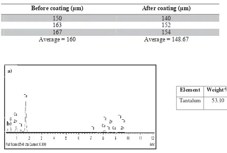
Scanning Electron Microscope (SEM) technique used to analyze the HA coating quality, and to evaluate the porous microstructure and morphology of the samples. Field Scanning Electron Microscope (FESEM) was also used to estimate the ratio of Calcium over Phosphate (Ca/P) at the Hydroxyapatite coated area to measure the weight element of each samples.
3.1. SEM and FESEM images
The result of SEM shows the average coating thickness of deposition HA on pure porous Ta substrate. Meanwhile, the pore size before and after coating also was investigated shown on Fig.1a, the average coating thickness obtained by using plasma spray coating technique is about 157μm.
Fig. 1 SEM Image of Average Coating Thickness on HA Coated Porous Ta (a) and SEM Image of Pore Size of Porous Tantalum before (b) and after (c) coating with HA
Fig. 1b shows the pore size thickness of porous Ta before and after coating with HA. The average of pore size for pure porous tantalum is 160 μm while after coating with HA it becomes smaller which is148.67 μm as shown in Table 1.
Table 1 Pore Size of Porous Tantalum before and after coating with HA
Before coating (
μ
m)
After coating (
μ
m)
150
140
163
152
167
154
Average = 160
Average = 148.67
Element Weight%
Tantalum 53.10
150 μm
140 μm
a)
b)
b)
Fig. 3 FESEM graph and weight element for porous Tantalum (a) Uncoated (b) HA coated
As shown in Fig. 3 (b), Ta is still present in the coated sample however the weight decline since it is not pure anymore. By using only pure porous tantalum can caused adverse effect to the surrounding tissue. It is occurred due to the released of metallic ion to the surrounding tissue. In order to make metal surfaces interact to the surrounding tissue (bioactive), porous tantalum was coating with the calcium phosphate that have been modified the surface of the implants [9].
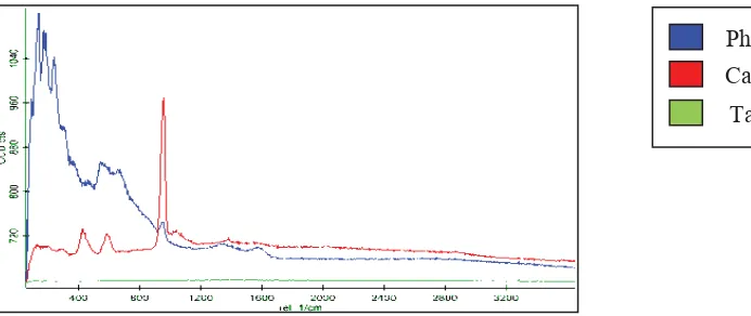
3.2. Confocal Raman Microscopy
Interface Raman microscopy was used to analyze the raman spectrum of porous tantalum coated HA. It used the concept of electromagnetic radiation due to the consequences at certain frequencies; it will correlate to the vibration of specific sets of chemical bonding within a molecule [12]. Therefore in Fig. 5, the green line showed on the graph was a constant because there was no absorption of electromagnetic radiation on the surface of porous tantalum sample. Besides, Confocal Raman Microscopy could not detect the present of pure metallic-based material such as Tantalum.
The blue line represented Phosphorus element which in the region of 10-200 cm-1 presented the lattice vibrations in crystals, LA modes. Thus, the Raman was strong, showed the highest peak at the beginning of testing. Therefore, it consumed the highest absorption of electromagnetic radiation initially towards Phosphorus elements.
While the red line in Fig. 5 indicated v(cc) alicyclic, aliphatic chain vibrations. The range of region is about 600-1300 cm-1 showed the medium peak and also indicating medium Raman for calcium elements based on the coating for the tested sample. Thus, it was proven that the coating was attached to the substrate.it shows that the green line on the graph is constant thus Confocal Raman Microscopy could not detect the present of pure metallic-based material such as Tantalum.
Fig. 5 Interface Raman Spectrums of Confocal Raman Microscopy Phosphorus Calcium
Tantalum
3.3 Corrosion Behavior Studies
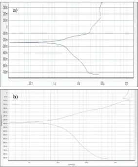
Potentiodynamic polarization used to analyze the corrosion behavior of tantalum material as shown in Fig. 6 (a) and (b).
Fig. 6 Polarization Curves of Porous Ta (a) Uncoated (b) HA Coated
Table 2 Mean values (standard deviation) of corrosion current densities and corrosion potentials of uncoated porous Ta and HA coated in SBF solution at ±37° C.
Porous Ta Ecorr (mV) Icorr (uA)
Uncoated -239.002 8.714
HA coated -381.428 6.820
Uncoated porous Ta obtained higher corrosion current density [Ecorr= -239.002mV, icorr= 8.714uA, Table 2] hence lower the corrosion resistance in SBF solution when compared to HA coated porous Ta [Ecorr= -381.428, icorr= 6.820, Table 2] that shows lower corrosion current density. Based on Fig. 6 (a), the pitting corrosion potential of the uncoated porous tantalum is lower than the coated porous tantalum. Previous study showed that coating the porous tantalum with HA reduced the corrosion current density however also increased the pitting corrosion potential of the porous tantalum. This showed that the coated
a)
as porous tantalum so that the human body will be safe from corrosion process that take place in the human body. Surface films like HA plays important part in corrosion and osseointegration process of implants. It helps by modifying the surface of alloy implants as well improving the corrosion resistance [13]. This explained the corrosion resistance is inversely proportional to the current density. As an example, if it had low current density indicates high corrosion resistance and consequently a low level of metals release towards the surrounding. It shows it was beneficial for the biocompatibility.
This study was conducted due to the corrosion resistance may affect the cell metabolism and also cell behavior from the corrosion occurred [13]. Currently, tantalum-based materials had gain attention as one of the promising bone implant materials with higher biocompatibility and corrosion resistance. However, if the porous tantalum in pure form, it is mechanically weak as poor bone integration with the surrounding cell. To enhance the usage of Ta in biomedical application, it was coated with bioceramics such as Hydroxyapatite. With the ability to bind the bond and its biocompatibility, Hydroxyapatite (HA) had been intensively studied for bone repair and replacement applications [14].
Previous in vitro research showed that if the metal coated with single HA reduced the corrosion current density of 316L SS substrate in physiological solutions. However this effect was not sufficiently complete. It is a fact that the surface morphology and structure of a single HA coating consists of a porous surface and some micro cracks. It will not become a complete barrier against the release of metallic ions from metallic implant to physiological solutions just by coating with HA. The HA coating act simply as a semi-mechanical barrier. On the other hand, previous in vivo study showed that the bone osteointegration around single HA coated Vitallium alloy and single HA coated SS were almost similar. Based on the point of clinical success and osteointegration, there was no significant difference was observed [15-16]. Vitallium was originally used in endodontic implants however the biocompatibility was questioned when it was shown to undergo surface corrosion [17]. Our result is also in a good agreement with other researchers [12, 18]. By coating the porous tantalum with HA we believe it can improve the corrosion resistance and thus the biocompatibility of the tantalum material [19]. From clinical study for porous tantalum implant, the results indicated the present of swelling besides there also fluid developed around the implant within the first week of surgery. Based on the cell culture perspective, the implant also caused tissue failure happened at or beyond the implant surface [20]. This may occurred due to the questioning of biocompatibility of metallic biomaterials which had considered for medical use in term of corrosion resistance. If the corrosion resistance was excellent hence it would help to minimize the released of toxic metal-ions in the human body [21]. Therefore, Hydroxyapatite (HA) coated porous tantalum would be the best way to help solving problems regarding
4. Conclusion
Based on SEM and FESEM results, the morphologies as well the weight element consists in the uncoated and hydroxyapatite coated porous tantalum were revealed. As for pure tantalum, 53.10% weight of element was present while in HA coated samples there was just only 30.65 % consist of pure tantalum followed by calcium 19.61 % and phosphorus 11.57 %. From this study, the average thickness of coating by plasma technique had also obtained which is 160 μm. The results based on electrochemical test indicated that the decrease in corrosion current density for HA coated porous Ta compared to the uncoated porous Ta. Thus, by coating porous tantalum with HA helps to reduce the corrosion rate of pure porous tantalum and the release of metallic ions to the surrounding tissue. By releasing the toxic metallic-ion towards surrounding environment may affect the growth of the cell and it is not good for people who
that has most characteristics similar to bone composition such as bone-bonding and mechanical properties, we had magnificently proven that by coating porous tantalum with bioceramic, Ca/P had improve the surface of the implants being optimum biocompatible. With this research, we hope that it be a helpful and contributes to the world of orthopedic field. In future research, there is needed to test in-vitro biocompatibility assessment of HA coated and uncoated porous tantalum by doing cell culture and animal studies. Besides, it also needed to study the cytotoxicity of the tantalum materials.
Acknowledgements
The authors thank to Dr. Hendra Hermawan (FBME UTM) and Dr. Jamal Siddiqui for their constructive comments. UTM Tier-1 (vote#03H12), MOHE-FRGS (vote#4F128) and UTM RU (vote#00G670) are also kindly acknowledged.
References
[1] National Osteoporosis Foundation. Available from: http://www.nof.org.
[2] Kim S. Changes in surgical loads and economic burden of hip and knee replacements in the US: 1997-2004. Arthritis Rheum 2008. 59(4). p. 481-8.
[3] D. H. Kohn, Curr. Opin. Solid St. M. 1998. (3). p. 309.
[9] Lyle D. Zardia Ckas, Douglas E. Parsell, Lance D. Dillom, Darrel W. Mitchell, Laura A. Nunnery, Robert Poggle. Structure, Metallurgy and Mechanical Properties of Porous Tantalum Foam. 2000.
p. 180-187.
[10] T. LI, J. Lee, T. Kobayashi and H. Aoki, J. Mater. Sci., Mater. Med. 1996. (7). p. 355.
[11] Williams David F.(ed.), 1981. Biomaterials an Biocompatibility: An introduction, in Fundamental Aspects of Biocompatibility, vol. 1, CRC press, Boca Raton, Fl, pp. 2-3
[12] Joan Coates, Interpretation of Infrared Spectra, A Practical Approach, Encyclopedia of Analytical Chemistry, R. A. Meyers (Ed.) 2000. p. 10815-10837
[13] M.H. Fathi, F. Azam, Materials Letters,Novel Hydroxyapatite/Tantalum Surface Coating for Metallic Dental Implant. 2007. 61. p. 1238-1241.
[14] M. Metikos-Hukovic, A. Kwokal, J. Piljac, Biomaterials. 2003. 24. p. 3765.
[15] X. Miao, Materials Letters. 2003. 57. p. 1848.
[16] M.H. Fathi, M. Salehi, A. Saatchi, V. Mortazavi, S. B. Mousavi, Dent. Mater. 2003. 19. p. 188.
[17] M.H. Fathi, M. Salehi, A. Saatchi, V. Mortazavi, S. B. Mousavi, Surface Engineering. 2001. 17. p. 459.
[18] Locci P, Marinucci L, Lilli C, Belcastro S, Staffolani N, Bellocchio S, et al. Biocompatibility of alloys used in orthodontics evaluated by cell culture tests.J Biomed Mater Res 2000. 51(4). p. 561- 68.
[19] W. Suchanek, M. Yoshimura, J. Mater. Res. 1998. 13. p. 94.
[20] S.A. Hacking,J.D. Bobyn,K.-K. Toh, M. Tanzer,J.J. Krygier, Fibrous tissue ingrowths and attachment to porous tantalum, 2000