MetriC evAluAtiOn Of PArtiAlly diSPlACed
teMPOrOMAndibulAr jOint diSC
Mirko laškarin1, tomislav badel2, josipa Kern3, ivana Savić Pavičin4 and dijana Zadravec5 1Prosthodontics Outpatient department, Šibenik Health Center, Šibenik; 2department of removable Prosthodontics, School of dental Medicine; 3department of Medical Statistics, epidemiology and Medical informatics, Andrija Štampar School of Public Health, School of Medicine, university of Zagreb; 4department of dental Anthropology, School of dental Medicine; 5department of diagnostic and interventional radiology,
Sestre milosrdnice university Hospital Center, Zagreb, Croatia
SuMMAry – The objective was to determine the quantitative relationship between the condyle and disc position in the glenoid fossa between two different slices of the same temporomandibular joints (tMjs) with partial anterior disc displacement (dd). The study was conducted on 40 patients with dd of tMjs (mean age, 35.5 years). The clinical diagnosis of dd was confirmed by magnetic resonance imaging. joints from the patient groups were analyzed according to the laterality and depending on disc displacement (a total of 80 joints). Comparison was made between two different
slices of 9 joints with partial dd with reduction: partial dd was analyzed in the
representati-ve centrolateral or centromedial parasagittal slice of the tMj (tMj partial dd – slice dd). The contralateral slice of the same joint was without dd (tMj partial dd – slice ndd). The analysis also included 34 healthy joints without dd (tMj ndd) of the same patients. The position of the condyle and disc was calculated using the Kurita et al. method on the parasagittal view of the tMj. A statistically significant difference was recorded for different slices of the same tMjs with partial dd (tMj partial dd – slice dd and tMj partial dd – slice ndd) (p<0.01), but no difference was found in condyle positions depending on the existence of partial dd (p>0.05). The compared values between slice tMj partial dd – slice ndd with the group of tMj ndd showed no significant difference in either disc position or condyle position (p>0.05). There were differences of disc position in various slices of the same joint with visually confirmed partial dd. The dorsocranial condyle position could not indicate partial anterior dd.
Key words: Magnetic resonance imaging; Temporomandibular joint disc – pathology; Temporomandi-bular joint disorders – pathology; Dislocations – pathology
Correspondence to: Assoc. Prof. Tomislav Badel, PhD, department of removable Prosthodontics, School of dental Medicine, uni-versity of Zagreb, Gundulićeva 5, Hr-10000 Zagreb, Croatia e-mail: [email protected]
received november 26, 2012, accepted july 17, 2014
Introduction
temporomandibular disorders (tMds) is a col-lective term that includes a number of clinical condi-tions and diagnosis of functional disorders involving the masticatory muscles, the temporomandibular joint (tMj) or both with the associated orofacial structures.
The two major clinical features of functional tMj problems are preauricular or auricular pain, noises (clicking and crepitation), and oral dysfunction (lim-ited mouth opening). tMj disorder is usually present as a disruption of the normal condyle-disc movement. This disc disorder is the most common tMd diagno-sis under the name anterior disc displacement (dd). Another common diagnosis is osteoarthritis, related to roughness of the articular surfaces1-4.
Magnetic resonance imaging (Mri) has been widely accepted as a ‘gold standard’ tool for diagnos-ing dd, which is the most common type of tMj
disorders. A cross-sectional image of the tMj in the individually angulated or parasagittal plane enables clear view of the glenoid fossa, disc and condyle as the most important parameter of tMj visual analysis5.
There is slight uncertainty in defining the term ‘partial dd’ when it is observed in closed mouth posi-tion. Many authors differentiate between partial and complete anterior dd (which is evaluated on the basis of three different parasagittal slices of the same joint) according to radiologic and/or anatomic analyses of the tMj, as well as on the basis of clinical diagno-sis6-9. However, numerous studies based on Mri and the diagnostic Criteria (dC) for tMd Axis i diag-nostic system do not mention the diagnosis of partial dd10-13. Apart from the metric evaluation of anterior dd in degrees according to the 12 o’clock method14, there are no studies on the quantitative evaluation of the position, that is, on the dd in joints with partial dd.
The aim of the study was to determine the rela-tionship between the position of the disc and condyle head in the glenoid fossa between two different paras-agittal slices (slice with dd and slice without dd) of the same tMjs with partial anterior dd. Slices with dd of partially displaced tMjs were compared with physiological disc position (without dd) differenti-ated from the same sample of patients with tMj dis-order in contralateral tMjs.
Materials and Methods
The study was conducted on 40 patients with dd of tMjs (aged 15-71, mean 35.5 years; 1:3 men to women ratio), who were collected consecutively be-cause they sought help due to the following clinical symptoms of tMj disorders: painful tMj and click-ing and/or limited mouth openclick-ing. The clinical diag-nosis of various dds (partial dd with reduction, total dd with and without reduction) was established
us-ing manual functional analysis (MfA)9,15. All patients were examined by Mri, which was used to confirm and establish definitive diagnosis of dd. All patients willingly agreed and gave their written consent to par-ticipate in the study which was approved by the ethics Committee of the School of dental Medicine.
Mri was performed with a 1 t Harmony scan-ner (Siemens, erlangen, Germany; with the following spin-echo-sequent parameters: t1 weighted image tr/te 450/12, t2 weighted image tr 3000/te 66, field of view of 160x160, matrix of 256x192 and 3-mm slice), and with a 1.5t Avanto scanner (Sie-mens, erlangen, Germany; with the following spin-echo-sequent parameters: t1 weighted image tr/te 410/9.4, t2 weighted image tr 460/te 15, field of view of 180x180, matrix of 410x512 and 2-mm slice).
Mr images in the parasagittal plane were obtained in the closed- and open-mouth position. The angle of the parasagittal imaging was individually determined by the angle shown on the individual angulated slices of the axial and coronal slice. The open mouth posi-tion was fixed with an inter-incisal individual fixator using the Optosil® P plus (Heraeus Kulzer, Hanau, Germany).
The disc physiological position in closed-mouth position was defined according to the placement of its inter-medial zone between the articular eminence and the shortest distance of the bone contours of the con-dyle ventrocranial part in the parasagittal plane in the closed mouth position according to Orsini et al.16 and bumann and lotzmann9.
Qualitative analysis was performed for each tMj to examine the three representative slices (centrolat-eral, central and centromedial) in the parasagittal slice on the basis of which partial or complete dd was determined in closed mouth position. Complete dd means that the disc is displaced in all slices. Par-tial dd is confirmed if the disc is in the physiological position in one (centrolateral or centromedial) slice, Table 1. Distribution of disc positions in TMJs and symptomatic TMJs of all patients
disc position
(n, %) Partial dd dd with reduction dd without reduction normal total
All tMjs 9 (11.25%) 15 (18.75%) 21 (26.25%) 35 (43.75%) 80 (100%)
Painful tMjs 9 (20.93%) 15 (34.88) 19 (44.19%) – 43 (100%)
and displaced in the opposite slice (centrolateral or centromedial) in the same tMj. Partial dd with re-ductionwas foundin the representative centrolateral or centromedial parasagittal slice of 9 tMjs (figs. 1 and 2) in closed-mouth position. Other categories of tMjs, i.e. complete (n=15) reducing dd and com-plete (n=19) nonreducing dd, were not included in this study (table 1).
Qualitative analysis was used to compare the dif-ference between the calculated disc and condyle posi-tion related to the following groups: at first, two dif-ferent slices of the same tMjs with partial dd were compared: slice without dd (‘tMj partial dd – slice ndd’) and slice with dd (‘tMj partial dd – slice dd’). Secondly, 34 healthy joints without dd (‘tMj ndd’) of the same patient group from this study
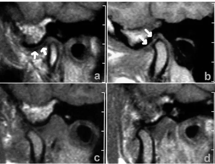
(Fig. 1. Magnetic resonance imaging (1 T scanner) of temporomandibular joint with disc displacement with partial re-duction. The disc is displaced in one parasagittal slice (a) and normally positioned in another slice (b). The arrows show disc position. The slices show normal disc position in the open mouth images (c, d).
gardless of the existence of partial or complete dd in the contralateral joint) served as a control group of tMjs compared with the ‘tMj partial dd – slice dd’ group. However, one joint without dd and with osteoarthritis and two joints with asymptomatic dd were excluded.
relative position of the condyle and disc was cal-culated using the method described by Kurita et al.17 (fig. 3). A line was drawn on the tangent between the lowest part of the articular eminence (t) and the highest edge of the external auditory canal (P), per-pendicular to the tangent, touching the back edge of the disc, and their intersection was marked as point d. Another perpendicular, touching the back edge of the condyle was also drawn and marked as point C. A lower value indicates a more anterior condyle or disc
position. Absolute values (tP, tC and td) were mea-sured using iSSA (iSSA network Station version 3.1, vAMSteC® d.o.o. 1994-2013, Zagreb). Millimeter values to one decimal place were calculated based on the measurement scale shown on Mr images. The disc and condyle positions were calculated as tC/tP and td/tP and expressed in one-hundredth of dis-tance between points t and P.
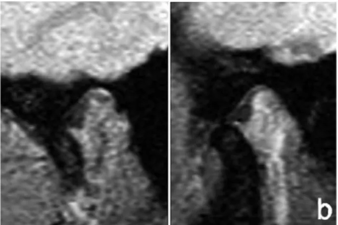
Kruskal-Wallis test and Wilcoxon paired test were used. On data analysis (performed by StAtiStiCA software), the left and right tMjs of the same pa-tient were presented as two entities. The measured values of metric evaluation were displayed by means of a box-and-whisker plot data display (the marked median encompassed the values between the 25%- to Fig. 2. Magnetic resonance imaging (1.5 T scanner) of
temporomandibular joint in the closed mouth position (a) with partially displaced disc with reduction (arrow shows displaced disc). The slices show normal disc position in the open mouth images (b).
Fig. 3. Measuring the position of the disc and condyles in the parasagittal plane by Kurita etal.17.
Fig. 4. The calculated disc (a) and condylar (b) positions comparison between the slices with disc displacement (DD) (left) and without DD (right) of the same TMJs with the diagnosis of partial DD (njoints=9).
a b 54 52 50 48 46 44 42 40 38 36 34 32 30 28 26 74 72 70 68 66 64 62 60 58 56 54 52 ca lc ul at ed c on dy la r p os iti on ca lc ul at ed d is c p os iti on
75%-quartile; all the measured values except for the outliers were shown within the whisker limits). dif-ferences were considered statistically significant at values of 0.05 and 0.01.
The reliability of Mri assessment was evaluated independently of the patient’s clinical signs on Mri images on the basis of two researchers’ (d.Z., radi-ologist and t.b., dentist) inspection and the Kappa index of reliability was between 0.8 and 1.0. The reli-ability of the measurements for metrical analysis was measured on 12 patients twice using the same Mris of both joints (24 measurements in all). The method error values according to dahlberg for all measured distances were between 0.10 and 0.0718.
Results
There was a statistically significant difference be-tween the position of the disc in slices ‘tMj partial dd – slice ndd’ and ‘tMj partial dd – slice ndd’ in the same tMjs with partial dd (Wilcoxon paired test: z=2.934; p=0.003) (fig. 4a). in the same group of joints with partial dd, there was no difference in con-dyle positions (dorsocranial displacement) depending on the existence of slice with dd (Wilcoxon paired test: z=1.378; p=0.168) (fig. 4b).
The following groups of patient joints were also compared and measured: ‘tMj partial dd – slice ndd’ of joints with partial dd and healthy joints
‘tMj ndd’ of other patients (with total dd in the contralateral tMj), which showed no significant dif-ference in either disc position (Kruskal-Wallis test KW (1.43) =0.514; p=0.474) (fig. 5a) or in condyle (Kruskal-Wallis test KW (1.43) =2.140; p=0.144) (fig. 5b).
Discussion
Although MfA as a method of manual medicine and physiatric treatment was presented in the early 1990s9, this classification of tissue-specific diagnoses has not been regularly used in the diagnosis of pa-tients with tMds. The existing rdC/tMd system requires revision and implementation of manual diag-nostic tests9,19-21. On the other hand, the useful pos-sibility of clinical diagnosing partial dd using MfA was very rarely used compared to axiography and tMj analysis of several parasagittal slices on Mri. in a recent review of topics regarding dd, Manfredini et al.22 only partially mention other authors who also stressed the diagnosis of partial dd in their studies.
Westesson et al.6 noted that the diagnostic limita-tion of arthrotomographic study of tMj was visual-ization of disc position or dd in one sagittal slice only. Contrary to them, partial dd can be visible on more than one slice of tMj. There are also some difficulties in Mri diagnostics of tMj disc: physiological posi-tion of the disc is difficult to determine in some cases Fig. 5. The calculated disc (a) and condylar (b) positions comparison between partial reduced temporomandibular joints (TMJs) – slice without disc displacement (DD) (left; (njoints=9) and healthy TMJ without DD of the same patient (right; njoints=34). a b 60 55 50 45 40 35 30 25 ca lc ul at ed d is c p os iti on 76 74 72 70 68 66 64 62 60 58 56 54 52 ca lc ul at ed c on dy la r p os iti on
in the intra-articular area with the same low signal as the cortical bone. Anteriorly displaced disc is situated around tissues with high signal, which enables easier detection in the relative small joint such as tMj.
larheim et al.7 found partial dd in 22.6% of patient joints and in 21.8% of asymptomatic volun-teer joints. tominaga et al.8 studied tMjs of healthy volunteers in closed mouth position only. The pre-dominant diagnosis of partial dd remained during the follow up in 13.6% of joints, normally positioned disc partly moved anteriorly in 20.5%, and only one (2.3%) joint with a normally positioned disc became a total anteriorly displaced disc. in our study, partial dd was found in 20.9% of 43 joints of patients who were diagnosed with any form of dd, i.e. in 15% of left and 7.5% of right tMjs out of all (n=80) joints of patients with dd19. foucart et al.23 report on the following types of tMj internal derangement found on Mri: anterior disc displacements without reduc-tion (52%), anterior disc displacements with reducreduc-tion (26%), partial anterior disc displacement (11%), pure sideways displacements (5%) and stuck discs (4%).
The term ‘partial dd’ is ambiguously used to de-termine the variations of anterior dd in visual anal-ysis. in a recent study, Maizlin et al.24 used the 12 o’clock position to determine disc displacement (dd), using the terms ‘mild’ and ‘significant’ disc displace-ment without differentiation in terms of partial dd. Mild dd (displacement between 10 and 11 o’clock) was more associated with reducible dd. On the other hand, in a recent study, Giraudeau et al.25 consider a disc position with a tendency to anterior displacement as partial dd, which creates uncertainty on compari-son, whereas other authors6-8,12 do not even mention this type of displacement.
using the 12 o’clock method, rammelsberg14 suc-ceeded to quantitatively differentiate partial and total dd using more than one parasagittal slice of the same joint, wherein partial dd occurred in 66% of joints with reciprocal clicking. furthermore, rammelsberg et al.26 found that condyle position was more poste-riorly dislocated in the glenoid fossa if the patient had bilateral dd with reduction. in joints with dd without reduction, the condyle is positioned centrally in the fossa. variability of the condyle position in the fossa was demonstrated in patients with unilateral dd. The relation between condyle positions (their
dorsocranial displacement) in joints with anterior dd is not completely elucidated; according to Kurita et al.17, the condyle is more posteriorly displaced at slight dd; contrary to them, dd without reduction is as-sociated with more concentric position of the con-dyle. Alexander et al.27 have reported that there was no prediction and significant association between the condyle position and dd of tMj and that it does not prove the relationship between them. incesu et al.28 were in favor of dd detection according to the ex-centrically, that is, dorsocranially displaced condyle, even on the basis of x-ray tMj diagnostics. Our study showed no change in condyle position in several slices of the same joint regardless of the existence of par-tial dd. The disc is a flexible structure and may take various positions (physiological or partially displaced only) in the sagittal plane, and an unchanged position of the condyle in different slices of the same joint is to be expected since it is a rigid, osseous structure whose position is partially determined by dental occlusion. in our study, there was no difference in condyle posi-tion of joints with or without dd.
in the intra-articular analysis, the anterior dd does not have to exist in all slices of the parasagit-tal plane (anterolateral or anteromedial) of the same joint in closed mouth position. differential diagnosis should consider fibrozation of the posterior edge of the disc, that is, pseudodisc creation. Also, visual detec-tion of anterior dd depends on the angle of the con-dylar pathway, which means that in a steep pathway the physiologically positioned disc could be charac-terized as anteriorly displaced9,16. On the other hand, analysis of only one mediosagittal slice creates uncer-tainty in the detection of pathological disc position (that is, of the displaced disc), which lowers the value of Mri as the gold standard, and can also have legal repercussions in the evaluation of Mri and clinical signs and symptoms of tMj disorders21,29. Ahmad et
al.30 introduced a new category of intermediate dd, in cases when the pars intermediate zone of the disc is displaced, and the posterior part of the disc is situated superiorly to the condyle. This echoes the previous definitions of partial dd (according to Giraudeau et al.25). However, bumann and lotzmann9 believe that as long as the disc with its posterior part is on the condyle, it is in the physiological position, and tMj
analysis has to encompass all three parasagittal slices, which was the methodology of our study.
A recent proposal for revision of the rdC/tMds diagnostic system suggests that in Group ii disc dis-placements the revised Axis i diagnostic algorithms be supplemented with the use of Mri and Ct imag-ing, respectively, but does not include the diagnosis of partial dd or clinical criteria for such a type of dis-placement30,31. Without supplementing clinical diag-nosis with techniques such as MfA, it is not possible to differentiate between partial and total dd. The re-liability of MfA in diagnosing particular clinical en-tities of partial dd in patients with Mri findings was demonstrated in 70% of joints9,20,21. in studies by other authors, the need of evaluating the inter- and intra-observer agreement is stressed, wherein the greatest matching was achieved for anterior dd without re-duction. it should be pointed out that anterior dd allows for better visual differentiating of cortical bone Mri signal from the cartilaginous structure of the disc, which can be difficult in the small intra-articular space in physiological disc position. in conclusion, the study confirmed significant differences of disc position in different parts of the same tMjs with partial dd. Partial dd can only be determined by analyzing sev-eral parasagittal slices, which was confirmed by quan-titative analysis in this study. insufficient diagnostics is based on only one parasagittal slice of the tMj im-age, which can reduce the effectiveness of Mri as the gold standard in dd of tMj. The dorsocranial con-dyle head position is not expected to indicate partial dd, which was compared with tMjs without dd. There was no difference in physiological disc position in joints without dd (‘tMj ndd’) and physiological disc position in the group of ‘tMj partial dd – slice ndd’ joints.
References
1. jerOliMOv v. temporomandibular disorders and orofa-cial pain. rad 504 Medical Sciences 2009;33:53-77. 2. bAdel t, SAvić PAvičin i, POdOreŠKi d,
MA-rOtti M, KrOlO i, GrbeŠA Đ. temporomandibular joint development and functional disorders related to clinical otologic symptomatology. Acta Clin Croat 2011;50:51-60. 3. bAdel t, KrAPAC l, MArOtti M, KerOS j,
rOSić d, Kern j. razne reumatske bolesti u bolesnika s poremećajem temporomandibularnog zgloba. reumatizam 2011;58:172-3. (in Croatian)
4. bAdel t, MArOtti M, KrOlO i, Kern j, KerOS j. Occlusion in patients with temporomandibular joint anterior disk displacement. Acta Clin Croat 2008;47:129-36. 5. PHArAbOZ C, CArPentier P. exploration en irM des
articulations temporo-mandibulaires. j radiol 2009;90:642-8. (in french)
6. WeSteSSOn Pl, KAtZberG rW, tAllentS rH, SAnCHeZ-WOOdWOrtH re, SvenSSOn SA. Ct and Mr of the temporomandibular joint: comparison with au-topsy specimens. Ajr Am j roentgenol 1987;148:1165-71. 7. lArHeiM tA, WeSteSSOn P, SAnO t.
temporo-mandibular joint disk displacement: comparison in asymp-tomatic volunteers and patients. radiology 2001;218:428-32. 8. tOMinAGA K, KOnOO t, MOriMOtO y, tAnAKA t, HAbu M, fuKudA j. Changes in temporomandibular disc position during growth in young japanese. dentomaxil-lofac radiol 2007;36:397-401.
9. buMAnn A, lOtZMAnn u. funktionsdiagnostik und Therapieprinzipien. Stuttgart: Thieme verlag, 2000. (in Ger-man)
10. leMKe Aj, GrietHe M, PerOZ i, lAnGe KP, felix r. Morphometrische Analyse des Kiefergelenkes anhand von 320 Gelenken mit der Mrt. rofo fortschr 2005;177:217-28. (in German)
11. vOGl tj, AbOlMAAli n. Magnetresonanztomographie des temporomandibular-gelenkes: untersuchungstechnik, ergebnisse, indikationsstellung. rofo fortschr 2001;173:969-79. (in German)
12. Müller leiSSe CH, AuGtHun M, rOtH A, bAuer W, GüntHer rW. diskusvorverlagerung des Kiefergelenks: Korrelation von Magnetresonanz-tomogra-phie und klinischem untersuchungsbefund. rofo fortschr 1996;165:264-9. (in German)
13. SCHiffMAn e, OHrbACH r, truelOve e, lOOK j, AnderSOn G, GOulet jP, liSt t, SvenSSOn P, GOnZAleZ y, lObbeZOO f, MiCHelOtti A, brOOKS Sl, CeuSterS W, drAnGSHOlt M, et-tlin d, GAul C, GOldberG lj, HAytHOrntH-WAite jA, HOllender l, jenSen r, jOHn Mt, de lAAt A, de leeuW r, MAixner W, van der Meulen M, MurrAy GM, nixdOrf dr, PAllA S, PeterSSOn A, PiOnCHOn P, SMitH b, viSS-CHer CM, ZAKrZeWSKA j, dWOrKin Sf. diagnos-tic Criteria for temporomandibular disorders (dC/tMd) for Clinical and research Applications: recommendations of the international rdC/tMd Consortium network* and Orofacial Pain Special interest Group†. j Oral facial Pain Headache 2014;28:6-27.
14. rAMMelSberG P. untersuchungen über Ätiologie, di-agnose und Therapie von diskopathien des Kiefergelenkes. berlin: Quintessenz, 1998. (in German)
15. bAdel t. temporomandibularni poremećaji i stomatološka protetika. Zagreb: Medicinska naklada, 2007. (in Croatian)
16. OrSini MG, KubOKi t, terAdA S, MAtSuKA y, yAMASHitA A, ClArK Gt. diagnostic value of 4 crite-ria to interpret temporomandibular joint normal disk position on magnetic resonance images. Oral Surg Oral Med Oral Pathol Oral radiol endod 1998;86:489-97.
17. KuritA H, OHtSuKA A, KObAyASHi H, KurASH-inA K. A study of the relationship between the position of the condylar head and displacement of the temporomandibu-lar joint disk. dentomaxillofac radiol 2001;30:162-5. 18. HOuStOn Wjb. The analysis of errors in orthodontic
mea-surements. Am j Orthod 1983;83;382-90.
19. bAdel t, SAvić PAvičin i, jAKOvAC M, Kern j, ZAdrAveC d. disc and Condylar head position in the temporomandibular joint with and without disc displace-ment. Coll Antropol 2013;37:901-6.
20. bAdel t, SAvić PAvičin i, bAŠić KeS v, ZA-vOreO i, ZAdrAveC d, Kern j. Orofacial pain caused by trigeminal neuralgia and/or temporomandibular disorders. Period biol 2013;115:185-9.
21. bAdel t, MArOtti M, SAvić PAvičin i, ZAdrAveC d, Kern j. radiographic validation of man-ual functional analysis of temporomandibular joint osteoar-thritis. Acta Clin Croat 2012;51:35-42.
22. MAnfredini d, buCCi Mb, MOntAGnA f, GuArdA-nArdini l. temporomandibular disorders as-sessment: medicolegal considerations in the evidence-based era. Oral rehabil 2011;38:101-19.
23. fOuCArt jM, CArPentier P, PAjOni d, MAr-GuelleS-bOnnet r, PHArAbOZ C. Mr of 732 tMjs: anterior, rotational, partial and sideways disc displace-ments. eur j radiol 1998;28:86-94.
24. MAiZlin Zv, nutiu n, dent Pb, vOS PM, fen-tOn dM, Kirby jM, vOrA P, GillieS jH, CleM-ent jj. displacemCleM-ent of the temporomandibular joint disk:
correlation between clinical findings and Mri characteris-tics. j Can dent Assoc 2010;76:a3.
25. GirAudeAu A, CHeynet f, MAntOut b, PHiliP e. Prevalence and distribution of intracapsular derangement of tMj in an asymptomatic and a symptomatic population. j Stomat Occ Med 2008;1:5-15.
26. rAMMelSberG P, jÄGer l, duC jM. Magnetic resonance imaging-based joint space measurements in tem-poromandibular joints with disk displacements and in con-trols. Oral Surg Oral Med Oral Pathol Oral radiol endod 2000;90:240-8.
27. AlexAnder, Sr, MOOre r.n, dubOiS lM. Man-dibular condyle position: comparison of articulator mount-ings and magnetic resonance imaging. Am j Orthod dento-facial Orthop 1993;104:230-9.
28. inCeSu l, tAşKAyA-yilMAZ n, ÖğütCen-tOller M, uZun e. relationship of condylar position to disc position and morphology. eur j radiol 2004;51:269-73. 29. SCHiffMAn el, OHrbACH r, truelOve el,
tAi f, AnderSOn GC, PAn W, GOnZAleZ yM, jOHn Mt, SOMMerS e, liSt t, velly AM, KAnG W, lOOK jO. The research diagnostic Criteria for tem-poromandibular disorders. v: Methods used to establish and validate revised Axis i diagnostic algorithms. j Orofac Pain 2010;24:63-78.
30. AHMAd M, HOllender l, AnderSOn Q , KArtHA K, OHrbACH r, truelOve el, jOHn Mt, SCHiffMAn el. research diagnostic criteria for temporomandibular disorders (rdC/tMd): development of image analysis criteria and examiner reliability for image analysis. Oral Surg Oral Med Oral Pathol Oral radiol en-dod 2009;107:844-60.
31. PeterSSOn A. What you can and cannot see in tMj im-aging – an overview related to the rdC/tMd diagnostic system. j Oral rehabil 2010;37:771-8.
Sažetak
MetričKA evAluACijA djelOMičnOG POMAKA diSKA teMPOrOMAndibulArnOG ZGlObA
M. Laškarin, T. Badel, J. Kern, I. Savić Pavičin i D. Zadravec
Predmet istraživanja bio je utvrditi kvantitativni odnos između položaja kondila i diska u glenoidnoj jamici između dva različita sloja istog temporomandibularnog zgloba (tMZ-a) s djelomičnim anteriornim pomakom diska. istraživanje je provedeno na 40 bolesnika s pomakom diska tMZ-a (prosječna dob 35,5 godina). Klinička dijagnoza pomaka diska potvrđena je magnetskom rezonancijom. lijevi i desni zglob pojedinog pacijenta bio je predstavljen kao kao dva entiteta (ukupno 80 zglobova). usporedba je učinjena između dva različita sloja 9 zglobova s djelomičnim pomakom diska s re-dukcijom: djelomični pomak diska analiziran je u reprezentativnom centrolateralnom ili centromedijalnom sloju tMZ-a (“tMZ djelomični pomak diska – sloj s pomakom”). Kontralateralni sloj istoga zgloba bio je bez pomaka diska (“tMZ djelomični pomak diska – sloj bez pomaka”). također, analiza je uključila 34 zdrava zgloba bez pomaka diska istih bolesni-ka. Položaj kondila i diska izračunat je metodom po Kuriti i sur. na parasagitalnom prikazu tMZ-a. utvrđena je statistički značajna razlika za različite slojeve istoga tMZ-a s djelomičnim pomakom diska (“tMZ djelomični pomak diska – sloj s pomakom”, “tMZ djelomični pomak diska – sloj bez pomaka”) (p<0,01), ali nije bilo razlike u položaju kondila ovisno o postojanju djelomičnog pomaka diska (p>0,05). uspoređene vrijednosti između sloja “tMZ djelomični pomak diska – sloj bez pomaka” sa skupinom zdravih zglobova bez pomaka diska pokazale su da nema značajne razlike ni za položaj diska niti za položaj kondila (p>0,05). Postoje razlike u položaju diska u različitim slojevima istoga zgloba kod kojih je vizualno potvrđen djelomični pomak diska. dorzokranijalni položaj kondila ne ukazuje na djelomični anteriorni pomak diska.
Ključne riječi: Magnetska rezonancija; Temporomandibularni disk – patologija; Temporomandibularni zglob, poremećaji – patologija; Dislokacije - patologija