UNIVERSITY OF ZAGREB
SCHOOL OF MEDICINE
Mia Staniši
ć
Brain Death
GRADUATE THESIS
Zagreb, 2017
This graduate thesis was made at the Clinic of Neurology, KBC Zagreb, mentored by
Prof. dr. sc. Zdravka Poljakovi
ć
and was submitted for evaluation in the academic year
of 2017.
ABBREVATIONS
AT = apnea testing
BAEPs = brainstem auditory evoked potentials BD = brain death
CPAP = continuous positive airway pressure CT = computed tomography
ECG = electrocardiogram EEG = electroencephalography EP = evoked potentials ERG = electroretinography GCS = Glasgow Coma Scale CNS = central nervous system ICP =intracranial pressure ICU = intensive care unit
MEPs = multimodal evoked potentials MRI = magnetic resonance imaging PaO2 = arterial partial pressure of oxygen
PaCO2 = arterial partial pressure of carbon dioxide PCO2 = partial pressure of carbon dioxide PEEP = peak end-expiratory pressure PO2 = partial pressure of oxygen
SEPs = somatosensory evoked potentials SIDS = sudden infant death syndrome
SPECT = single photon emission computed tomography TCD = transcranial Doppler ultrasonography
UDDA = Uniform Determination of Death Act in USA VEPs = visual evoked potentials
CONTENTS
1. Summary 2. Sažetak
3. Preface………..……..1
4. Current definition of human death and its legal determination...………….2
5. Historical perspective of the brain death concept………..……...3
6. Clinical diagnosis of brain death..…..………..………..7
6.1. Causes of brain damage and irreversible loss of brain function….……….7
6.2. Confounding factors to be excluded………..………...8
6.2.1 Unresuscitated shock……….……8
6.2.2 Hypothermia ………8
6.2.3 Severe metabolic disorder capable of causing a potentially reversible coma………..8
6.2.4 Peripheral nerve or muscle dysfunction……….10
6.2.5 Clinically significant drug intoxications (e.g. alcohol, barbiturates, sedatives, hypnotics)……….10
6.3 Criteria of brain death: Loss of cerebral hemispheric function (Deep Unresponsive Coma) ………..10
6.4 Criteria of brain death: Loss of brain stem function………12
6.4.1. Pupillary tests………..12
6.4.2. Corneal reflex………..14
6.4.3. Oculocephalic reflex………14
6.4.4. Oculovestibular reflex……….14
6.4.5. Gag (pharyngeal) and Cough reflexes……….15
6.4.6. Atropine test……….15
6.4.7. Apnea testing……….15
6.4.7.1.Procedure of Apnea Test………17
6.4.7.2. Complications of apnea test………..19
6.4.7.3. Timing of second exam………..20
7. Confirmatory tests in brain death declaration process……….………...21
8. Brain death in children……….………….22
9. Patient’s family care………..24
11. Case report 2………28
12. References………32
13. Annex……….36
1. SUMMARY
Before modern medical advances, the predominant standard in determination of human death was the traditional cardiopulmonary standard: the irreversible loss of heart and lung function. However, as a response to certain advances in intensive care medicine, since the late 20th century, a new standard for determination of human death has been introduced and accepted in both the medical and legal communities in many parts of the world. This new standard, which took its place alongside the traditional one, is based on the irreversible loss of all bilateral brain hemispheric and brain stem dependent functions in a heart-beating and ventilator-supported patient, known as brain death (BD).
In most human deaths, irreversible cessation of all functions of the entire brain, including the brain stem, is accompanied by the traditional, familiar signs of death: the patient stops breathing, his or her heart stops beating, and the body starts to decompose. In relatively rare cases, however, the irreversible loss of brain-dependent functions occurs while the body, with medical and mechanical assistance, continues to circulate blood and to show other signs of life. Thus, BD is a byproduct of the modern intensive care, which became defined by the Harvard Medical School ad Hoc Committee (1968). However, both the definition of BD and diagnostic criteria have undergone a substantial evolution. Despite the fact that BD, as a concept, is now widely accepted all over the world and the certainty of its diagnosis, when carefully formulated, is still being doubted. It’s definition as well as diagnostic criteria are far from being perfect and require further adjustments.
This review presents the historical development of the concept of brain death, current clinical and ancillary tests used during attempt to determine a diagnosis of BD, responsibility of declaring dead a “corpse with a beating heart”, as well as two typical clinical cases, which illustrate the clinical process of legal determination and certification of human death in the Republic of Croatia.
1. SA
Ž
ETAK
Prije ere suvremenih medicinskih dostignuća, standardno određivanje smrti kod čovjeka se baziralo na tradicionalnom kardio-respiratornom konceptu: nepovratan prestanak fukcije srca i pluća. Međutim, zahvaljujući razvoju medicinskog tretmana u jedinicama intenzivne njege i rehabilitacije od druge polovice 20-og stoljeća definiran je novi koncept za
određivanje smrti kod pacijenata, koji je medikolegalno postepeno prihvaćen širom svijeta. Taj novi koncept za definiciju smrti, baziran na nepovratnom gubitku funkcija mozga i moždanog debla kod pacijenata koji imaju očuvanu spontanu srčanu funkciju, ali su bez adekvatne repiratorne funkcije i zbog toga su tretirani sa mehaničkom plućnom ventilacijom, poznata je kao moždana smrt.
U većine smrtnih slučajeva, nepovratni prestanak svih moždanih funkcija, uključujući funkcije moždanog debla, udružen je s tradicionalnim znacima smrti: prestanak disanja sa prestankom srčane radnje i multiorganskim zatajenjem. Međutim, u relativno rijetkim slučajevima, može se desiti nepovratni gubitak moždanih funkcija u pacijenata koji zahvaljujući modernom intenzivnom tretmanu imaju druge znakove života kao na primjer očuvanu kardiocirkulatornu funkciju. Drugim riječima to znači da je moždana smrt jedan popratni efekt moderne intenzivne terapije kako je definirano od Harvard Medical School ad Hoc Committee (1968). Ipak, definicija moždane smrti i dijagnostički kriteriji su tijekom vremena doživjeli bitnu evoluciju. Unatoč tome što je moždana smrt danas prihvaćen koncept širom svijeta i što je dijagnoza jasna, oboje, i njena definicija i dijagnostički kriteriji ne se mogu smatrati savršenim I zahtijevaju dodatna unaprijeđenja.
Ovaj rad prikazuje povijesni razvoj koncepta moždane smrti, aktualne kliničke i parakliničke dijagnostičke testove, odgovornost u procesu proglašavanja smrti u pacijenata sa očuvanom kardiocirkulatornom funkcijom koji su bez respiratorne funkcije, kao i dvije tipične kliničke situacije u dva pacijenta koje ilustriraju klinički proces legalnog proglašavanja i potvrde smrti u Republici Hrvatskoj.
Ključne riječi: Intenzivna njega, klinički dijagnostički testovi, moždana smrt, paraklinički dijagnostički testovi.
1. PREFACE
As a response to certain advances in intensive care medicine, the late 20th century introduced a new standard for determining death, which became accepted in both the medical and legal communities in many parts of the world. Until then, the predominant standard in determination of human death was the traditional cardiopulmonary standard: the irreversible loss of heart and lung functions. The new standard, which took its place
alongside the traditional one, is based on the irreversible loss of all brain-dependent
functions. In most human deaths, the loss of these neurological functions is accompanied by the traditional, familiar signs of death: the patient stops breathing, his or her heart stops beating, and the body starts to decompose. In relatively rare cases, however, the irreversible loss of brain-dependent functions occurs while the body, with medical and mechanical assistance, continues to circulate blood and to show other signs of life.
When physicians suspect an irreversible loss of brain function in a heart-beating and ventilator supported patient, BD diagnostic criteria are applied. This review presents the historical development of the concept of brain death, current clinical and ancillary tests used during attempt to determine a diagnosis of brain death, as well as two typical clinical cases which illustrate the clinical process of legal determination and certification of human death in Croatia.
2. Current definition of human death and its legal determination
Long before the advent of modern medical technology in the middle of the 20th century, death was considered to have occurred when the heartbeat and breathing irreversibly ceased. This cardiorespiratory view of life and death was supported when the French physician, Rene Laёnnec, invented the stethoscope in 1819 (Adler 1981), and another French physician, Eugene Bouchut, first applied this device to the diagnosis of death in 1846 (Bouchut 1849). This advance contributed to the public’s fear of premature burial to decrease to some extent (Pernick 1988).
Currently, cardiac arrest is determined in the field by the absence of a spontaneous pulse palpated at a carotid, and by the auscultation of the main four auscultation points on the chest (aortic, pulmonic, tricuspid and mitral valves). It can also be determined by the presence of ventricular fibrillation or asystole using an electrocardiographic monitor. Respiratory arrest is determined in the field by the absence of effective, spontaneous ventilation, assessed by inspection and auscultation. The diagnosis of death can be made if a cardiorespiratory arrest of at least 5 minutes is demonstrated, excluding hypothermia. Nonetheless, if an effective mechanical cardiorespiratory reanimation is applied, or neuroprotective methods are used (such as controlled hypothermia) this 5-minute period could be increasingly prolonged.
By the end of the 19th century, listed drugs that could induce apparent death, included atropine, curare, cyanide, amylnitrate, chloral hydrate and pure oxygen (Richardson 1879,1888). This evidence together with identified disorders and states, including suffocation, stroke, head injury, hypothermia, intoxication by alcohol, opiates or hypnotic drugs, catalepsy or hypnotic trance, that mimic death challenged the traditional concept of death (Machado 2007a). Moreover, new inventions, developing and successful use of effective techniques of cardiopulmonary resuscitation, such as the cardiac defibrillator to recover heartbeat after cardiac arrest (Pernick 1988, Meyer 1988, Zoll et al. 1955), and the mechanical respiration device to support ventilation (Edmonson 1982, Maxwell 1986) undermined the physician’s ability to diagnose death using the traditional concept (Machado 2007).
At the end of the 1950s and during the 1960s, increased use of artificial cardiopulmonary support as well as improvements in critical care and growing need for transplantable organs radically changed the course of the practical and the ethical debates about continued support for patients without brain function (Machado 2007a). In the meantime, some reports appeared about a condition in which the brain was massively damaged and nonfunctional while other organs remain functioning (Machado 2007a). This
drove the efforts to define death based on neurological criteria and brain death (BD) was gradually accepted as death of the individual (Pernick 1988, Wijdicks 2001a, b). Currently, when physicians suspect an irreversible loss of brain function in a heart-beating and ventilator supported patient, BD diagnostic criteria are applied. To assess brain function during BD diagnostics, the clinical neurological examination is the accepted standard (Table 1).
In 1980, The Uniform Determination of Death Act (UDDA) in USA proposed a legal definition of human death, which has since been widely accepted. The act reads: “An individual, who has sustained either irreversible cessation of circulatory and respiratory functions, or irreversible cessation of all functions of the entire brain, including the brain stem, is dead.” The latter is defined as BD. Thus, a patient determined to be brain dead is clinically and legally dead (Spinello 2015). However, diagnosing of human death, based on findings of irreversible loss of respiratory, cardiocirculatory and brain functions is not related to the concept that there are different types of death. The irreversible loss of respiratory and cardiocirculatory functions can only cause death when ischemia and anoxia are prolonged enough to produce an irreversible destruction of the brain. According to this concept there is only one kind of death, based on the irreversible loss of brain function (Machado 2007b).
The signs of human death related to forensic medicine circumstances, algor mortis (postmortem coldness), livor mortis (postmortem lividity), rigor mortis (postmortem
rigidity), cadaveric spasm, loss of muscle contractions and putrefaction, are beyond the scope of this review.
3. Historical perspective of the brain death concept
At the end of the 19th century and beginning of the 20th century, several neurosurgeons reported that there was provoked respiratory arrest with continuation of heart beats, in patients having an increase of intracranial pressure (ICP), e.g. in those with brain abscesses, cerebral hemorrhage, brain tumors and depressed fractures of the skull. This state in fact resembles what would today be recognized as BD (Machado 2007a). Although these reports provide early descriptions of the brain death syndrome, these neurosurgeons still considered cessation of respiration or heartbeat to be the sign of death.
Thus, the concept of BD and its determination remained unclear for almost a half-century. During that time, many important discoveries were made describing states of irreversible coma after severe brain injury, and advances in life-sustaining therapies were
used to keep patients with irreversible coma after severe brain injury “alive” for extended periods of time (Schwab et al. 1963).
After the discovery of the electroencephalogram by Hans Berger in 1924 in Germany (Gloor 1969), George Washington Crile proposed in 1930 a definition of death as a drop in electric potentials between the brain and other organs and tissues (Crile et al. 1930) and in 1938 electroencephalography (EEG) was used to show loss of brain potentials after anoxia in animals (Sugar & Gerard 1938).
After the introduction of the first cerebral angiography technique in 1927 by Egas Moniz and Almeida Lima from Lisbon (Tandreau 1985), neuroradiologists and neurosurgeons at the end of the 1950s recognized cessation of cerebral blood flow, referred to as cerebral circulatory arrest, by angiography. This angiographic imaging was identified in apneic or comatose patients after head injury, intracranial hemorrhage, or other space occupying brain lesions (Settergren 2013).
In 1959, Mollaret and Goulon coined the term coma dépassé for an irreversible state
of coma and apnea (Mollaret & Goulon 1959). Based on 23 cases, they described a condition of deep coma with no spontaneous respiration, no reflexes, polyuria, hypotension if norepinephrine was not given continuously, and the absence of all EEG activity. They stated that if the ventilation or the infusion of norepinephrine were stopped, the cardiac arrest would follow and the patient would rapidly die. Despite treatment the patients died from cardiac arrest usually within days after the deep coma had been established. Although this article was a breakthrough contribution to the description of brain death, these authors did not believe in the death of the nervous system. They considered the concept of cardiac death to be the defining phenomenon, generating a major controversy (Machado 2007a).
First in 1959, Pierre Wertheimer’s group characterized the “death of the nervous system” and went further to propose stopping the ventilator if death of the nervous system was diagnosed clinically and by “the repeatedly verified absence of EEG activity both in the cortex and in the diencephalon, and if resuscitative efforts have been given enough time, 18 to 24 hours” (Wertheimer et al. 1959).
In 1963, Robert Schwab et al. (Schwab et al. 1963) stated that “the total absence of EEG activity after 30 min of recording is the most important evidence of death of the nervous system.” This was also among the first accepted diagnostic criteria of human death on neurological grounds in the presence of a beating heart, and the authors went further than just EEG findings, as an ancillary test, would be incorporated into diagnostic criteria. The Schwab’s proposed criteria for establishing brain death are presented in Table 1.
The 1968 report of the Ad Hoc Committee of the Harvard Medical School to Examine the Definition of Brain Death was the first attempt to define brain death as the irreversible loss of all brain function, both cerebral and brain stem, and to present the criteria required for the diagnosis (Beechler 1968). The criteria included irreversible coma, as unresponsiveness and lack of receptivity, whose cause has been identified, absence of movement and breathing, absence of brain stem reflexes, with an isoelectric EEG, and no changes in these findings after at least 24 hours of observation, as presented in Table 1. In the same year, the Harvard criteria were presented at the World Medical Assembly in Sydney, and essential statements on human death were released. The Harvard Report had momentous repercussions and constituted a breakthrough account, establishing a paradigm for defining death by neurological criteria that were widely accepted, and later somewhat improved by Wijdicks (Wijdicks 2001a, b) as presented in Table 1. However, despite the fact that the definition of human death based on neurological criteria, BD, was accepted worldwide, there was no global consensus in diagnostic criteria (Wijdicks 2002).
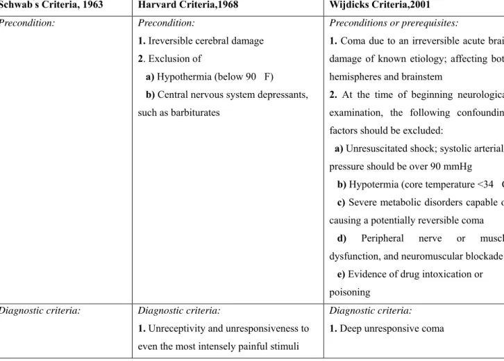
Table 1. Comparison of Schwab’s criteria, Harvard criteria and Wijdicks criteria for diagnosing of brain death in adults (for explanation, see text).
Schwab’s Criteria, 1963 Harvard Criteria,1968 Wijdicks Criteria,2001
Precondition: Precondition: 1. Ireversible cerebral damage 2. Exclusion of
a) Hypothermia (below 90 F) b) Central nervous system depressants, such as barbiturates
Preconditions or prerequisites:
1. Coma due to an irreversible acute brain damage of known etiology; affecting both hemispheres and brainstem
2. At the time of beginning neurological examination, the following confounding factors should be excluded:
a) Unresuscitated shock; systolic arterial pressure should be over 90 mmHg
b) Hypotermia (core temperature <34 C c) Severe metabolic disorders capable of causing a potentially reversible coma
d) Peripheral nerve or muscle
dysfunction, and neuromuscular blockade e) Evidence of drug intoxication or poisoning
Diagnostic criteria: Diagnostic criteria:
1. Unreceptivity and unresponsiveness to even the most intensely painful stimuli
Diagnostic criteria: 1. Deep unresponsive coma
1. No pupillary reflexes and the pupils must be dilated 2. Eyeball pressure must not change the heart rate
3. Spontaneous respiration must be absent for at least 30 minutes,
4. There must be no tendon reflexes of any type
5. Confirmatory tests
●Theelectroencephalography (EEG) should show flat lines without any rhythms in all leads over a 30-minute period ● A loud noise must not produce any detectible discharge in the EEG
2. Absent brainstem reflexes ● Corneal response
● Pupil fixed and dilated and will not respond to a direct source of bright light ● Oculocaphalic and oculovestibular responses
● pharyngeal reflexes
3. Swallowing, yawning, vocalizations are in abeyance
4. No movement or breathing
5. There is no evidence of postural activity (decerebrate or other)
6. As a rule, the stretch of (sic) tendon reflexes cannot be elicited
7. Plantar or noxious stimulation gives no response
8. Observation covering a period of at least 1 hour by physicians are adequate to satisfy the criteria of no spontaneous muscular movements or spontaneous respiration (established by turning off the respirator for 3 minutes) or response to stimuli such as pain, touch, sound, or light) 9. All of the above tests shall be repeated at least 24 hours later with no change 10. Confirmatory tests
●Flat electroencephalography (EEG) (when available it should be utilized)
2. Absent brainstem reflexes ● Corneal responses
● Pupillary responses to light with pupils at mid-size or greater
● Oculocaphalic and oculovestibular responses
● Gag and cough responses 3. Negative atropine test
4. Absent respiratory effort confirmed by the apnea test
5. Periods of observation. 6 hours; in case of acute hypoxic-ischemic brain injury, clinical evaluation should be delayed for 24 hours subsequent to the
cardiorespiratory arrest or an ancillary test could be performed
6. Confirmatory tests
● Tests to demonstrate a complete cessation of brain circulation: Transcranial Doppler (TCD) ultrasonography
● Confirmatory tests to demonstrate loss of bioelectrical activity:
electroencephalography (EEG), evoked potentials (EP), and electroretinography (ERG)
Schwab’s Criteria, 1963 (Schwab et al. 1963), as stated in the review (Machado 2007b); Harvard Criteria, 1968 (Beechler 1968) ; and Wijdicks Criteria, 2001 (Wijdicks 2001a, b)
4. Clinical diagnosis of brain death
As has already been emphasized, when a physician suspects an irreversible loss of brain function in a heart-beating and ventilator supported patient, BD diagnostic criteria are applied. To assess brain function during BD diagnostics, the clinical neurological examination is the accepted standard (Table 1). While any physician should be able to diagnose BD, in practice, the physician who performs the exam and renders the diagnosis must adhere to strict professional standards. The physician must be competent to perform the clinical exam and interpret the results, as well as be free of even the appearance of conflict of interest. Physicians, who procure either harvest organs of brain dead donor or are otherwise actively involved in an organ transplant program, must not determine BD.
The physician must determine the cause of coma and whether the symptoms are irreversible. If one cannot determine the initiating cause, one must know how to establish that the patient has passed into an irreversible condition. Bilateral cerebral hemispheric and brain stem function must be absent. One must know if and when to repeat the examination and when to obtain confirmatory exams. Since most institutions have clinical practice guidelines that define BD and detail the requirements for determination of BD, physicians must know the policy of their individual hospitals. If a policy proves old and no longer matches current understanding, one should work to update it. Finally, the physician must know the pitfalls in rendering the diagnosis.
6.1 Causes of brain damage and irreversible loss of brain function
The diagnosis of BD requires demonstration of the irreversible cessation of cerebral hemispheric and brain stem function. Determination of irreversibility requires knowledge of the cause of coma and the likelihood that it will result in BD. Such causes include severe brain injury, anoxic encephalopathy, and metabolic disease, which can generate uncontrollable cerebral edema.
Hence, clinical and radiological evidence and history of non-survivable brain injury must be documented. Radiological examinations, such as computed tomography (CT) and magnetic resonance imaging (MRI), of the brain can rapidly reveal devastating structural lesions and pathological changes causing irreversible loss of consciousness with terminal
brain herniation. When the cause of the coma is unknown or potentially reversible, the irreversible decline to BD can be demonstrated by clinical exam, appropriate period of observation, and confirmatory tests.
6.2 Confounding factors to be excluded
Clinical states that may mimic brain death, and potentially reversible causes of apparent marked depression of consciousness or responsiveness(Table 1 and Table 2), must be ruled out (Wijdicks 2001a, Wijdicks et al. 2010, Morenski et al. 2003, Hills 2010, Arbour 2013).
6.2.1. Unresuscitated shock
Neurological assessments may be unreliable in the acute post-resuscitation phase after cardiorespiratory arrest. Moreover, the presence of unstable blood pressure has also been reported in BD. Hence, it is indispensible to apply BD criteria only with a systolic blood pressure minimum value of 90 mmHg. The use of hydration fluids or vasopressor drugs might reestablish adequate blood pressure values.
6.2.2. Hypothermia
An inability to regulate temperature, or poikilothermia, is often present in BD. Core temperature results should be obtained through central blood, rectal or esophageal gastric measurement. Most sets of criteria for BD diagnosis require a body temperature of at least 32.2 C. It was stated that hypothermia below 32 C is one of the best signs of total BD. The correction of hypothermia before applying criteria of BD is appropriate, especially in children and alcoholic patients who become hypothermic after mild brain injuries. Moreover, it is important for physicians to be aware of subjects undergoing accidental hypothermia (immersion/submersion in cold water, snow avalanche or prolonged exposure to cold surroundings).
In contrast, it was stated that hyperthermia during the post-resuscitation period after cardiac arrest is an early indicator of brain damage and poor prognosis, highly correlated with an evolution to BD.
6.2.3. Severe metabolic disorder capable of causing a potentially reversible coma
Severe metabolic or endocrine abnormalities make BD unreliable. Metabolic or endocrine derangements, including glucose, electrolytes (phosphate, calcium and magnesium), inborn
errors of metabolism, and liver or renal dysfunction, may cause a potentially reversible coma. If in the treating physician’s judgment the metabolic abnormality may play a role, it should be corrected.
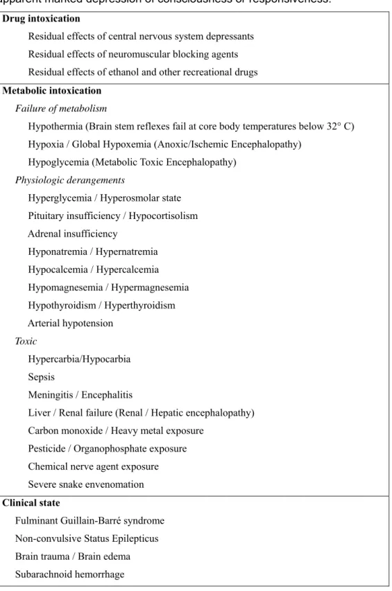
Table 2. Clinical states that may mimic brain death, and potentially reversible causes of apparent marked depression of consciousness or responsiveness.
Drug intoxication
Residual effects of central nervous system depressants Residual effects of neuromuscular blocking agents Residual effects of ethanol and other recreational drugs Metabolic intoxication
Failure of metabolism
Hypothermia (Brain stem reflexes fail at core body temperatures below 32° C) Hypoxia / Global Hypoxemia (Anoxic/Ischemic Encephalopathy)
Hypoglycemia (Metabolic Toxic Encephalopathy) Physiologic derangements
Hyperglycemia / Hyperosmolar state Pituitary insufficiency / Hypocortisolism Adrenal insufficiency Hyponatremia / Hypernatremia Hypocalcemia / Hypercalcemia Hypomagnesemia / Hypermagnesemia Hypothyroidism / Hyperthyroidism Arterial hypotension Toxic Hypercarbia/Hypocarbia Sepsis Meningitis / Encephalitis
Liver / Renal failure (Renal / Hepatic encephalopathy) Carbon monoxide / Heavy metal exposure
Pesticide / Organophosphate exposure Chemical nerve agent exposure Severe snake envenomation Clinical state
Fulminant Guillain-Barré syndrome Non-convulsive Status Epilepticus Brain trauma / Brain edema Subarachnoid hemorrhage
Hydrocephalus / Brain abscess
Direct / Indirect compression of the brain stem
Traumatic / Hemorrhagic lesion affecting the ascending Reticular Activating System Post-cardiac arrest syndrome
Psychogenic coma / Catatonia / Hypoactive delirium
Based on information from Morenski et al. 2003, Hills 2010, Wijdicks et al. 2010 and Arbour 2013.
6.2.4. Peripheral nerve or muscle dysfunction
Peripheral nerve or muscle dysfunction and neuromuscular blockade potentially play a role in inducing unresponsiveness in patient, and it could be a confounding factor in BD diagnosis.
6.2.5. Clinically significant drug intoxications (e.g. alcohol, barbiturates, sedatives,
hypnotics)
Central nervous system-depressant drugs should be ruled out if the clinical history is indicative. It is therefore mandatory that the drug history be carefully reviewed, and if there is any possibility of intoxication being the cause of, or contributing to, the patient’s comatose state, it precludes a diagnosis of BD. In this case, drug screens are only supportive when tests are performed for a specific drug. Depressing drugs may have prolonged action, particularly when hypothermia coexists, or in the context of renal or hepatic failure. Nonetheless, therapeutic levels or therapeutic dosing of anticonvulsants, sedatives, and analgesics do not preclude the diagnosis. Regarding barbiturates, BD diagnosis most likely can be determined with sub therapeutic levels, even though reliable research in this area is lacking. In brain dead neonates and children, an isoelectric EEG remains, even decrementing from therapeutic to sub therapeutic levels of barbiturates.
6.3. Criteria of brain death: Loss of cerebral hemispheric function (Deep
unresponsive coma)
Consciousness and responsiveness (wakefulness) reflects intact function of neurotransmitters and the ascending reticular activating system connecting the pons, thalamus, and midbrain with the diencephalon and cerebral cortex (De Ribaupierre 2011). As mentioned above, neurological evaluation must be done first to determine the cause of depressed consciousness. Taking a through history and identifying brain trauma,
hemorrhage, ischemia, and/or hypoxic or anoxic injury is appropriate as soon as possible, even before a brain death protocol is started.
Assessment of consciousness and responsiveness may be done initially by using the Glasgow Coma Scale (GCS) score. A score of 3 (no eye opening, no verbal response and no movement either spontaneously or in response to stimulation) indicates complete loss of consciousness and responsiveness to noxious stimuli, which reflects severe brain dysfunction.
The diagnosis of deep unresponsive coma requires that a comatose patient show a lack of spontaneous movements in addition to an absence of motor responses mediated by stimuli applied within the cranial nerve distribution (Wijdicks 2001a, Truog & Flacker 2006). Thus, unresponsiveness to noxious stimuli should be assessed by application of central pain stimuli, such as pressure on the supraorbital notch or temporo-mandibular joint or both. Findings consistent with BD are no facial movements or grimacing in response to noxious stimuli (Hills 2010, Wijdicks et al. 2010).
A loss of cerebral hemispheric function is evidenced by total loss of consciousness and responsiveness to all stimulation mediated above the spinal cord, and lack of arousal, awareness, such as cognition, perception, speech, voluntary movements and facial expressions as well as lack of sleep-wake cycles (Hills 2010, Wijdicks et al. 2010).
An unresponsive state of coma implies a complete lack of the above-mentioned CNS response to any kind of external and internal excitation (Wijdicks et al. 2010). A brain-dead patient is, by definition, in an irreversible coma with total absence of awareness of self and environment despite external stimuli. This state differs from stupor, in which the patient requires continued stimuli to maintain consciousness. Quite vigorous stimuli might be required to reveal this difference. Furthermore, coma differs from a persistent vegetative state, in which the patient achieves sleep-wake cycles with eye opening but lacks cognitive awareness (Plum & Posner 1989).
If noxious stimulation elicits a purposeful motor response or posturing, BD is promptly ruled out. The brain dead can neither exhibit a purposeful motor response, such as localization, nor posture. Both the decorticate “flexion” and decerebrate “extension” postures originate in the brain stem, and their presence rules out complete brain stem failure and impedes BD diagnosis (Morenski et al. 2003).
In the original Harvard Committee Report, absent spinal reflexes was one of the diagnostic criteria of BD. However, later it was documented that some BD patients may present spinal reflexes or motor responses, confined to spinal distribution, which, also, do not preclude the BD diagnosis (Morenski et al. 2003, Wijdicks et al. 2010). The spinal cord
generates a number of complex movements, such as rapid withdrawal of the lower extremities, in response to peripheral pain stimuli. While these responses may seem purposeful, careful examination reveals their reflexive nature, and their presence does not exclude the possibility of BD. Furthermore, one cannot rely on peripheral pain when the patient has suffered a spinal cord injury. In BD, plantar responses, finger jerks, undulating toe flexion, muscle stretch reflexes, abdominal reflexes, triple flexion response, pronation-extension reflex, Lazarus sign and facial myokymia have been described (Saposnik et al. 2005).
6.4.
Criteria of brain death: Loss of brain stem function
The next step in determining BD is assessing brain stem and cranial nerve function. As mentioned above, both the decorticate “flexion” and decerebrate “extension” postures originate in the brain stem, and their presence rules out complete brain stem failure (Morenski et al. 2003).
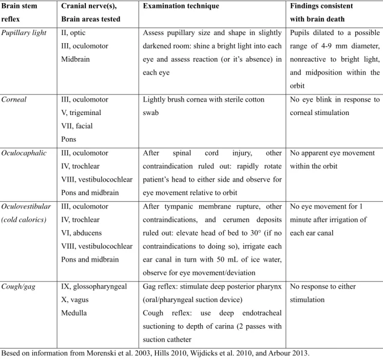
In order to assess brain stem function, most of the clinical exam evaluates the brain stem reflexes. Cranial nerve dysfunction occurs later in the progression of severe intracranial hypertension, because of increasing pressure on the brain stem, tissue distortion and deformation of cranial nerve roots (Arbour 2013). Table 3 summarizes brain stem assessment and findings consistent with BD.
6.4.1. Pupillary tests
Examination of the pupils of the eye is critical in clinical determination of BD (Arbour 2013). Facial and ocular trauma may compromise the ability to perform an optimal bilateral pupillary examination. Pupillary assessment must also take into account medications the patient has received, such as topical atropine for eye examination or intravenous atropine during cardiac arrest. Drugs may influence the size of pupils but generally do not eliminate the reflex, although the clinician may need a magnifying glass to perceive the reaction (Morenski et al. 2003).
The pupillary light reflex involves cranial nerves II and III. It localizes anatomically to the midbrain. Normally, both pupils constrict in response to light. “Fixed and dilated” pupils result from intact sympathetic fibres that arise from the cervical spinal cord and innervate the pupillary dilators. Damage to the midbrain often results in “midposition” (4-6 mm) pupils from damage to the parasympathetic Edinger-Westphal nucleus. Damage to the pons results in “pin-point” pupils from damage to the descending sympathetic fibres, which
allows the midbrain parasympathetic Edinger-Westphal nucleus to control size. Since completely pin-point pupils imply a functional Edinger-Westphal nucleus, their presence is not compatible with brain death. The pupil size of a brain-dead patient may therefore vary depending on the sequence of damage. The pupillary reflex, both direct and consensual, should be explored in both eyes using a bright light. The crucial part of the test is whether the pupil reacts to light; the brain-dead patient will have nonreactive pupils.
Table 3. Brain stem assessment during determination of brain death. Brain stem
reflex
Cranial nerve(s), Brain areas tested
Examination technique Findings consistent
with brain death Pupillary light II, optic
III, oculomotor Midbrain
Assess pupillary size and shape in slightly darkened room: shine a bright light into each eye and assess reaction (or it’s absence) in each eye
Pupils dilated to a possible range of 4-9 mm diameter, nonreactive to bright light, and midposition within the orbit
Corneal III, oculomotor V, trigeminal VII, facial Pons
Lightly brush cornea with sterile cotton swab
No eye blink in response to corneal stimulation
Oculocaphalic III, oculomotor IV, trochlear
VIII, vestibulocochlear Pons and midbrain
After spinal cord injury, other contraindication ruled out: rapidly rotate patient’s head to either side and observe for eye movement relative to orbit
No apparent eye movement within the orbit
Oculovestibular (cold calorics) III, oculomotor IV, trochlear VI, abducens VIII, vestibulocochlear Pons and midbrain
After tympanic membrane rupture, other contraindications, and cerumen deposits ruled out: elevate head of bed to 30° (if no contraindications to doing so), irrigate each ear canal in turn with 50 mL of ice water, observe for eye movement/deviation
No eye movement for 1 minute after irrigation of each ear canal
Cough/gag IX, glossopharyngeal X, vagus
Medulla
Gag reflex: stimulate deep posterior pharynx (oral/pharyngeal suction device)
Cough reflex: use deep endotracheal suctioning to depth of carina (2 passes with suction catheter
No response to either stimulation
Besed on information from Morenski et al. 2003, Hills 2010, Wijdicks et al. 2010, and Arbour 2013.
6.4.2. Corneal reflex
The corneal reflex involves cranial nerves III, V and VII and localizes anatomically to the entire pons. In the intact patient, unilaterally touching the cornea with a cotton swab (stimulation) causes a bilateral closure of the eyelids. This is also one of the most discriminant reflexes in BD diagnosis. A bilateral or unilateral response of eyelid closure and upward deviation of the eye (Bell’s phenomenon) indicates preserved brainstem functioning. The brain-dead patient demonstrates no response. However, edema or drying of the cornea and severe facial and ocular trauma may preclude a satisfactory stimulus for this reflex.
6.4.3. Oculocephalic reflex
Often confusingly referred to as the “doll’s eyes” response, this reflex involves cranial nerves III, IV and VIII and interneurons within the pons and midbrain. In an intact patient, the eyes remain “fixed” on a point during head movement. Rotation to the right requires right-eye adduction and left-eye abduction. Head flexion and extension tests cranial nerve IV. Prior to performing the test, the physician must rule out a cervical spine fracture and instability, or use a rotation bed. Damage to the relevant areas result in fixed eye position. In the brain-dead patient, the eyes move with the head.
6.4.4. Oculovestibular reflex
The oculovestibular reflex involves cranial nerves III, IV, VI and VIII. It involves the entire pons and midbrain. While an interesting test, it does not really add to the exam since the pupillary, corneal and oculocephalic reflexes cover the same areas anatomically. It also presents a potential for confusion. Although medical students and physicians learn the acronym COWS for “cold opposite, warm same” to remember the direction of the apparent nystagmus, this is not the response seen in a comatose or brain-dead patient. With the use of cold water as stimulus, the eyes tonically deviate toward the side of the stimulus. The “nystagmus” – the rapid correction to the opposite side – comes from area 8 of the ipsilateral frontal lobe, the frontal eye fields, as it attempts to counteract the deviation. Since comatose patients do not have functional frontal lobes, only the tonic deviation toward the cold stimulus or away from a warm stimulus is seen. Absence of any eye movement is consistent with brain stem failure at the level of the pons and midbrain.
The following are steps to perform the test: 1. Elevate the head 30°.
2. Fill 50-cc syringe with iced water or saline.
3. Attach 16-gauge IV catheter with the needle removed.
4. Irrigate tympanic membrane. 5. Wait 1 minute for response.
6. Wait 5 minutes before testing other side.
6.4.5. Gag (pharyngeal) and Cough reflexes
The gag and cough reflexes test cranial nerves IX and X, and require a functional medulla. These reflexes are often difficult to explore because of the presence of tubes in the throat and dryness of the mucosa. To test the gag reflex, the physician must stimulate the posterior pharynx with a tongue blade or cotton-tip applicator. The cough reflex requires stimulation of the carina by suction through the endotracheal tube. Brain-dead patient will not have a gag or cough reflex. A potential pitfall results from failure to suction the patient to his carina. Merely manipulating the endotracheal tube may fail to generate a reflex.
6.4.6. Atropine test
The atropine test assesses bulbar parasympathetic impact on heart activity in a brain dead patient. This test proposes using atropine to explore bulbar activity for differentiating coma from so-called brainstem death. The test consists of injecting 2 mg of atropine under continuous monitoring with the electrocardiogram (ECG) for 10 minutes. The atropine test is considered negative, confirming BD, if the heart rate does not augment by more than 3% compared with based ECG records. It should be applied after the patient meets other clinical BD criteria.
There are, however, some limitations to the atropine test. An infratentorial lesion or a brainstem lesion may cause damage to the dorsal vagal nucleus, resulting in a negative test result independent of BD. Moreover, the test is not applicable in case of autonomic neuropathy and in patients who have undergone cardiac transplantation with denervation of autonomic fibers to the heart. Nonetheless, as this test explores parasympathetic arcs through the medulla oblongata, and it is simple to apply, its inclusion is recommended in BD diagnostic protocols (Wijdicks 2001a).
6.4.7. Apnea testing
The apnea testing (AT) is the final clinical test. The rationale behind the AT is to demonstrate failure of the medullary centers to drive ventilation. It is an essential sign of a definitive loss of brainstem function and implies that breathing has become extinct. Nonetheless, it is the most difficult, potentially harmful and lengthy clinical test in BD protocols. This test may induce hypotension, excessive hypercarbia, hypoxia, acidosis, cardiac arrhythmia or asystole, but it
is the decisive clinical test in brain death diagnosis. An examiner with experience can apply this test in 99.9% of all cases; the remaining ones may be studied using other ancillary tests (Machado 2007b).
Apnea testing is generally performed after the cause of coma, unresponsiveness, and lack of brain stem reflexes is established. Of interest, it was noted that “No report has been published of either an adult patient or child who, with otherwise absent brainstem reflexes but spontaneous breathing, ’recovers’ to a persistent vegetative state or better” (Wijdicks 2001a). More appropriately, no report has been published of either an adult or pediatric patient who demonstrates absent brain stem reflexes but spontaneous breathing able to maintain adequate ventilation without mechanical support (Morenski et al. 2003).
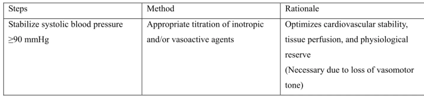
Prior to the test, the physician ensures the patient has met the other clinical criteria, which include adequate hemodynamic status, core temperature and absence of drugs and metabolic derangements. Thus, before applying AT some prerequisites should be considered to minimize risk of complications (Table 4):
● A pretest systolic blood pressure of at least 90 mmHg is recommended,
● Body temperature at 36 or 36.5 C is recommended. Hypothermia correction facilitates CO2 production and reduces the changes of hypotension by stabilizing blood pressure and the hemodynamic state,
● Normal arterial PCO2 or PaCO2 range of 35-45 mm Hg is recommended. It was remarked that the main issue is to consider starting 20 mmHg above the normal baseline level. The combination of hypoxia with hypercarbia provides a greater ventilator drive (Morenski et al. 2003). However, the resulting acidosis from extreme hypercarbia may cause hemodynamic compromise, while hypoxic stimulus risks generation of hypoxic brain injury in a patient who is not brain dead, as well as hypoxic-ischemic injury to potentially transplantable organs. In cases with a higher PaCO2 than normal baseline level, as seen in patients suffering from chronic obstructive pulmonary disease, guidelines suggest a value of 20 mmHg higher than the baseline (the Quality Standards Subcommittee of the American Academy of Neurology 1995),
● A normal pH or a value in the low basic range is recommended,
● Fluid balance: euvolemia or a positive fluid balance during the previous 6 hours is recommended,
● There are no clear PaO2 recommendations except preoxygenation with 100% O2 for roughly 10 minutes, and avoidance of hypoxia. Preoxygenation helps to correct possible hypocapnia, often due to hyperventilation or provoked by high tidal volumes of the
mechanical ventilator, or due to hypothermia. It will eliminate alveolar nitrogen stores and facilitate oxygen transport (Wijdicks et al. 2008). Preoxygenation also helps to avoid desaturation during the AT and its associated cardiovascular effects. In addition, the level of preoxygenation may be adjusted to obtain a starting PaCO2 of approximately 40 mmHg to shorten the period of apnea. A normal PO2 or preoxygenation to obtain an arterial PO2 ≥ 200 mmHG is recommended,
● Medication: apnea testing must not be performed under the influence of drugs that can paralyze respiratory muscles, such as relaxants.
6.4.7.1. Procedure
of
Apnea
Test
Two techniques for ascertaining sufficient oxygenation during apnea testing are used. The physician may either disconnect the mechanical ventilator and place a large bore catheter down the endotracheal tube to deliver 100% oxygen, or maintain the patient on continuous positive airway pressure (CPAP) through the respirator circuit. Both methods have their advantages and disadvantages, and the choice depends on the experience of the physician and respiratory therapy staff. In addition, these techniques may be used to obtain a starting PaCO2 of approximately 40 mmHg to shorten the period of apnea.
Hence, the first technique is disconnection of the patient from the respirator and insertion of a catheter or cannula into the endotracheal tube down to the level of the carina, providing pure oxygen at a flow rate of 6 to 10 L/minute during 4 and 10 minutes. This ensures sufficient ventilation into the alveoli and transporting oxygen to the blood even without any respiratory movements (Wijdicks 2001a, Wijdicks et al. 2008).
The diameter of the insufflation on the catheter should be approximately 14F to 16F or slightly larger and should not occlude the airway. While use of large bore insufflation catheter may help avoid inadequate oxygenation, use of a too large insufflation catheter may lead to inadequate egress of air and may lead to barotraumas, pneumothorax or subcutaneous emphysema.
Table 4. Preparatory steps before apnea testing.
Steps Method Rationale
Stabilize systolic blood pressure ≥90 mmHg
Appropriate titration of inotropic and/or vasoactive agents
Optimizes cardiovascular stability, tissue perfusion, and physiological reserve
(Necessary due to loss of vasomotor tone)
Stabilize body temperature ≥36.5 C (preferred).
(Apnea testing can be done at lower temperatures, but additional time required for an increase in CO2, risking cardiovascular instability)
Use of heating blankets, infusion of warmed intravenous fluid, heat lamps
Hypothermia suppresses neuronal activity, including at brain stem level, and prolongs duration of action for central nervous system depressants and neuromuscular blockers
Normalize PaCO2 to range of 35-45 mmHg
Downward titration of controlled ventilation (tidal volume/ventilator set rate), unless contraindicated, to attain normocarbia/baseline PaCO2
Baseline arterial CO2 level establishes a starting point for CO2 measurement at intervals.
Beginning with hypocarbic state will require additional time for CO2 increase and increase risk for cardiovascular instability Maintain adequate circulating blood
volume
Appropriate administration of crystalloid, colloid, and/or blood products to replace volume losses and correct absolute/relative hypovolemia
Stabilizes cardiovascular/blood pressure during the stress of apnea testing
Administer 100% oxygen for ≥10 minutes to PaO2 ≥200 mm Hg
Fraction of inspired oxygen 1.0 with positive end-expiratory pressure at 5 cm H2O.
When pulse oximetry indicates stable oxygen saturation, sample arterial blood gases to document baseline level
Facilitates oxygenation, eliminates nitrogen stores, and avoids oxygen desaturation and cardiovascular consequences
Based on information from Wijdicks 2001a, Hills 2010 , Wijdicks et al. 2010, Wijdicks et al. 2008, and Scott et al. 2013.
In the second technique for ascertaining sufficient oxygenation during AT, the patient is not disconnected from the respirator but the volume is decreased to a very low level (controlled hypoventilation from 0.5 to 2 L/minute), using synchronized intermittent mandatory volume ventilation and providing pure oxygen for inspiration; in this way, the patient is not disconnected until the required PaCO2 is achieved. This method prevents tracheopulmonary complications and still allows the examinator to detect any spontaneous breathing movement (Machado 2007b). It was considered that it is safer to test the apnea by leaving patients on a continuous flow of 100% oxygen and low peak end-expiratory pressure (PEEP) than to disconnect them from the respirator (Machado 2007b). However, maintaining
the patient on CPAP can give the false impression that the patient is breathing if the sensitivity of the ventilator is not appropriately adjusted. Hence, examples were noted with significant false readings of spontaneous respiratory rates of 20 to 30 breaths/min by the mechanical ventilator sensors, with CPAP settings as low as 2 (Wijdicks 2001a, Wijdicks et al. 2008).
The AT result is positive, supporting a BD diagnosis, when no breathing effort is observed at a PaCO2 of 60 mmHg or with a 20 mmHg increase from baseline. A true spontaneous breath consists of abdominal or chest excursions that produce adequate tidal volumes (Wijdicks 2001a, Wijdicks 1995). In contrast, if the patient demonstrates spontaneous respiration at any time, the apnea test is considered “negative”, not supporting a BD diagnosis, and the patient should be connected again to the mechanical respirator.
If arterial pressure becomes <90 mmHg, or a noticeable desaturation is detected by the oximeter, or cardiac arrhythmias take place, the AT procedure should be stopped and the mechanical respirator should be re-connected. Fixed durations of AT are not considered standard anymore, and arterial blood gas determinations are used to formally document PaCO2 to prescribed thresholds and the absence of intrinsic respiratory drive (Wijdicks 2001a, Morenski et al. 2003, Hills 2010, Wijdicks et al. 2010, Scott et al. 2013). The first baseline arterial blood gas is drawn after about 10 minutes. Should the initial PaCO2 not equal, or exceed 60 mmHg or 20 mmHg above the patient’s baseline, the physician can then obtain another baseline arterial blood gas. The patient’s failure to demonstrate spontaneous breathing with a PaCO2 equal to or greater than 60 mmHg or 20 mmHg above his baseline constitutes a “positive” test and is consistent with BD. There is “little scientific evidence to support raising the target value for PaCO2 beyond 60 mmHg or 20 mmHg above the patient’s baseline (Morenski et al. 2003).
6.4.7.2. Complications of apnea test
Inadequate preoxygenation has a clear association with an increased number of complications. Hypotension is the most frequent complication in 24% of cases, while cardiac arrhythmias with the potential for ventricular fibrillation or arrest is much less common (1%) (Machado 2007b).
The following are the main reported complications:
● Excessive hypercarbia. CO2 values over 120 mmHg cause necrosis and should be avoided, although general recommendation do not exist, and there are no generally accepted PaCO2 values for patients whose natural respiration is adapted to a PaCO2 of more than 45 mmHg. In these cases, other ancillary tests are recommended (Lang & Heckmann 2004).
●Hypoxia. O2 values less than 70 or 60 mmHg should be avoided. If pulse oximetry is used, values should not drop below 80%. It has been suggested that hypoxemia contributes to cardiac arrhythmia or hypotension during AT (Ropper et al. 1981).
● Respiratory acidosis. There are no precise recommendations for the pH, although pH values less than 7.2 or 7.0 should be avoided. Its fall is highly correlated with the increase of PaCO2 and it is rapidly restored with normoventilation or mild hyperventilation. Lower values of pH (as low as 6.8) during AT should not be corrected by buffer or alkaline solutions, because alkalosis will result after normoventilation; as an alternative, a normal pH or value in the low basic range should be established at the onset of AT (Lang & Heckmann 2005).
● Hypotension. Blood pressure values of less than 60 or 70 mmHg systolic, 40 mmHg diastolic, and mean arterial pressure of 50 mmHg should be avoided. The presence of a blood pressure of 90 mmHg before testing is acceptable as a rule. Usually there is a mild increase of blood pressure with hyperoxygenation and a somewhat more marked decrease with hypercapnia. Persistent hypotension may be corrected using intravenous fluids, 5% albumin, or an increase of intravenous dopamine or (nor)epinephrine.
●Cardiac arrhythmia and arrest. Cardiac arrhythmias induced by AT are rare (Goudreau et al. 2000) and cardiac arrest occurring only in two of 63 cases (Saposnik et al. 2004). It should be avoided at any rate. Arrhythmias are mostly due to excessive acidosis or to hypoxia, which can be suspected by the onset of new cardiac arrhythmias or a noticeable drop in heart rate (Lang & Heckmann 2005).
● Barotrauma. Barotrauma is considered extremely rare (Saposnik et al. 2004). Tension pneumothrorax and pneumoperitoneum may ensue after massive oxygen insufflations into the endotracheal tube with an oxygen supply that obliterates the tube or a valve mechanism that makes the escape of the insufflated gas impossible (Vivien et al. 2001).
● Increase of intracranial pressure. Intracranial pressure (ICP) is usually not monitored during AT except in some neurosurgical intensive care units. Since theoretically AT may increase ICP via local hypercapnic vasodilatation and the ensuing increase of cerebral blood volume, this test should be the last of all clinical tests (Lang & Heckmann 2004).
When a reliable AT is impossible to achieve, due to the unfeasibility of reaching the target PaCO2, or when an unsafe drop in PaO2 is inevitable, or in patients with severe thorax trauma or other pulmonary problems, other ancillary tests should be used.
6.4.7.3. Timing of second exam
The period of observation between BD examinations depends on the clinical aspects, the use of confirmatory tests and the age of the patient. As presented in Table 1. an observation
period of 6 hours in adults is recommended (Wijdicks 2001a). The repeat exam includes the brain stem reflexes as in the first examination but does not require repeat apnea testing. As discussed in the section on the apnea trial, no report exists of either an adult or pediatric patient who met the criteria of brain death clinically and received an appropriately performed apnea test, who then went on to achieve any neurological recovery. Furthermore, a patient may not tolerate a second apnea testing. However, individual physicians and institutions are free to perform a repeat apnea testing, as they are free to obtain confirmatory tests.
7. Confirmatory tests in brain death declaration process
It is widely accepted that BD is a clinical diagnosis, and that confirmatory tests are not mandatory in most situations. However, in certain countries, including Croatia, the law requires confirmatory tests. Finally, individual institutions may require confirmatory tests as part of their protocol for establishing a BD diagnosis (Machado 2007b, c].
The confirmatory tests provide invaluable assistance with the complex medical and social aspects of BD. Properly chosen and performed, confirmatory tests are essential when doubt exists about the clinical findings, conditions preclude an apnea test or suspicion of confounding conditions exists. The confirmatory tests can shorten the interval before the second test based on clinical judgment (Wijdicks 1995). However, despite this there is no global consensus in choice of confirmatory tests in BD declaration process. An ideal confirmatory study for BD should be safe, extremely accurate and reliable, available, quick and inexpensive (Machado 2007c).
Clinical judgment must enter into situations that include the hemodynamically unstable patient who does not respond to resuscitation efforts. Such patients may not tolerate an apnea test and may not survive even the 6-hour period. A similar clinical picture can follow acute overdoses of benzodiazepines, barbiturates and opiates. Furthermore, less certain remains the case of the patient who has not passed the period necessary for elimination of the drugs. Patients, who receive “pentobarbital coma” therapy to achieve electroencephalographic burst suppression, in a final effort to control ICP, present a similar problem. The physician faces a possible waiting period of days before the serum pentobarbital level decreases sufficiently. In cases of severe head injury, the clinical history, exams, and an inability to maintain ICP at levels compatible with life, even with institution of a pharmacologic coma, justify the addition of confirmatory tests.
Confirmatory tests in BD can be divided into those that prove absent cerebral and brain stem blood perfusion, and those that demonstrate loss of brain bioelectrical activity.
Among tests that demonstrate a complete cessation of brain blood flow, the following are included:
● bilateral carotid and vertebral contrast angiography (cerebral four-vessels angiography),
● cerebral intravenous digital subtraction angiography,
● intravenous radionuclide angiography using a gamma camera,
● single photon emission computed tomography (SPECT),
● transcranial doppler (TCD) ultrasonography,
● computed tomographic (CT) angiography, and
● magnetic resonance (MR) angiography.
Among tests that demonstrate a loss of brain bioelectrical activity, the following are included:
● digital continuous EEG monitoring,
● multimodal evoked potentials (MEPs), such as visual evoked potentials (VEPs) and electroretinography (ERG), brainstem auditory evoked potentials (BAEPs) and short latency somatosensory evoked potentials (SEPs) have been used.
In fact, confirmatory tests that are widely accepted are conventional cerebral four-vessels angiography and EEG. Instead of that, combining of transcranial doppler (TCD) ultrasonography and a neurophysiological test battery (EEG, MEPs and electroretinography (ERG) (Table 1) have been recommended recently (Machado 2007c).
8. Brain death in children
Although pediatric BD diagnosis encloses the same concept and similar diagnostic procedure as in adults, it is a more difficult task for physicians and nurses because of several ethical and psycho-emotional issues. Parents and the general population are more reluctant to accept death in children, and find it difficult to understand an explanation that a child with preserved heartbeats but no brain activity is dead (Ashwal 2011).
Moreover, the clinical exam of suspected brain-dead pediatric patients is more difficult to carry out because of the smaller size of the patient, the immaturity of the reflexes that change with central nervous system (CNS) maturation, and the existence of fontanels and open sutures in neonates and infants.
The most frequent etiology of BD in children is trauma, due to motor vehicle accidents, shaken baby syndrome and abuse. Asphyxia producing anoxia is also a common cause, in home accidents such as near drowning, suffocation, and from the so-called sudden
infant death syndrome (SIDS). Some massive infections of the brain can also be the origin of permanent destruction of CNS structures (Ashwal 2011).
The current guidelines (Table 5) derive from the 1987 report of a special task force composed of representatives of the pediatric, neurologic and legal disciplines (Task Force for the Determination of Brain Death in Children 1987a, b]. These guidelines which have been widely introduced in pediatric hospitals are presented in Table 5.
Table 5. Guidelines for brain death determination in children A. History: determine the cause of coma to eliminate reversible conditions B. Physical examination criteria:
1. Coma and apnea.
2. Absence of brainstem function. a. Mid-position or fully dilated pupils.
b. Absence of spontaneous oculocephalic (doll’s eye) and caloric-induced eye movements.
c. Absence of movement of bulbar musculature, corneal, gag, cough, sucking and rooting reflexes.
d. Absence of respiratory effort with standardized testing for apnea. 3. Patient must not be hypothermic or hypotensive.
4. Flaccid tone and absence of spontaneous or induced movements, excluding activity mediated at spinal cord level.
5. Examination should remain consistent for brain death throughout the pre-determined period of observation.
C. Observation period according to age: 1.age of7 days to 2 months:
Two examinations and EEGs 48 hours apart 2. age of 2 months to 1 year:
Two examinations and EEGs 24 hours apart, although the repeat exam and EEG is not necessary with an initial EEG showing electrocerebral silence (ECS) combined with a radionuclide angiography showing no cerebral blood flow (CBF). 3. older than 1 year:
Two examinations 12 to 24 hours apart, EEG and radionuclide angiography are optional; multimodality evoked potentials (MEPs) and transcranial Doppler ultrasonography (TCD) can be used as confirmatory tests to shorten periods of observation.
Compared with BD diagnostic criteria in adults, these guidelines do not differ in any important way. The main differences are the age related periods of observation and requirement of particular confirmatory tests in children under 1 year of age. As in adults, in children over 1 year of age, the diagnosis is based on clinical examination and confirmatory tests are not mandatory. However, the commission has also proposed the use of confirmatory tests to shorten the period of observation in children older than 1 year (Task Force for the Determination of Brain Death in Children 1987b). Ashwall has proposed BD criteria for term infants younger than 7 days of age (Ashwal & Serna-Fonseca 2006). One of the differences with adult BD protocols is the exploration of brainstem reflexes, because the maturation of the nervous system entails different periods of time for a specific reflex to be fully developed. The pupil light reflex is only present after 30 weeks gestation and the oculocephalic response is absent before 32 weeks.
9. Patient’s family care
Advocacy, honesty and the development of trust between the patient’s family and personnel on the health care team markedly promote communication and rapport during a stressful and difficult time. Family members of a patient with a devastating brain injury are under stress and need empathy, teaching and an environment of trust. It is crucial to establish trust and help the family to understand the torrent of information from multiple health care providers. Taking time and building on the trust established upon ICU admission can help the family understand brain injury, progression of the injury and what BD means. All these considerations are vital, with or without organ recovery, organ donation and transplantation as possible end points.
Allowing time and resources, including spiritual support or chaplains, when formal brain death protocols are being contemplated or initiated, gives a patient’s family members an opportunity to process their experiences. Encouraging family members to speak and ask questions during meetings is associated with families’ satisfaction, whether or not organ donation is considered in end-of-life discussion. Facilitating early visiting with the patient and providing realistic and honest communication about his or her condition and prognosis, in ways a family can understand, are vital. When brain injury is refractory to aggressive management to control the consequences of injury, such as ICP elevation, and the patient progresses toward BD, empathy and honesty are important in communicating this decline to the patient’s family. At this stage, with a worsening neurological status, specific clinical triggers may be met to initiate referral of a potential donor to the organ procurement
organization. The triggers may include a decrease in the GCS score, or loss of one or more brainstem reflexes, or both. The representative of the procurement organization has no direct involvement in clinical management of the patient until BD is formally declared and consent has been obtained for organ donation. A conflict of interest can occur if direct involvement by an organ procurement organization is involved in the management of the patient, before the declaration of BD is confirmed. Any family discussion should start with a thorough explanation of the patient’s grave condition and should include definition of terms. Providing a clear definition of BD and ensuring the family’s understanding before organ donation is discussed, are vital to avoid later misunderstanding (Murthy 2009). Communication with family members after completion of a BD protocol needs to be unequivocal that BD has occurred and that the patient is in fact dead (Murthy 2009). The dead-donor rule requiring declaration of death before organ recovery for transplant can then apply (Truog & Robinson 2003).
Any discussions with a patient’s family should take into account cultural and religious perspectives and how family members define death. BD may have variable meanings for individuals. One meaning may be severe brain injury. Another may be unawareness of surroundings. Establishing how family members perceive death is an optimal starting point for exploring organ donation. Religious issues are of utmost importance in discussing BD. Most religions, including Christianity, Islam, Judaism and Hinduism support organ donation as a charitable and helping act. Understanding the religious perspectives of the family of a potential donor can better prepare health care providers for discussions about BD. The need for careful choice of words and use of the same terminology by all members of the health care team in discussing brain death with the patient’s family cannot be overstated (Savel & Munro 2012).
10. Case report 1
Mr. P.Š., a 73-year old male, suddenly developed left sided facial paresis, along with sweating and weakness, at 10.45 a.m. on the 2016.09.16, witnessed by his wife. He proceeded to take a shower but collapsed in the washing room. His wife activated the emergency medical system. After transport to the emergency department, evaluation revealed dyspnea, decreased consciousness and left sided hemiplegia. His medical history included recently diagnosed and untreated diabetes mellitus type 2, arterial hypertension and atrial fibrillation. A non-contrast CT scan of the head revealed the right sided zone of
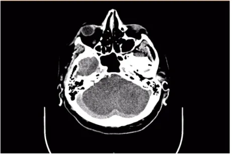
acute occipital ischemia (Figure 1), along with a cerebral CT angiography, which confirmed a finding consistent with basilar artery occlusion.
Figure 1. A non-contrast CT scan showing extensive ischemic lesions in the brainstem, as well as the entire area supplied by the right posterior cerebral artery.
Firstly, the patient urgently underwent intravenous thrombolysis with tissue plasminogen activator and after that, a mechanical thrombectomy was attempted. At this time, P.Š. was in deep coma (GCS 3) with miosis of symmetrical sized pupils, decerebrate posturing on painful stimuli, tachyarrhytmic, breathing spontaneously with airway in place, but multiple crackles on lung auscultation and regurgitated content in the oral cavity. A cerebral angiography showed sub-occlusive stenosis at the transition from the proximal onto middle third of the basilar artery, at a length of 3 mm, proximal to the origin of the anterior inferior cerebellar artery, through which the contrast had a delayed flow and marked out the thrombi distally in the middle third of the basilar artery (Figure 2). Multiple attempts were made to pass through the point of stenosis, but ultimately failed. Due to the same cause, the point of stenosis could neither be dilated with a balloon catheter, which resulted in a failed attempt and the patient was transferred back to the neurological ICU for medical management.
Figure 2. Digital subtraction angiography of the vertebrobasilar circulation, showing basilar artery stenosis with basilar artery occlusion in proximal parts and migrated thrombus distally.
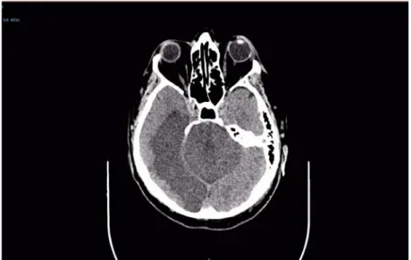
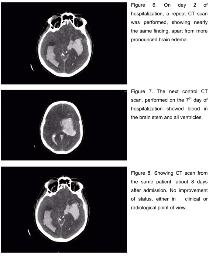
A control CT was performed 3 days later, which showed an extensive ischemic lesion affecting the entire cerebellum and brain stem, as well as the entire area supplied by the right posterior cerebral artery, including the right thalamus (Figure 3). In addition, the territory of the left posterior cerebral artery was affected, as well as both anterior cerebral arteries. The brain edema caused narrowing of the IV ventricle and tonsillar herniation of the brain parenchyma, with consequent development of obstructive hydrocephalus.
Figure 3. Same CT scan, also showing ischemia of complete cerebellum.
The patient had diffuse ischemic lesions of both the brain and brain stem. He remained in a comatose state on the fifth day of admission, with dilated, non-reactive pupils,