Bambang Udji Djoko Rianto, Department of Ear, Nose, and Throat, Faculty of Medicine Gadjah Mada University/Dr. Sardjito Hospital, Yogyakarta
Human papillomavirus (HPV) as the causal
factor of tympanal cholesteatoma in
malignant-type chronic suppurative
otitis media
Bambang Udji Djoko Rianto
Department of Ear, Nose, and Throat
RSUP Dr. Sardjito/Faculty of Medicine Gadjah Mada University, Yogyakarta
ABSTRACT
Bambang Udji Djoko Rianto - Human papillomavirus (HPV) as the causal factor of tympanal cholesteatoma in malignant-type chronic suppurative otitis media
Background: Tympanal cholesteatoma is an uncoordinated, uncontrolled hyperproliferative epidermic epithelial cells keratinized with aggressive, erosive and destructive development to the mucous membrane and the bone of external auditory canal and tympanic cavity. This process is invasive and may migrate to the surrounding tissue. Ethiopathogenesis of tympanic cholesteatoma is unknown. Histological examination shows that there are hyperproliferative development, papillomatous, koilocyte clusters and destruction of the bone. These abnormalities are characteristics of papillomavirus.
Objective: To know the whether HPV-18 is the risk factor in the occurrence of tympanic cholesteatoma ini malignant-type chronic suupurative otitis media.
Methods: A case-control study was conducted on 31 tympanal cholesteatoma specimens from malignant-type chronic suppurative otitis media as the case group, and 31 mucosal tympanic cavity specimens from benign-type chronic suppurative otitis media as the control group. For detecting Polymerase chain reaction (PCR) examination was used to detect DNA HPV-18, that performed in Parasitology Laboratorium Faculty of Medicine, Gadjah Mada University.
Results: Polymerase chain reaction (PCR) examination showed that there were 21 (67.7%) DNA HPV-18 positive specimens in the case group, while in the control group there were 5 (16.1%) DNA HPV-18 positive specimens. This difference was statistically significant (p= 0.0001; Odds ratio:10.92; 95% confidence interval = 2.83-45.29). Immunohistochemistry examination was used to identify host immune response to HPV, by observing the interleukin-1 (IL-1) and interferon g (IFN g) expressions. Both IL-1 and IFN g cytokines were highly expressed in tympanic cholesteatoma samples, compared to both in the control group. The difference was statistically significant (p< 0.05, Odds ratio 14.29; 95% confidence interval: 3.64-60.5 and p< 0.05, Odds ratio:10.2; 95% confidence interval: 2.74-40.35, respectively). Conclusion: It can be concluded that HPV-18 DNA was iidentified, and was one of the multiple risk factors in the occurence of tympanic cholesteatoma in malignant-type chronic suppurative otitis media..
Key words: tympanal cholesteatoma - human papillomavirus - malignant-type chronic suppurative otitis media
ABSTRAK
Bambang Udji Djoko Rianto – Human papillomavirus (HPV) sebagai faktor penyebab kolesteatoma timpani pada otitis media suppurativa kronik tipe maligna
diferensiasi dengan manifestasi klinis berupa invasi dan migrasi ke jaringan sekitarnya. Etiopatogenesis kholesteatoma timpani hingga saat ini masih belum dapat diketahui secara pasti. Gambaran histologis menunjukkan adanya pertumbuhan sel epitel skuamus kompleks yang hiperproliferatif, papillomatosus dan klaster koilosit yang agresif, disertai destruksi tulang, sehingga besar kemungkinan melibatkan peran virus papilloma.
Tujuan: membuktikan bahwa HPV-18 merupakan faktor risiko kejadian kolesteatoma timpani pada otitis media suppurativa kronis tipe maligna.
Metode: Penelitian ini menggunakan desain kasus kontrol terhadap 31 spesimen kelompok kasus otitis media supuratif kronik maligna (kholesteatoma +), sedangkan kelompok kontrol adalah 31 spesimen otitis media supuratif kronik benigna (kholesteatoma -). Pemeriksaan polymerase chain reaction (PCR) digunakan untuk mendeteksi DNA HPV-18 yang dilakukan di Laboratorium Parasitologi Fakultas Kedokteran, Universitas Gadjah Mada.
Hasil: Berdasarkan pemeriksaan menggunakan polymerase chain reaction (PCR) untuk mengidentifikasi adanya DNA HPV, didapatkan 21 (67,7%) sampel positif DNA HPV-18 pada kelompok kasus, sedangkan pada kelompok kontrol sebanyak 5 (16,1%) sampel positif DNA HPV-18. Berdasarkan analisis statistik tes
Chi square, perbedaan jumlah sampel DNA HPV-18 tersebut beda bermakna (p= 0,0001). Rasio Odds: 10,92 (interval kepercayaan 95%= 2,83-45,29). Identifikasi respon imun inang terhadap HPV berupa ekspresi sitokin yaitu: interleukin-1 (IL-1) dan interferon g (IFN g) menggunakan metode pemeriksaan imunohistokimiawi menunjukkan bahwa ekspresi sitokin IL-1 dan IFN g pada kelompok sampel kholesteatoma timpani lebih tinggi secara bermakna dibanding kelompok kontrol ((p< 0,05, rasio Odds14,29; interval kepercayaan 95%: 3,64-60,5 untuk ekspresi IL-1, dan p< 0,05, rasio Odds:10,2; interval kepercayaan 95%: 2,74-40,35).
Simpulan: Hasil penelitian tersebut menyimpulkan bahwa terdapat DNA HPV-18 dalam kholesteatoma timpani dan merupakan salah satu faktor risiko pada kejadian kholesteatoma timpani pada penderita otitis media supuratif kronis tipe maligna.
INTRODUCTION
Tympanic cholesteatoma is an uncontrolled, uncoordinated, and differentiated hyperproliferative process of keratinized squamous epithelial cells with aggressive and erosive growth, and destructs the mucosal layer and bone of external auditory canal and tympanic cavity. It can be followed by granulat-ion tissue formatgranulat-ion. These hyperproliferative cells invade and migrate to surrounding tissues.1, 2
Malignant-type chronic suppurative otitis media (MtCSOM) is a chronic inflammatory condition with tympanic cholesteatoma formation. The number of MtCSOM patient in Department of Ear, Nose and Throat of Dr. Sardjito Hospital between 1998 and 1999 were 40 patients. Only 25 of 40 patients underwent surgery procedure, and 3 of these patient died. Deric et al. (1998) reported that 84% MtCSOM were suffered from cerebral abscess and 80% suffered from cerebellum abscess complication, with mortality rate 18% and 29%, respectively.3 Until now, surgery procedure is the
treatment of choice, without consideration of the causal factor and ethiopathogenesis of tympanic cholesteatoma.4 The etiopathogenesis of tympanic
cavity is still debatable. Even though there were theory about it, none are based on valid evidence.
Principally, these theories can be divided to two periods: before and during biomolecular periods.
Habberman (1989) stated that tympanic cholesteatoma was formed from epidermal epithe-lial migration of tympanic membrane remnant or external auditory canal through tympanic membrane perforation, and then it entered the tympanic cavity. This theory was supported by Palva, who suggested that the epithelial migration would be increased by certain conditions.5 Ruedi (1958) cit Sanna & Zini
(1982) stated that tympanic cholesteatoma was developed from embryonal ectoderm remnant in tympanic cavity after stimulated by certain conditions. Attick’s cholestea-toma type resulted from invagination process of flaccid part of tympanic membrane (ex vacuo theory).6 The other authors stated that
tympanic cavity epithelial layer was resulted from the metaplasia of cuboid cells turned into squamous complex type of tympanic cavity epithelial layer.7,8,9
choles-teastoma.10,11 Based on the research results, Broekaert
et al. (1982) showed that there was an overexpression of cytokeratin in tympanic cavity mass, and was the characteristic of hyperproliferative epithelial cells.12
The hyperproliferative characteristic was supported by the research by Bujia et al. (1993) which showed that cytokeratin overexpression in tympanic cavity was 3 times higher than that in normal canal skin, and there were hyperproliferatve and metaplasia cells.13
Histological aspect of tympanic cholesteatoma showed the papillomatous and koilosit cluster, aggressive growth, and bone destruction. These feature was the same as the characteristic of abnormality caused by papillomavirus. Bergmann et al. (1994) and Stremlau et al. (1995) stated that there was DNA of human-papilloma virus 11 (HPV 11) in the tympanic cholesteatoma mass, while Chao et al. (2000) had identified the DNA of human-papilloma virus 6 (HPV 6).14,15,16 Based on these
data, research questions were formulated: Can HPV be detected in tympanic cholesteatoma in MtCSOM patients, and is HPV one of multiple risk factors in the etiopathogenesis of tympanic cholesteatoma in MtCSOM patients? The aim of this study was to determine the role of HPV in etiopathogenesis of tympanic cholesteatoma in MtCSOM patients by identifying the DNA, type of HPV, and cytokine (IL-1 and IFNã) expression as the host immune response against HPV using Polymerase Chain Reaction (PCR) and immuno-histochemistry examination.
Until recently, there was no studies discussing the role of HPV and host immunity response in tympanic cholesteatoma, using appropriate, valid and reliable methodological research. Based on this fact, these hypotesis were formulated: HPV DNA can be identified in the tympanic cholesteatoma mass of MtCSOM patients, and HPV is one of the multiple risk factors in the occurrence of tympanic cholesteatoma in MtCSOM patients.
The role of Human-papillomavirus (HPV)
The accumulation of keratin cell debris in tympanic cavity showed that there is an increased in keratin cell death process, that is stimulated by keratin cell differentiation. This condition also
showed that it is related with the apoptosis in the term of keratinized and cornification process, which is is a part of essential cell differentiation process.17
This pathological feature is an appropriate condition for HPV replication. The inflammation process in tympanic cavity stimulates the HPV replication, and the tympanic cholesteatoma growth will be aggressive.15
Herber et al. (1996) stated that HPV is a small DNA virus type that may infect every epithelial tissue. HPV 16 and HPV 18 are the high risk HPV type that related to the incidence of 90% cervix carcinoma.18 The genome of HPV DNA is
integrated with the host chromosomes (Schwarz et al., 1985; Yee et al., 1985), and the expression of E6 and E7 viral genes.19,20 These genes is related
to the change of E6 and E7 mRNAs stability, and the cell growth.21,22,23 The E6 and E7 play important
role in the cervix carcinoma development. Herber et al (1996) stated that those proteins play role in the hyperproliferative process in the carcinogenesis and maintain the malignant condition.24
The life cycles of high and low risk HPV types are related to the epithelial cells differentiation. The multiplication of viral genes in the suprabasal layer needs the protein to inhibit and exit from the normal cell cycle during the differentiation state. This process is controlled by protein pRb (protein Retinoblastoma) and p53 (protein p53). The E7 of low risk HPV changes the cell differentiation in HPV-infected cell.25
The HPV 16 and HPV 18 play role in the induction and the progressive hyperproliferative cell growth. The E6 and E7 oncoproteins were identified and had roles in the malignant cell growth in vitro.26
The HPV cell infection process was consisted of the HPV particle absorption by cell in the basal layer that suffered from local surface cell trauma, followed by the expansion of HPV clone genome as the extra chromosome element.27,28,29
Those HPV infected cells can be detected in the superficial cell layer.18
Certain HPV type plays a role in the induction of keratinized epithelial cells remnant proliferation in the tympanic cavity. The increased cell proliferation is needed for HPV replication. The tympanic cholesteatoma cell growth induces and stimulates the inflammation response in the tympa-nic cavity. The cellular immunity infiltrates and expresses certain cytokines, including the growth factor. Furthermore, the interaction between epithe-lial cells and cellular immune response product will be occurred, causing apoptosis inhibition, abnormal cell cycle regulation, and cell hyperproliferation.18
Host immune response to HPV infection
HPV is an intracellular parasite who needs biochemical substance in the host for protein and carbohydrate synthesis to maintain the viral life cycle. There are DNA (for example, HPV) and RNA virus type.30
The HPV infects the host cell by binding to the specific receptor in cell host. The HPV particle (virion) penetrates host cell without virion mantle, and then liberates its nucleic acid, followed by transcription process, and production of viral protein. The HPV genome will be replicated, followed by virion rearrangement, and it will be released and infects surrounding cells. HPV has capability to infect, persist, and initiate the manifestation in infected cells.21,30,32
The interaction between HPV and host immune system is a very complex process which determines the effect of infection and the strategy of prevention. Qualitatively, HPV is an immunogen and pathogen complex which induces cellular and humoral immune response.32
The host infected by HPV produces interferon (IFN) a and b which stimulate antiviral mechanisms in the surrounding infected cells to prevent the invading of infection. Cytokine IFN-g is expressed to directly inhibit the viral replication, and increase the efficiency of adaptive immune response by increasing the class I and II Major Histocompati-bility Complex (MHC), beside macrophages and Natural killer (NK) cell activation.28,29,30
The defect in T cell function causes an increase in HPV progressiveness, which related to the
incidence of cell hyperproliferation/cancer. The cytolytic T lymphocyte (CTL) do not respond to E7 HPV 16 gene in cervix carcinoma patients, event if there are humoral and CD4+ cell responses. Until now,
there was no known exact mechanism of T cell response.26
The microorganism infection in the tympanic cavity mucosal layer worsens local inflammation, and increases the cytokine expression. This condition induces non- aggressive tympanic cholesteatoma to become aggressive.33
METHODS
This case control study was conducted in ENT Department, Faculty of Medicine, Gadjah Mada University (GMU)/Dr. Sardjito Hospital Yogyakarta during September 2001 until August 2004. The case group sample was Malignant type Chronic Suppura-tive Otitis Media (MtCSOM: posiSuppura-tive tympanic cholesteatoma), while the control group was Benign-type Chronic Suppurative Otitis Media (BtCOM: negative tympanic cholesteatoma).
Consecutive sampling was used to recruite and obtain the samples. The inclusion criteria for case group was patient suffered from MtCSOM, while the control group was patient suffered from BtCOM. Both groups underwent surgery procedure. The exclussion criteria: patient was not agree participated in this researh. Specimen that was underwent PCR (Polymerase Crain Reaction): cholesteatoma mass (case group), and mucous tissue or granuloma (control group), from midle ear that obtained during surgery.
The independent variables as risk factor were: sex, site of ear, age, and HPV DNA, while the dependent variable (outcome variable): cholesteatoma mass. Measurement that was used were routine ENT, PCR examination, and immunohistochemistry.
RESULT AND DISCUSSION
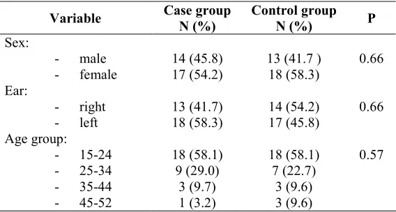
Based on the sample size calculation formula (a: 5%; b: 20%, 95% CI, and Odds ratio: 4), there were 62 samples included in this study, consisted of 31 samples in case group and 31 samples in control group. The age range of samples was 4 to 52 year
old, consisted of 11 (45.8%) male and 13 (54.2%) female in the case group, 10 (41.7%) male and 14 (58.4%) female in the control group (TABLE 1).
Chi-square and Mantel-Haensel test showed that both groups were not significantly different for the each variable, or in other terms, both groups were in the homogeneous condition.
TABLE 1. Characteristic of samples
HPV DNA detection
PCR and agar electrophoresis examinations detected HPV DNA 18 in 21 (67%) samples in the case group and 5 (16%) HPV DNA 18 positive
samples in the control group (TABLE 2). Statistical analysis using Chi-square test revealed a significant difference between case group and control group (p=0.0001, Odds ratio 10.92, 95 % CI =2.83–45.29).
TABLE 2. HPV DNA 18 in case group and control group
Evidences of epidemiological and experimental studies showed that HPV had an important role in the pathogenesis of warts, displasia and cancer. HPV infection as reported by some authors to be detected in the lesion of oral cavity, nasal cavity, conjunctiva, paranasal sinuses, larynx, tracheo-bronchial mucosa, oesophagus, urethra, anogenital tract, and skin. There were different prevalences at those locations, and factors causing different prevalences are demographic factor and patient habit.34
Terai et al (1999) studied 30 specimens of normal oral cavity mucosa and 7 specimens of skin diseases (condyloma acuminata, verruca vulgaris and seborrhoic keratosis). The study showed 9 (30%) of normal samples were HPV 18 positive. Even though the HPV has been detected in specimen of displasia and oral cavity cancer, its prevalence and role in the pathogenesis of displasia and cancer has not been known exactly.34
There were different prevalence ratios of positive HPV between 12% to 60% for HPV 6, 11, 16, 18, 31, 33 and 57 (Sugerman & Shillitoe,
! " ! " ! "" "! !
" #"!$ % & #!"$ %
#" $&% #! $ %
#! $ % ' # '$ %
#'$&% # $ %
#" $& % #! $ %
" #!"$ % & #"!$ %
#! $ % & # $&%
#'$ % #'$ %
$
$
1997). The study did not mention about the differences between latent, sub-clinical and clinical period. Even though the HPV 16 and 68 were usually be considered in high risk type, based on its presentation in displasia lesion and oral cancer, those HPV also could detected in normal oral cavity with high frequency 51.3%.36 HPV 18 had a normal
keratinocyte transformation action in human and showed carcinogenic effect in vitro.37,38 Based on
epidemiological studies on oral cancer patients, there were other factors that influence the pathogenesis of oral cavity carcinogenesis of the tumor. They were cigarette smoking, tobacco chewing, alcohol, old age, HIV infection, and sexual habit.39
HPV DNA detected in oral mucosa showed that persistent or temporary HPV infection might occur in human. Usually, HPV were transmitted by sexual contacts or non-sexual means, for
example, by autoinoculation from a region to other region in the same individual, or by vertical transmission during perinatal period.40,41
Interleukin-1 (IL-1) identifications
Based on IHC examination, there was a significant difference (p < 0.001; CI95%: 4.94 – 14.23) in IL-1 expressions between positive DNA HPV 18 case group (23.62 ± 6.9) and control group (11.73 ± 8.24).
If all samples were classified into positive and negative DNA HPV 18, there was no significant difference (p: 0.275; CI95%: -3.1-10.3) in IL-1 expressions between positive DNA HPV 18 case group (23.62 ± 6.9) and control group (20.0 ± 4.06), as well as negative DNA HPV 18 negative case group (8.4 ±3.9) and control group (11.73 ± 8.24) (TABLE 3).
TABLE 3. IL-1 expressions in both groups
In case group of samples with complication, the IL-1 expression (37 ± 4.63) was significant statistically higher (p < 0.001; CI95%: 9.86 – 21.36) than samples without complications (21.39 ± 4.38). Bujia et al. (1996) studied 20 samples of extract of tympanic cholesteatoma tissue using bicinchonic acid protein assay and enzyme-linked immunoabsorbent assay (ELISA).42 They found a
higher expression of IL 1a in tympanic cholesteatoma compared to the normal auditory canal skin, 34.9 ±19.5 ng/g and 6.7±2.8 ng/g, respectively. Statistically, this result was significantly different. (p < 0.001). In the skin samples, there wa a higher concentration of IL-1 RA (248.3±30.2 ng/g) compared to that in the tympanic cholesteatoma (80.8±13.5 ng/g), and statistically they were significantly different (p < 0.01). IL-1 activity could not be detected in the skin samples. Biologic activity of IL-1 was analyzed by the cell line LBRM-33. It could be concluded that there
was an increase of IL-1 expression along with the decrease of IL-1-RA. Furthermore, imbalance of IL-1 RA production and total IL-1 production caused IL-1 to take an active position. The presence of network between cytokines, and between IL-1 and other interleukin or other cytokines suggest that the network has an important role in the pathogenesis of tympanic cholesteatoma Interleukin-1a (IL-1a) has an important role by stimulating thef keratin cell proliferation and inducing bone resorption.
Some authors stated that the presence of microorganism infection in MtCSOM would increase the progressiveness of tympanic chole-steatoma In this study, the type of microorganism of the samples were not detected, because of no differences in microorganism types between MtCSOM and BtCSOM in the previous studies, so that the response immune of the host was not significantly different.43,44
$ ( $' $ ( "$ $ &! #'!) *+ $ $ %
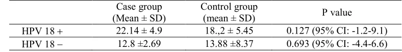
Interferon g (IFN-ggggg) identification
Based on IHC examination, there was a significant difference (p < 0.,001; CI95%: 4.09 – 12.42) in IFN-g expression between positive DNA HPV 18 case group (22.14 ± 4.9) and control group (13.88 ± 8.37).
If all samples were classified into positive and negative DNA HPV 18, there was no significant
difference (p: 0.275; CI95%: -3.1-10.3) in IFN-g expressions between positive DNA HPV 18 case group (23.62 ± 6.9) and control group (20.0 ± 4.06), as well as negative DNA HPV 18 negative case group (8.4 ± 3.9) and control group (11.73 ± 8.24) (TABLE 4).
TABLE 4. IFN-g expressions in both groups
MtCSOM (positive tympanic cholesteatoma) was commonly followed by bone destruction that cause a decrease in the hearing acuity, vestibular disturbance and intracranial complications. This inflammation was associated with the cytokine production; IL-1b, TNF-a, and IFN-g. These cytokines play an important role in the regulation and activation of osteoclast, and in bone resorption process. Many kinds of inflammatory cells, including macrophages and neutrophiles; were found in MtCSOM. Those cells would produce nitrous oxide (NO) after exposed and activated by cytokines. NO had an effect on osteoclast activation.45
CONCLUSION
Based on the research resuls it could be concluded that the HPV 18 DNA were identified in the tympanic cholesteatoma mass of MtCSOM patients, and HPV 18 was one of the multiple risk factors in the occurrence of tympanic chole-steatoma in MtCSOM patients.
Experimental research in animal samples is needed to determine and support the exact role of HPV 18 and other risk factors on the occurrence of tympanic cholesteatoma. The role of HPV as a risk factor should be considered in the persistent otitis media treated with conservative treatment.
REFERENCES
1. Bluestone CD and Klein CM. Intratemporal Complicat-ions and Sequelae of Otitis Medis: Etiology and Management. In: Bluestone CD, Stool SE, eds. Pediatr Otolaryngol 1983; I: 513-64.
2. Albino AP, Kimmelman CP and Parisier SC. Cholestea-tom: a molecular and cellular puzzle. Am J Otol 1998; 19 (1): 7-19.
3. Deric D, Arcosic N and Dordevic V. Pathogenesis and methods of treatment of otogenic brain abscess. Med-Pregl 1998; 51(1-2): 51-55.
4. Portmann M. Definition of success and failure in cholesteatoma surgery. In: Sade J, editor. Cholesteatoma and mastoid surgery 1982: 431-38.
5. Palva, T. Surgery related to histopathology in chronic inflammatory middle-ear disease. J Laryngol Otol 1988; 102: 851-56.
6. Sanna M, Zini C. Congenital cholesteatoma of the middle ear. In: Sade J, editor. Cholesteatoma and mastoid surgery 1982: 29-36.
7. Sade J, Babyatzki A, Pinkus G. The metaplastic and congenital origin of cholesteatoma. In: Sade J, editor. Cholesteatoma and mastoid surgery 1982: 305-319. 8. Michael L. The metaplastic process in epithelial strata.
In: Sade J, editor. Cholesteatoma and mastoid surgery 1982: 299-303.
9. Palva T, Karma P, Makinen J. The invasion theory. Cholesteatoma and mastoid surgery 1982: 249-64. 10. Cheshire IM, Blight A, Proops DW. An in vitro growth
study on cholesteatoma and normal skin. Clin Oto-laryngol 1996; 20(5): 453-60.
11. Sudhoff H, Bujia J, Holly A, Kim C, Fisseler-Eckhoff A. Functional Characterization of middle ear mucosa residues in cholesteatoma sam-ples. Am J Otol 1995; 15(2): 217-21.
* ,
#- . ( %
* .
# . ( % /
$ " ( "$' $0 ( !$"! $ & #'!) *+ $ '$ %
12. Broekaert D, Cooreman K, Coucke P, Nsabumukunzi, S, Reyniers P, Kluyskens P, Gillis E. Keratinization of aural cholesteatoma (keratoma): A quantitative histophotometric study of the sulphydryl and disul-phide content. In: Sade J, editor. Cholesteatoma and mastoid surgery 1982: 161-73.
13. Bujia J, Holly A, Kim C, Schilling V, Kasten-bauer. New aspects on the pathogenesis of cholesteatoma: the possible role of immune cell-induced keratinocyte hyperproliferation. Laryngootologie 1993; 72(6): 279-83.
14. Bergmann K, Hoppe F, Helms J, He J, Muller-Hermelink HK, Stremlau. A.Human-papillomavirus DNA in cholesteatomas. Int J Cancer 1994; 59(4): 463-66. 15. Stremlau A, Helms J, Muller-Hermelink HK, Hoppe F,
de-Villiers EM. Detection of DNA of humman-papillomavirus (HPV) in an ‘aggre-ssively’ growing kholesteatom. Is kholesteatom a virus-induced tumor? HNO 1995; 43(1):1-2.
16. Chao WY, Chang SJ, Jin YT. Detection of human papillomavirus in cholesteatomas. Eur Arch Otorhino-laryngol 2000; 257: 120-23
17. Shinode H, Huang CC. Heat shock protein in middle ear cholesteatoma.Otolaryngol. Head-Neck Surg 1996; 114(1): 77-83.
18. Herber R, Liem A, Pitot H, Lambert PF. Squamous epithelial hyperplasi and carcinoma in mice transgenic for the human papillomavirus type 16 E7 oncogene. J Virol 1996; 70(3): 1873-81.
19. Schwarz E, Freese UK, Gissmann L, Mayer W, Roggen-buck B, Stremlau A, zur Hausen H. Structure and transcription of human papillomavirus sequences in cervical carcinoma cells. Nature 1985;314(6006):111-14. 20. Yee C, Krishnan HI, Baker CC, Schiegel R, Howley, PM. Presence and expression oh human papillomavirus sequences in human cervical carcinoma cell lines. Am J Pathol 1985; 119: 361-66.
21. Arbeit JM, Munger K, Howley PM, Hanahan D. Neuroepithelial carcinoma in mice transgenic with human papillomavirus type 16 E6/E7 ORFs. Am J Pathol 1993; 142: 1187-97.
22. Griep A, Lambert PF. Role of papillomavirus oncogenes in human cervical cancer: transgenic animal studies. Proc Soc Exp Biol Med 1994; 206: 24-34.
23. Jeon S, Lambert PF. Integration of human papillomavirus type 16 DNA into human genome leads to increased stability of E6 and E7 mRNAs: implication for cervical carcinogenesis. Proc Natl Acad Sci USA 1995; 92: 1654-58.
24. Liu Y, Chen JJ, Gao Q, Dalal S, Hong Y, Mansur CP, Band V, Androphy EJ. Multiple functions of human papillomavirus type 16 E6 contribute to the immorta-lization of mammary epithelial cells. J Virol 1999;73(9): 7297-307
25. Thomas JT, Oh ST, Terhune SS, and Laimins LA. Celluler changes induced by low-risk human papillomavirus type 11 in keratinocytes that stably maintain viral episomes. J Virol 2001; 75 (16): 7564-71.
26. Melero I, Singhal MC, McGowan P, Haugen HS, Blake, J, Hellstrom KE. Immunological ignorance of an E7-encoded cytolytic T-lym-phocyte epitope in transgenic mice expressing the E7 and E6 oncogenes of human papillomavirus type 16. J Virol 1997; 71: 3998-4004. 27. Howley PM. Papillomavirinae. The viruses and their
replication. In: Fields BN, Knipe DM, Howley PM, editors. Virology 3rd (ed). 1996: 2045-75.
28. Sell S. Immunology, immunopathology and immunity. 6th (ed). Washington DC: ASM Press., 2001.
29. Janeway CA, Travers P, Walport M, and Schlo-michik, M. Immunobiology. 5th (ed.). New York: Garland Publishing, 2001: 1-35.
30. Roittt I, Brostoff J, Male D. Immunology. 5rd (ed.). London: Mosby, 1998.
31. Alberts B, Bray D, Lewis J, Raff M, Robert K, Watson JD. Molecular biology of cell. 3rd (ed). Garland Pub.Inc. New York & London; 1995: 273-287.
32. Mills J. Viral infection. In Stites DP, Terr AI and Parslow, T.G. (eds). Basic & clinical immunology. 8th (ed), 1994: 637-48.
33. Grote JJ, Hesseling SC, and Koopmann JP. Effect of endotoxin on the advancing front between cultured middle ear mucosa and epidermis. A preliminary study. Acta Otolaryngol Stockh 1995; 115(2): 286-90. 34. Terai M, Hashimoto K, Sata T. High prevalence of human
papillomaviruses in the normal oral cavity of adult. Oral Microbiol Immunol 1999; 14: 201-205.
35. Sugerman PB, Shillitoe EJ. The high risk human papillomavirus and oral cancer: evidence for and against a causal relationship. Oral Dis 1997; 3: 130-147. 36. Miller CS, White DK. Human papilloma-virus
expression in oral mucosa, premalignant conditions and squamous cell carcinoma: a retrospective review of the literature. Oral Surg Oral Med Oral Pathol Oral Radiol Endod 1996; 82: 57-68.
37. Shin KH, Min BM, Cherrick HM, Park NH. Combined effects of human papillomavirus-18 and N-methyl-N’-nitro-N-nitrosoguanisine on the transformat-ion of normal human oral keratinocytes. Mol Carcinog 1994; 9: 76-86.
38. Lakshmi S, Nair SA, Pillai MR. Oral cancer and human papillomavirus: is there a link? J Surg Oncol 1993; 52: 193-96.
39. Coutlee F, Trottier AM, Ghattas G. Risk factors for oral human papillomavirus in adults infected and not infected with human immunodeficiency virus. Sex Transm Dis 1997; 24: 23-31.
40. Koch A, Hansen SV, Nielsen NM, Palefsky J, Melbye M. HPV detection in children prior to sexual debut. Int J Cancer 1997; 73: 621-24.
41. Puranen M, Yliskoski M, Syrjanen K, Syrjanen S. Vertical transmission of human papillomavirus from infected mothers to their newborn babies and persistence of the virus in childhood. Am J Obstet Gynecol 1996; 174: 694-99.
analysis of protein production and biological activity. Eur Arch Otorhinolaryngol 1996; 253(4-5): 252-55. 43. Rianto BUD. Hasil guna tetes siprofloksasin 0,3%
dibandingkan khloramfenikol 3% pada terapi otitis media kronis benigna aktif. Maj Kedokt Indon 2001; 51 (3): 95-90.
44. Helmi. Penggunaan antimikroba secara rasional untuk pengobatan infeksi telinga tengah. Maj Kedokt Indon 2001; 51 (3): 91- 96.