2
RINGKASAN
A.
Pendahuluan
Sindrom nefrotik (SN) dihubungkan dengan risiko penyakit kardiovaskuler (PKV) terutama aterosklerosis karena adanya hipoalbuminemia, hiperlipidemia, hiperoksidatif stres,
hiperkoagulasi, inflamasi dan pemakaian steroid jangka panjang.1, 2
Hiperlipidemia pada SN ditandai dengan peningkatan kadar kolesterol total, kolesterol low density lipoprotein (K_LDL), trigliserida, atau kolesterol high density lipoprotein (K_HDL) normal atau meningkat. Hiperlipidemia merangsang timbulnya spesies oksigen
reaktif (SOR) yang dapat menimbulkan kerusakan pada lipid, karbohidrat dan DNA.3
SOR memicu reaksi oksidasi LDL, disfungsi endotel dan inflamasi yang mengawali
terjadinya aterosklerosis.4 Terjadinya aterosklerosis pada anak SN terbukti melalui studi
kohort, otopsi, hasil nekropsi dan laporan kasus. Penelitian kohort selama 16 tahun di Kalifornia Utara menemukan 11/142 anak SN (7,7%) infark miokard dengan risiko relatif (RR) 5,5 (IK 95%: 1,6 – 18,3) dan risiko kematian akibat penyakit koroner 2,8 kali (KI 95%
0,7–11,3).5 Prevalensi advanced vascular age pada SN lebih tinggi secara signifikan
dibanding kontrol (29,4% vs 10,7%).6 Keadaan yang sering kambuh dan persistensi lipid
pada 40–50% kasus SN menyebabkan paparan terus menerus faktor risiko PKV yang akan
menyebabkan disfungsi endotel sehingga terjadi aterosklerosis dini.7, 8
Pencegahan aterosklerosis sebaiknya dimulai pada anak sejak dini, karena pada masa ini proses aterosklerosis telah dimulai. Restriksi diet, memberikan hasil yang kurang memuaskan, dan penggunaan obat penurun lipid mengganggu pertumbauhan dan
menyebabkan miopati berat, sehingga membahayakan bagi anak.9
Konsensus di Bagian Ilmu Kesehatan Anak (IKA) melalui unit kerja koordinasi (UKK) nefrologi anak belum memberikan rekomendasi nyata untuk mengatasi hiperlipidemia yang berkaitan dengan pencegahan aterosklerosis secara dini karena terbatasnya penelitian. Tanpa ada penanganan yang serius akan meningkatkan mortalitas, morbiditas dan progresivitas
penyakit glomerulosklerosis dan aterosklerosis.10, 11
Penelitian pemberian anti oksidan α-tokoferol, β-karoten dan asam askorbat yang dilakukan pada hewan coba tikus Sprague-Dawley hiperlipidemia, memberi efek
menurunkan indeks aterogenik pada kelompok dengan asam askorbat.12 Penelitian pada
orang dewasa dengan risiko aterosklerosis dan yang sudah mengalami aterosklerosis memberi
3
dl-α-tokoferol dan asam askorbat dengan dosis 10–15 mg/kgBB/hari selama 12 minggu pada anak SN usia >1–<15 tahun berpengaruh terhadap stres oksidatif yang diukur dengan kadar LDL-ox, ekspresi reseptor penyapu makrofag dalam serum yang diukur melalui kadar sCD36), dan marker disfungsi endotel yang diukur dengan kadar sVCAM-1 dan NO sebagai
petanda dini aterosklerosis.14-17 (Hubungan antar variabel dijelaskan dalam kerangka teori
terlampir).
B. Metode penelitian
Penelitian ini menggunakan rancangan Randomized Pretest-posttest Control Group
Design. Subyek penelitian adalah penderita sindrom nefrotik yang mengalami hiperlipidemia berumur >1– <15 tahun, dirawat di RSUP dr.Kariadi Semarang/ RSUD/RS swasta sekitarnya yang bersedia kontrol di RSUP dr. Kariadi Semarang serta memenuhi kriteria
inklusi-eksklusi. Randomisasi dilakukan dengan cara simple block random sampling.18 Kelompok
perlakuan mendapat terapi standar (steroid) dan kombinasi dl-α-tokoferol dan asam askorbat dengan dosis sama 10–15 mg/kgBB/hari dibagi dua dosis diberikan secara oral bersama makan selama 12 minggu. Kelompok kontrol mendapat terapi standar dan plasebo. Variabel bebas: dl--tokoferol dan asam askorbat. Variabel perantara: kadar LDL-ox. Variabel tergantung: sVCAM-1, sCD36, dan NO. Sampel subyek diambil dari serum darah puasa dan diperiksa dengan cara ELISA kecuali kadar NO dengan kolorimetri. Variabel perancu: asupan makanan (lipid dan vitamin), kadar albumin, hipertensi, steroid.
C.
Hasil penelitian
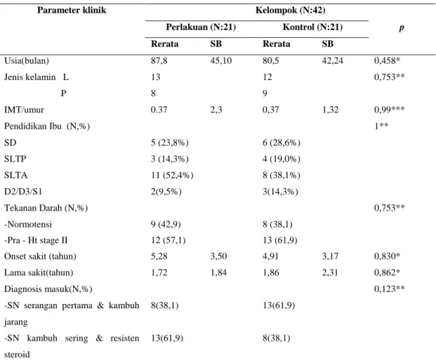
Sebanyak 42 dari 46 anak SN memenuhi kriteria inklusi dan eksklusi. Selama 12 minggu perlakuan 36 subyek (85,71%) dapat menyelesaikan penelitian masing-masing kelompok 18 subyek. Sembilan belas subyek (52,7%) mengalami remisi di akhir perlakuan, dengan 10 subyek dari kelompok perlakuan.
4
Tabel 1.Karakteristik klinik subyek sebelum intervensi
Parameter klinik Kelompok (N:42)
Perlakuan (N:21) Kontrol (N:21) p Rerata SB Rerata SB Usia(bulan) 87,8 45,10 80,5 42,24 0,458* Jenis kelamin L 13 12 0,753** P 8 9 IMT/umur 0.37 2,3 0,37 1,32 0,99*** Pendidikan Ibu (N,%) 1** SD 5 (23,8%) 6 (28,6%) SLTP 3 (14,3%) 4 (19,0%) SLTA 11 (52,4%) 8 (38,1%) D2/D3/S1 2(9,5%) 3(14,3%) Tekanan Darah (N,%) 0,753** -Normotensi 9 (42,9) 8 (38,1) -Pra - Ht stage II 12 (57,1) 13 (61,9)
Onset sakit (tahun) 5,28 3,50 4,91 3,17 0,830*
Lama sakit(tahun) 1,72 1,84 1,86 2,31 0,862*
Diagnosis masuk(N,%) 0,123** -SN serangan pertama & kambuh
jarang
8(38,1) 13(61,9)
-SN kambuh sering & resisten steroid
13(61,9) 8(38,1)
Keterangan: IMT/umur: indeks masa tubuh/umur; Ht: hipertensi; *= Mann-Whitney U, **= uji Chi square; ***= uji t ; *) Uji Kolmogorov-Smirnov;SB: Simpang Baku; TD: Tekanan Darah; Ht: hipertensi
Tabel 2. Gambaran klinik subyek sebelum dan sesudah intervensi
Gambaran klinik Kelompok (N:36)
Perlakuan (N:18) Kontrol (N:18) Nilai p
Sebelum Sesudah Sebelum Sesudah
Subyek remisi (N,%) - 10 (55,6%) - 9 (50%) 0,738** Tekanan Darah (mmHg)
-Normotensi (N,%) 8(44,4) 15(83,3) 8(44,4) 9(50) 0,075*) -PraHt-Ht stage II (N,%) 10(55,6) 3(16,7) 10(55,6) 9(50)
Morbiditas* - 3a - 2b
Total subyek akhir 18 18 18 18
Keterangan: *): Uji Fisher exact ; Morbiditas:aSatu anak cacar air, satu anak infeksi saluran kencing 19, satu anak kambuh saat perlakuan. :b Kambuh karena infeksi saluran pernafasan akut (ISPA). Persentase yang ditampilkan per kelompok
5 Perlakuan
Kontrol
Hasil pre test-post test kelompok perlakuan: kadar LDL-ox:127,2 menjadi 89,5 U/L;
sCD36:72,5 menjadi 123,5 ng/mL; sVCAM-1:929,4 menjadi 868,4 ng/mL; NO: 8,9 menjadi 8,5 μM/L. Kelompok kontrol: LDL-ox:125,2 menjadi 76,7U/L; sCD36:56,9 menjadi 61,7
ng/mL; sVCAM-1: 1231,2 menjadi 1008,0 ng/mL ; NO: 8,2 menjadi 7,9μM. Uji Wilcoxon
kelompok perlakuan dan kontrol berturut-turut untuk kadar LDL-ox p=0,039 dan p=0,001
(Gambar 1). Kadar sCD36 p=0,163 & p=0,088, kadar sVCAM-1 p=0,306 & 0,122; Kadar
NO p=0,983 & p=0,760. Kadar Δ diantara dua kelompok perlakuan dan kontrol
berturut-turut Δ LDL-ox -37,7vs - 48.6U / L ; Δ sCD36: 51,1vs 17,2 ng /mL; Δ sVCAM-1: -61,0 vs –
223ng /mL ; Δ NO -0.4 vs- 0,6 mg/dL. Uji t tidak berpasangan Δ LDL-ox, ΔsVCAM-1
p=0,594dan 0,327, dan uji Mann Whitney U untuk Δ sCD36 dan Δ NO p berturut-turut
=0,883 dan 0,864.
Gambar 1. Diagram box plot kadar LDL-ox, sebelum, sesudah intervensi dan Δ kelompok perlakuan dan kontrol
Sebelum perlakuan Sesudah perlakuan
Sesudah perlakuan Δ Δ Kadar LDL-ox Kadar LDL-ox p=0,039 p=0,001 P=0,913 P=0,424
6
Tabel 3. Uji beda rerata dan Δ kadar LDL-ox, sCD36, sVCAM-1, NO berdasarkan status remisi kelompok perlakuan
Variabel
Remisi (N:10) Tidak remisi (N:8) p
Rerata+ SB Median Min Maks Rerata+ SB Median Min Maks
LDL-ox (U/L) 58,3+ 50,36 40,05 19,2 181,9 128,4+ 59,3 130,9 37,1 202,6 0,016* sCD36 (ng/mL) 153,7+ 205,2 33,7 18,1 652,1 85,8+ 150,0 19,8 12,3 451,6 0,274* sVCAM-1 (ng/mL) 714,8+ 109,8 735,5 507,8 843,5 1060,3+ 199,2 1024,9 861,4 1474,4 0,000# NO (μM) 8,7+ 6,4 6,6 2,5 19,4 8,2+ 5,6 7,4 2,8 20,7 1,000* ΔLDL-ox -66,6+ 67,6 -81,0 -136,0 49,2 -1,6+ 58,0 -1,4 -84,2 106,5 0,047# ΔsCD36 75,5+ 111,3 5,4 -8,8 255,2 20,3+ 102,1 0,44 -126.2 241,2 0,696* ΔsVCAM-1 -129,9+ 207,9 -65,8 -549,7 48,5 25,1+ 267,6 88,3 -430,3 343,3 203* ΔNO 0,11+ 4,4 0,1 -7,0 7,4 -1,05+ 10,2 0,3 -22,4 14,4 0,748**
Keterangan: * Uji Mann-Whitney U ; # Uji t tidak berpasangan;
Berdasarkan status remisi maka uji beda kadar LDL-ox post test, Δ kadar LDL-ox
dan kadar sVCAM-1 post test pada kelompok perlakuan berbeda bermakna dengan
berturut-turut p=0,016; 0,047 dan 0,000 (Tabel 3)
D.
Bahasan
1. Pengaruh dl-α-tokoferol dan asam askorbat terhadap kadar LDL-ox.
Penelitian ini menemukan bahwa kadar LDL-ox pada kedua kelompok perlakuan dan kontrol menurun setelah intervensi, namun tidak berbeda bermakna di antara dua kelompok. Hasil berbeda bermakna ditemukan pada Δ kadar LDL-ox kelompok perlakuan berdasarkan status remisi, yang tidak ditemukan pada kelompok kontrol. Hal ini menunjukkan bahwa
pemberian dl-α-tokoferol dan asam askorbat memperkuat kerja prednison. Prednison
menurunkan SOR melalui hambatan pembentukan radikal superoksida sehingga menurunkan peroksidasi lipid. Penelitian ini dibuktikan oleh Marumo, dkk melalui isolasi sel otot polos
aorta yang diinduksi dengan deksametason.19 Fase remisi akibat respon steroid juga memberi
peluang untuk menurunkan kadar LDL-ox karena peningkatan serum albumin sebagai
7
Albumin mengangkut ion Cu2+ bebas +15%, dimana pengangkut ion Cu2+ lainnya
diperankan terutama oleh seruloplasmin (70%) dan sisanya diangkut oleh macroglobulin
(15%). Kadar seruloplasmin ditemukan menurun pada sindrom nefrotik saat kambuh.20
Penelitian Engler menunjukkan pemberian dl-α- tokoferol (400 IU/hari) dan asam askorbat (500mg/hari) pada 15 anak hiperlipidemia familial tidak berefek pada biomarker
stress oksidatif (LDL-ox, dll) tetapi memperbaiki disfungsi endotel yang diukur dengan flow
mediated dilatation arteri brakialis (FMA).21 Devaraj, dkk memberikan 1200 IU α-tokoferol selama 2 tahun untuk penderita penyakit arteri koroner stabil usia 40–70 tahun memberikan hasil bermakna.22 Penelitian oleh Boushehri, dkk dilakukan pada orang laki-laki dewasa dengan faktor risiko PKV. Pemberian 400 IU Vitamin E, 500 mg Vitamin C, 15 mg β-karoten atau kombinasi ketiganya selama 12 minggu menunjukkan penurunan kadar LDL-ox
secara bermakna.23
Penambahan anti oksidan kurang memberikan pengaruh terhadap penurunan LDL-ox pada kelompok perlakuan yang tidak remisi. Beberapa kemungkinan yang bisa menjadi faktor penyebab adalah: subyek tidak respon terhadap steroid menyebabkan stress oksidatif terus berlangsung, variasi absorpsi α-tokoferol cukup besar antara 20–60%,
hipoalbuminemia mengurangi jumlah ion Cu2+ yang terikat, proteinuria masif menyebabkan
berkurangnya pembentukan protein dan α-tokoferol transfer protein (α-TTP).24
Penelitian Sato, dkk menyebutkan bahwa pemakaian glukokortikoid dapat menurunkan
aktifitas antioksidan enzimatik seperti superoxide dismutase (SOD), katalase (Kat),
glutathione peroxidase (GPx). Penurunan anti oksidan enzimatik menyebabkan terganggunya pembentukan asam askorbat. Asam askorbat dapat mengubah bentuk radikal tokoferoksil menjadi α-tokoferol secara langsung dan anti oksidan thiol seperti glutation dan asam lipoat
dapat menghasilkan α-tokoferol secara tak langsung melalui pembentukan asam askorbat.25
2. Pengaruh dl-α-tokoferol dan asam askorbat terhadap kadar sCD 36.
Rerata kadar sCD36 sebelum intervensi pada 40 subyek adalah 62,6+ 81,5 dengan median 28,6 (13,1- 396,9) ng/mL (Rujukan normal menurut kit pemeriksaan antara 3,12-200 ng/mL, rujukan untuk anak dewasa muda 18 – 25 tahun: 32,3 ng/mL (16,8 – 102,4 ng/mL). Hasil penelitian ini menunjukkan bahwa kadar sCD36 cenderung meningkat pada kedua kelompok, meskipun berbeda tidak bermakna (p>0,05). Penelitian ini berbeda dengan hasil penelitian Devaraj, dkk yang memberikan alfa tokoferol 50μM (2,3 mg) dan 100 μM (4,5
mg) pada makrofag derivat monosit volunteer normal ternyata dapat menurunkan ekspresi
8
Kemungkinan peningkatan ini berkaitan dengan mikropartikel yang berasal dari trombosit, lekosit dan sel endotel. Mikropartikel ini ditemukan pada kasus resistensi insulin
dan obesitas dengan diabetes mellitus tipe-2 karena adanya inflamasi derajat rendah.27
Suryohudoyo mengatakan bahwa sekali reseptor penyapu sCD36 terangsang, maka tak
akan mengalami down regulation sehingga makrofag terus menerus memproduksi sCD36.28
Kadar sCD36 tinggi dapat ditemukan pada penderita dengan aterosklerosis yang simtomatik, hiperinsulinisme, DM tipe-2, obesitas dan polikistik ovarii dan perlemakan
hati.11 Analisis regresi menunjukkan bahwa kadar sCD36 dipengaruhi oleh K_HDL,
IMT/umur, dan berat badan sebanyak 30%. Hasil penenlitian ini menunjukkan kadar HDL yang tinggi pada kelompok perlakuan (70mg/dL) dibanding kontrol (58,7mg/dL).
3. Pengaruh dl-α-tokoferol dan asam askorbat terhadap kadar sVCAM-1
Penurunan kadar rerata dan Δ kadar sVCAM-1 di akhir intervensi tidak berbeda bermakna. Hal ini bisa disebabkan oleh pengaruh steroid yang mungkin lebih dominan sebagai anti inflamasi yang kuat.
Glukokortikoid memiliki efek menurunkan gen transkripsi sitokin (interleukin, TNF-α),
kemokin (antara lain macrophage inflammatory protein-1(MIP-1)α, monocyte
chemoattracted protein-1(MCP-1)dan 2), enzim (inducible nitrix oxide synthase (iNOS), siklooksigenase2, Cytoplasmic phospholipase A), dan molekul adhesi (ICAM-1 dan VCAM-1). Penelitian yang dilakukan oleh Upritchard, dkk pada orang dewasa dengan DM tipe II menunjukkan bahwa suplementasi vitamin E 800 IU/hari dan vitamin C 500 mg/hari dan jus tomat 500 ml/hari selama 4 minggu ternyata tidak memberi perubahan bermakna pada kadar
sVCAM-1 maupun sICAM-1.29
Hasil analisis regresi menyebutkan bahwa prediktor sVCAM-1 yaitu HDL memberikan kontribusi 16,1%, sedangkan faktor-faktor lain masih banyak berpengaruh diantaranya adanya endotoksinemia, atau respon inflamasi. Pengaruh infeksi sudah disingkirkan dengan pemeriksaan CRP. Respon inflamasi masih mungkin terjadi pada penderita SN terutama yang tidak remisi, oleh karena adanya hiperlipidemia, hipertensi dan hipoalbuminemia. Kadar sVCAM-1 sangat tinggi (2664,3;2480,6;2258,9 ng/mL) memiliki kadar HDL yang sangat rendah yaitu 18,27 dan 36 mg/dL ditemukan pada kelompok kontrol.
Rendahnya kadar NO akibat hiperlipidemia dan hipertensi (55,6% kasus) pada penelitian ini mendukung untuk terjadinya peningkatan aktivitas molekul adhesi sVCAM-1.
9
Berdasarkan status remisi di akhir perlakuan, kadar sVCAM-1 menurun secara bermakna pada kelompok intervensi. Perbedaan bermakna ini tidak ditemukan pada kelompok kontrol, sehingga perubahan yang terjadi pada kadar sVCAM-1 kemungkinan besar oleh adanya pengaruh steroid dan kombinasi dl-α-tokoferol serta asam askorbat. Efektivitas alfa tokoferol dan asam askorbat terjadi pada saat remisi kemungkinan karena kadar albumin yang sudah
membaik sehingga tercukupinya α-tocopherol transfer protein (α-TTP) yang mengangkut α
-tokoferol menuju lipoprotein plasma (HDL & LDL) agar terlindung dari proses oksidasi.30
4. Pengaruh pemberian dl-α tokoferol dan asam askorbat terhadap kadar NO
Kadar NO yang rendah saat awal intervensi kemungkinan dipengaruhi oleh hiperlipidemia, peningkatan kadar LDL-ox (72% kasus) dan hipertensi (55,6% kasus). Δ kadar NO mengalami penurunan yaitu 0,35 μM pada kelompok perlakuan dan 0,39 μM pada kelompok kontrol.
Pemberian glukokortikoid (prednisone/metil prednisolon) menghambat pembentukan ion superoksida, seharusnya dapat meningkatkan kadar NO. Stres oksidatif dan peroksinitrit (ONOO-) yang berlebihan karena terjadinya reaksi NO dengan superoksida (O2-) menyebabkan timbulnya perubahan fisiologis dan serologis. Perubahan ini berkaitan dengan peningkatan tekanan darah, proteinuria, disfungsi trombosit, peningkatan tromboksan (TXA)
dan endotelin serta penurunan prostasiklin (PGI2).31
Penelitian Engler menyebutkan bahwa pemberian vitamin E dan C memperbaiki
fungsi endotel yang ditunjukkan dengan pemeriksaan dilatasi arteri brakialis (flow mediated
dilatation, FMD), namun tidak mempengaruhi petanda stres oksidatif.21
Penelitian oleh Shouman menunjukkan bahwa penderita SN kelainan minimal (SNKM) mempu nyai kadar nitrit plasma sebesar 45,87+ 20,85 μmol/L dan nitrat 45,58+ 19,7 μmol/L
sedangkan pada anak normal sebesar 12,33+ 7 μmol/L.32
Penyebab lain penurunan produksi NO adalah menurunnya kadar L-arginin yang diinduksi oleh peningkatan lipoprotein dan rendahnya L-arginin menyebabkan produksi yang berlebihan dari superoksida. Kadar NO juga dipengaruhi oleh kadar kalsium, dan asupan makanan yang kaya akan NO seperti daun seledri, bit, sayur-sayuran berdaun seperti bayam, dan lettuce. 33
Asam askorbat juga mengurangi aktivitas NAD(P)H oksidase dan produksi superoksida
secara invitro dan in vivo. NAD(P)H oksidase adalah sumber utama dari superoksida endotel
yang bertanggung jawab untuk uncoupling eNOS dini, yang dapat diinaktivasi oleh α-
10
Penelitian Heller menyebutkan bahwa pemberian α-tokoferol meningkatkan
pembentukan NO dan diperkuat oleh adanya asam askorbat. Penelitian ini dilakukan melalui
isolasi sel endotel vena umbilikalis manusia. 34
Temuan yang menarik dari penelitian ini adalah didapatkan perbaikan secara klinis tekanan darah pada 40 % kasus (7/18 subyek) dari kelompok perlakuan. Hal ini
membuktikan bahwa terdapat peran kombinasi dl-α-tokoferol yang diperkuat oleh asam
askorbat dalam meningkatkan kadar NO sebagai vasodilator vaskuler, meskipun belum terdapat bukti peningkatan kadar NO dan tidak bermakna secara statistik. Kadar NO tetap rendah kemungkinan terjadinya peningkatan pemakaian NO dalam jaringan, atau diperlukan waktu tertentu untuk meningkatkan kadar NO dalam darah sebagai proses adaptasi dalam
tubuh, sehingga perlu penelitian lebih lanjut. Penelitian ini menunjukkan efek saling
memperkuat antara dl-α-tokoferol dan asam askorbat memperbaiki fungsi vaskuler baik di membran sel maupun dalam sel.
E.
Simpulan dan Saran
Simpulan : Pemberian kombinasi dl-α-tokoferol dan asam askorbat dengan dosis masing- masing 10-15 mg/kgBB/hari yang diberikan selama 12 minggu pada anak SN :
1. Menurunkan kadar LDL-ox secara tidak bermakna. Kadar LDL-ox, Δ LDL-ox
menurun secara bermakna pada kelompok perlakuan yang mengalami remisi dibanding tidak remisi.
2. Meningkatkan kadar sCD36 secara tidak bermakna.
3. Menurunkan kadar sVCAM-1 secara tidak bermakna. Kadar sVCAM-1 menurun
secara bermakna pada kelompok perlakuan yang mengalami remisi dibanding tidak remisi
4. Menurunkan kadar NO secara tidak bermakna.
Saran:
1. Perlu penelitian lebih lanjut untuk memisahkan subyek penelitian SN tanpa hipertensi
atau dengan hipertensi.
2. Perlu penelitian pemantauan kadar biomarker secara serial selama penelitian untuk
melihat kecenderungan peningkatan atau penurunan selama remisi.
3. Pemberian kombinasi dl-α-tokoferol dan asam askorbat lebih lama minimal 6 bulan
untuk melihat respon obat terhadap biomarker stress oksidatif dan disfungsi endotel selama remisi
11
SUMMARY
A.
Introduction
Nephrotic syndrome (NS ) in children is associated with a risk of cardiovascular disease (CVD) especially atherosclerosis due to hypoalbuminemia, hyperlipidemia, hyperoxidative
stress, hypercoagulation, inflammation and long-term steroid use.1,2
Hyperlipidemia in NS is characterized by elevated levels of total lipids (cholesterol, triglycerides), and enormous increase in cholesterol in the low-density lipoprotein (LDL) and very low-density lipoprotein (VLDL)-cholesterol fraction, whereas the concentration of high-density lipoprotein (HDL)-cholesterol is normal or even decreased. Hyperlipidemia induce reactive species oxygen (ROS) and cause damage of lipid, carbohydrate and deoxyribo
nucleic acid (DNA).3
Reactive oxygen species lead to oxidized LDL, endothelial dysfunction and
inflammation that initiate atherosclerosis.4 Atherosclerosis in NS was proven through cohort
studies, autopsy , necropsy and case reports. One of the studies from North California was a 16 years cohort study. Eleven of 142 children (7.7%) with NS were found to have myocardial infarction with a relative risk (RR) 5.5 (95% CI:1.6-18.3) and coronary mortality risk 2.8
(95%CI:0.7 –11.3).5 Prevalence of advance vascular age in NS was higher significantly
compared with control (29.4% vs 10.7%).6 Continuous exposure because of the risk of
recurrence and persistence of lipids (40-50 % of NS) increases the risk of these events.7,8
Prevention of atherosclerosis should begin early in childhood because this process has already begun. Diet restriction gives less real results, and use of lipid-lowering drugs have the
risk of impaired growth and development and severe myopathy, so it is harmful to children.9
There has been no agreement or consensus in Child Health Department through Pediatric Nephrology Working Group to manage hyperlipidemia associated with early atherosclerosis prevention. Without any serious treatment, this would increase mortality, morbidity,
glomerulosclerosis and atherosclerosis progressivity.10,11
Previous studies showed that the use of anti-oxidant α-tocopherol , β-carotene and ascorbic acid in hyperlipidemic Sprague Dawley rats reduced the atherogenic index in the group with ascorbic acid.12 Studies in adults with the risk and who had experienced of
12
atherosclerosis gave variable results.13 This study aimed to prove that the oral combination
of 10-15mg/kgBW/day dl-α-tocopherol and ascorbic acid in children with NS aged >1- <15 years had any effect on biomarkers of oxidative stress ( oxidized low density lipoprotein, ox-LDL), scavenger receptor of macrophage (soluble CD36, sCD36), disfunction of endothelial
(soluble vascular cell adhesion molecule, sVCAM –1) and nitric oxide (NO).14-17
B.
Method
A Randomized Pretest-Posttest Control Group Design was conducted. Subjects who fulfilled inclusion and exclusion criteria were enrolled in the study. Subject with idiopathic nephrotic syndrome (INS) who had hyperlipidemia aged >1 - <15 years and were treated in the outpatient or inpatient clinic in the Department of Child Health/Dr.Kariadi Hospital Semarang or other hospitals willing to be followed in Dr. Kariadi Hospital were included in
the study. Randomization was done by simple block random sampling.18 The treatment group
received standard steroid therapy and combination of oral dl-α-tocopherol and ascorbic acid of 10-15 mg / kg/day divided into two doses daily, orally and the control group received
standard therapy and placebo. Independent variables were dl--tocopherol and ascorbic acid,
intermediate variables was ox- LDL and dependent variables were sVCAM-1, sCD36, NO. The levels of oxLDL, sVCAM-1, and sCD36 were measured by enzyme linked immunosorbent assay (ELISA), NO was measured by colorimetric assay. Confounding variables were food intake that measured by 3 days food recall & record (lipids and vitamins), and albumin serum, hypertension, and steroid treatment.
C.
Results
Forty two of 46 children were included. During the 12-week treatment, 36 subjects (85.71%) completed the study (18 subjects of the each group). A total of 19 subjects in remission (52.8%) at the end of treatment (10 subjects from the treatment group).
The mean of pretest and posttest level of ox-LDL serum in treatment group were 127.2 to 89.5 U/L; sCD36:72.5 to 123.5ng/mL; sVCAM-1:929.4 to 868.4ng/mL; NO: 8.9 to 8.5μM/L. In the control group: ox-LDL:125.2-76.7U/L ; sCD36:56.9-61.7ng/mL; sVCAM-1: 1231.2–1008.0 ng/mL; NO: 8.2-7.9μM. The Wilcoxon test of ox-LDL levels were showed
significant difference in each group by p value =0.039 in the treatment group and p=0.001 in
the control group. There were no significant difference of sCD36 levels, sVCAM-1 levels and NO levels. In treatment and control group sCD36 levels showed p value = 0.163 and
13
0.760. The Δ level of ox-LDL on treatment group compared to control was -37.7 vs -
48.6U/L ; Δ sCD36: 51.1vs 17.2ng/mL; Δ sVCAM-1: -61.0 vs -223ng/mL ; Δ NO -0.4 vs -0,6
mg/dL. Independent t test of Δ ox-LDL, ΔsVCAM-1 and Mann Whitney U test of Δ sCD36
and ΔNO were not significantly different with P value of 0.594, 0.327, 0.883, 0.864, respectively. According to remission state at the end of the study the level of LDL, Δ ox-LDL and level of sVCAM-1 in treatment group were significanly different p=0,016,0,047 and 0,000 respectively.
D.
Discussion
1. The effect of dl-α-tocopherol and ascorbic acid on ox-LDL
In this study we found that ox-LDL tend to decrease in both groups, although not significantly difference (P>0.05). There was a reduction on ox-LDL level at remission state on both group. The remission of treatment group showed a significantly different in reduced Δ ox-LDL. This fact should be taken into special consideration. This result was likely caused by the effect of combined dl-α-tocopherol and ascorbic acid in synergism with steroid to induce remission and lowering the lipid peroxidation. Prednisone was known to lower reactive oxygen species (ROS) via inhibition of superoxide radical generation and the consequences of the reduction on lipid peroxidation. The study was done by Marumo, et al in
dexamethasone-induced isolated aortic smooth muscle.19 The remission phase on steroid
therapy were also considered to lead the reduction in ox-LDL level in relation with the
albumin’s role as the Cu2+
binding agent which would reduce the free metallic ion capacity to
trigger oxidation process. Albumin bind to 15% free Cu2+ while the rest will be bound to both
macroglobulin and ceruloplasmin. The last is a major copper-binding protein in human plasma (70%), while albumin bind the other 15% and the rest is bound to macroglobulin.
Ceruloplasmin level will be decreased in relaps nephrotic syndrome.20
Previous studies in 15 children with familial hypercholesterolemia and familial combined hyperlipidemia, treatment by vitamin C (500 mg/day) and E (400 IU/day) for 6 weeks did not have an effect on biomarkers of oxidative stress (autoantibodies to epitopes of oxidized LDL, F2-isoprostanes, 8-hydroxy-2'-deoxyguanosine), but improve the brachial artery dilatation.21 Two other studies among adult revealed a significant results. A study conducted by Devaraj , et al which administered 1200 IU α-tocopherol for 2 years to subject
with stable coronary artery disease aged 40–70,22 and other study was conducted by
14
supplementation of 400 IU Vitamin E, 500 mg Vitamin C, 15 mg β-carotene as a
combination or isolated treatment were showed a significant result in ox-LDL reduction.23
Antioxidant addition not effectively induced the ox-LDL reduction in non-remission treatment group. There are several possibilities which likely to be the etiologic factors: unresponsiveness to steroid, wide absorption spectrum dose of α-tocopherol (20–60%), elevation of oxidative stress level in the non-remission period, hypoalbuminemia reduces the Cu2+ bound, reduction of α-tocopherol transfer protein (α-TTP).24
Study conducted by Sato, et al revealed that glucocorticoid administration reduced enzymatic antioxidant activity such as superoxide dismutase (SOD), catalase (Cat), and glutathione peroxidase (GPx). This would lead to reduced ascorbic acid production which directly converts tocopheroxyl radical into α-tocopherol. While thiolic compound-antioxidant like glutathione and lipoic acid have the capacity of producing α-tocopherol
indirectly from ascorbic acid production pathway.25
2. The effect of dl-α-tocopherol and ascorbic acid on sCD 36 level
The mean of serum level of sCD36 of 40 subjects was 62.6+ 81.5 ng/mL, the median was 28.6 (13.1- 396.9) ng/mL. Normal reference for young adult 18 – 25 years was 32.3 (16.8 – 1024)ng/mL. The result of this study showed that sCD36 tend to increase in both groups, although it was not significantly different. The combination of dl-α-tocopherol and ascorbic acid administration were unable to lower the sCD36 expression. Devaraj et al showed a different declined where 50 and 100 μM α-tocopherol caused the 47% and 77%
sCD36 expression in monocyte-derived macrophage isolation of normal volunteer.26
Elevation on sCD36 was more likely related to microparticle of platelets, leukocytes, and endothelial either stimulation or apoptosis.27
According to Suryohudoyo, once the sCD36 scavenger receptor is being induced, it
never be down-regulated so the macrophages keep producing the sCD36.28 High level of
sCD36 is found in symptomatic atherosclerosis, hyperinsulinism, type-2 Diabetes Mellitus (DM), obesity, polycystic ovaries, and fatty liver.11
Regression analysis showed that 30% serum level of sCD36 was influence by HDL cholesterol, basal metabolism index for age (BMI/age) and body weight. The serum level of HDL cholesterol in treatment group (70 mg/dL) was higher than control group (58 mg/dL).
3. The effect ofdl-α-tocopherol and ascorbic acid on sVCAM-1 level
Combined regiment of dl-α-tocopherol and ascorbic acid had no effect on the mean and Δ sVCAM-1 post treatment. This might be due to the strong anti-inflammation property of
15
steroid more superior than dl-α-tocopherol and ascorbic acic, which reduced the sVCAM-1 level.
While glucocorticoid showed the lowering effect on cytokine transcription gene (interleukin, TNF-α), chemokines (MIP-1α, MCP-1 and 2), enzymes (iNOS, cyclooxygenase-2, Cytoplasmic phospholipase A), adhesion molecules (ICAM-1 and VCAM-1), and receptors (IL-2 receptor, tachykinin 1(NK-1) receptor). Upritchard, et al study revealed supplementation of Vitamin E, Vitamin C, and tomato juice with a dose 800 IU, 500 mg, 500 ml given daily for 4 weeks among type-2 DM subjects had no effect on sVCAM-1 and
sICAM-1 level.29
Regression analysis was used to determine the sVCAM-1 predictor, and HDL contributed 16,1%, meanwhile other contributing factors were endotoxinemia and inflammatory responses. Infection were excluded by CRP examination. There might be a sustaining inflammation especially among the non remission nephrotic syndrome patients, hyperlipidemia, hypertension and other factors stimulating adhesion molecules expression. Low NO level in this study supports the increase in activity of adhesion molecule sVCAM-1.
Based on the remission status at the end of treatment, there was a remarkable decline in sVCAM-1 level among the treatment group. On the other hand, there was no difference in sVCAM-1 level among the control, so this fact showed that combination supplementation dl-α-tocopherol and ascorbic acid sthrenghten steroid activity in the remission state. This was a possibility which likely increasing albumin at remission provide α-tocopherol transfer protein (α-TTP). α-TTP will pick α-tocopherol up to cholesterol LDL and HDL in order to prevent oxidation process.30
4. The effect of dl-α tocopherol and ascorbic acid on NO
In this study, there was no increase in NO level as expected after the administration of dl-α tocopherol and ascorbic acid. Low level of serum NO before intervension maybe caused by hyperlipidemia, high level of ox-LDL serum (72% of cases) and hypertension (55.6% of cases). Moreover, the Δ NO level was decreasing 0,35 μM and 0,39 μM in the treatment and control group respectively. Among the treatment group, the Δ NO level was increasing 0,11 during remission period and decreasing to the value of -1,05 among the non remission sub group.
Glucocorticoid administration blocked the superoxide production, so there must be an increase in NO level. Oxidative stress and accumulation of nitrite peroxide (ONOO-) as a
16
result of NO and superoxide (O2-) reaction cause changes in physiological and serological
environment. These changes include an increase blood pressure, proteinuria, platelet dysfunction, increase thromboxane (TXA) and endotheline, and also a decrease in
prostacycline (PGI2) level.31
A decrease in NO production was likely caused by reduction in L-arginine level induced by increased lipoprotein. Moreover, the low L-arginine level lead a superoxide over production. NO level is also affected by calcium, NO-rich food intake such as celery, bit,
leavy vegetables like spinach and lettuce.32
Engler stated that vitamin E and C administration improved endothelial function,
determined by brachialis artery dilatation evaluation (flow mediated dilatation arteria
brachialis, FMA), with no effect on oxidative stress markers.21
Shouman revealed a different result where plasma nitrite level was found high in minimal change nephrotic syndrome patient 45,87+ 20,85 μmol/L and nitrate level 45,58+
19,7 μmol/L whereas normal value is 12,33+ 7 μmol/L.33
Ascorbic acid assists NO synthesis by supporting redox system and stabilizing tetrahydrobiopterin (BH4) level. It also reduces NAD(P)H oxidase activity and superoxide production in vitro and in vivo. NAD(P)H oxidase plays as a major superoxide endothelial
responsible for early eNOS uncoupling, inactivated by α-tocopherol and ascorbic acid.17
Heller revealed that α-tocopherol improved NO production and being intensified by
ascorbic acid. This study was conducted in isolated human endothelial umbilical vein.34
The importance clinical finding in this study was improvement of blood pressure on 40 % cases (7/18 subjects) of treatment group. This proved that combined supplementation of dl-α-tocopherol and ascorbic acid had synergistic action increasing vascular dilatation, although the serum level of NO did not increase and it was not significantly different between two groups. The serum level of NO remained low because increasing level consumption of NO in cellular or needed time to adapt as physiological condition and then reach optimal level of NO. This study showed sinergistic effect between dl-α-tocopherol and ascorbic acid to improve vascular function in membrane and inside of the cell.
E.
Conclusion and Suggestion
Conclusion: Combined supplementation of dl -α-tocopherol and ascorbic acid with a dose of 10-15 mg kg/ day administered for 12 weeks in children with NS:
17
1. Decreased the serum levels of ox-LDL , but it was not significantly different. Mean levels of ox-LDL, delta of ox- LDL level decreased significantly in the treatment group on remission state.
2. Increased the serum levels of sCD36, but it was not significantly different .
3. Decreased the serum levels of sVCAM-1, but it was not significantly different. SVCAM –1 decreased significantly only in the treatment group on remission state. 4. Decreased the levels of NO, but it was not significantly different.
Suggestions:
1. Further research was needed to separate subject of NS with hypertension.
2. Research with serial examination to confirm trend of biomarker.
3. Administration of dl -α-tocopherol and ascorbic acid to overcome oxidative stress
minimally 6 months to maintain of remission state.
DAFTAR PUSTAKA
1. Shouman M, Abdallah N, Tablawy NE, Rashed L. Biochemical markers of endothelial dysfunction in pediatric nephrotic syndrome. Arch Med Sci. 2009;5:415-21.
2. Leichleitner M. Dyslipidemia and renal disease-pathophysiology and lipid lowering therapy in patients with impaired renal function. J Clin Basic Cardiol. 2000;3.
3. Al-Benna S, Hamilton CA, McClure JD, Rogers PN, Berg GA, Ford I, et al. Low-density lipoprotein cholesterol determines oxidative stress and endothelial dysfunction in saphenous veins from patients with coronary artery disease. Arterioscler Thromb Vasc Biol. 2006 Jan;26(1):218-23. PubMed PMID: 16254199. Epub 2005/10/29. eng.
4. Sargowo D, editor. Disfungsi endotel pada penyakit kardiovaskuler. 1 ed. Malang-Jawa Timur: Bayumedia Publishing; 2003.
5. Ordonez JD, Hiatt RA, Killebrew EJ, Fireman BH. The increased risk of coronary heart disease associated with nephrotic syndrome. Kidney international. 1993;44:638-42.
6. Hooman N, Isa-Tafreshi R, Otukesh H, Mostafavi SH, Hallaji F. Carotid artery function in children with idiopathic nephrotic syndrome. Nefrologia. 2013;3:650-6.
7. Lawang SA, Rauf S, Lisal JS, Albar H, Daud D. Plasma lipid as risk factors in relapsing nephrotic syndrome. Pediatr Indones. 2008;48:322-6.
8. Hulthe J, Fagerberg B. Circulating oxidized LDL is associated with subclinical atherosclerosis development and inflammatory cytokines (AIR study). Atheroscler Thromb Vasc Biol. 2002;22:1162-7.
9. McGill HC, McMahan CA, Herderick EE, Malcom GT, Tracy RE, Strong JP. Origin of atherosclerosis in childhood and adolescence. Am J Clin Nutr. 2000;72:1307S-15S.
10. Trihono PP, Alatas H, Tambunan T, Pardede SO, editors. Konsensus tata laksana sindrom nefrotik idiopatik pada anak. 2 ed. Jakarta: UKK Nefrologi IDAI; 2012.
11. Hanberg A, Hojlund K, Gastaldelli A, Flyvbjerg A, Dekker JM, Petrie J, et al. Plasma sCD36 is associated with markers of atherosclerosis, insulin resistance and fatty liver in a nondiabetic healthy population. J Intern Med. 2012;271:294-304.
18 12. Salem SA, Hassan DR, Mowafy AR. A comparative impact of different types of a single antioxidant supplementation (B-carotene,alfa tocopherols and ascorbic acids) on lipid profile in hyperlipidemic rats. Middle-East Journal of Scientific Research. 2009;4:354-60.
13. Steinberg D. The LDL modification hypothesis of atherogenesis:an update. J lipid res. 2009:S376-81.
14. Ricciarelli R, Zingg J, Azzi A. Vitamin E reduces the uptake of oxidized LDL by inhibiting CD36 scavenger receptor expression in cultured aortic smooth muscle cells. Circulation. 2000;102:82-7. 15. van_Dam B, Hinsbergh VWMv, Stehouwer CDA, Versteilen A, Dekker H, Buytenhek R, et al. Vitamin E inhibits lipid peroxidation -induced adhesion molecule expression in endothelial cells and decreases soluble cell adhesion molecules in healthy subjects. Cardiovascular research. 2003;57:563-71.
16. Huang H Y , Caballero B, Chang S, Alberg A J, Semba R D, Schneyer C, et al. Multivitamin/mineral supplements and prevention of chronic disease: executive summary. Am J Clin Nutr. 2007;85:265S- 8S.
17. Carr AC, Zhu B, Frei B. Potential antiatherogenic mechanisms of ascorbate (vitamin C) and alfa tocopherol (vitamin E). Circulation. 2000;87:349-54.
18. Sastroasmoro S. Dasar-dasar metodologi penelitian klinis. In: S S, S I, editors. Dasar-dasar metodologi penelitian klinis. 3 ed. Jakarta: CV Sagung Seto; 2010. p. 78-90.
19. Marumo T, Schini-Kerth VB, Brandes RP, Busse R. Glucocorticoids inhibit superoxide anion production and p22 Phox mRNA expression in human aortic smooth muscle cells. Hypertension. 1998;32:1083-8.
21. Mohamed H, Moneim A, Mohamed NA. Oxidant stress and antioxidant balance in nephrotic syndrome in children. AAMJ 2005;3.
22. Engler MM EM, Malloy MJ, Chiu EY, Schloetter MC, Paul SM,et al. Antioxidant vitamin C and E improve endothelial function in children with hyperlipidemia. Circulation. 2003;108:1059-63. 23. Devaraj S, Tang R, Adams-Huet B, Haris A, Seenivasan T, Lemos JAd, et al. Effect of high dose alpha-tocopherol supplementation on biomarkers of oxidative stress and inflammation and carotid atherosclerosis in patients with coronary artery disease. Am J Clin Nutr. 2007;86:1392-8.
24. Boushehri SN, Yusof RM, Taib MNM, Mirzaei K, Yazdekhasti N, Akbarzadeh S. Effect of vitamin supplementation on serum oxidized LDL levels in male subjects with cardiovascular disease risk factors. Iranian Journal of basic medical sciences. 2012;15:958-64.
25. Westergren T, Kalikstad B. Dosage and formulation issues:oral vitamin E therapy in children. Eur J Clin Pharmacol. 2010;66:109-18.
26. Sato H, Takahashi T, Sumitani K, Takatsu H, Urano S. Glucocoticoid generates ROS to induce oxidative injury in the hippocampus,leading to impairment of cognitive function of rats. J Clin Biochem Nutr. 2010;47:224-32.
27. Devaraj S, Hugou I, Jialal I. Alfa-tocopherol decreases CD36 expression in human monocyte-derived macrophages. J Lipid Res. 2001;42:521-7.
28. Silverstein RL. Inflammation, atherosclerosis and arterial thrombosis:role of the scavenger receptor CD36. Cleveland clinic journal of medicine. 2009;76:s27-s30.
29. Suryohudoyo P. Kapita selekta ilmu kedokteran molekuler. 2 ed. Jakarta: CV sagung Seto; 2007.
30. Uprichard JE, Sutherland WHE, Mann JL. Effect of supplementation with tomato juice, vitamin E and vitamin C on LDL oxidation and product of inflammatory activity in type 2 diabetes. Diabetes care. 2000;23:733-8.
31. Kirmizis D, Chatzidimitriou D. Antiatherogenic effects of vitamin E: the search for the Holy Grail. Vascular Health and Risk Management. 2009;5:767-74.
32. Wikstrom AC. Glucocorticoid action and novel mechanisms of steroid resistance:role of glucocorticoid receptor-interacting protein for glucocorticoid responsiveness. J Endocrinol,. 2003;178:331-7.
19 33. Souman M, Abdallah N, Tablawy NE, Rashed L. Biochemical markers of endothelial dysfunction in pediatric nephrotic syndrome. Arch Med Sci. 2009;5:415 -21.
34. Ellis G, Adatia I, Yazdanpanah M, Makela SK. Nitrite and nitrate analysis: a clinical biochemistry perspective. Clinical biochemistry. 1998;31:195-220.
35. Heller R, Werner-Felmayer G, Werner ER. Alpha-tocopherol and endothelial nitrix oxide synthesis (abstrak). Ann N Y Acad Sci. 2004;1031:74-85.
20
Input Proses Output
Kortikosteroid
Kadar Peroksidasi
lipid
Kadar albumin
Kadar Angiotensin I-II
Tekanan Darah Kadar Renin
Kadar anti oksidan: Enzimatik Non enzimatik:
dl-α-tokoferol & asam askorbat Asupan makanan Kadar LDL-ox Kadar NO NFK-β Shear stress, aktifitas fisik, bradikinin, asetil kolin, adenosin difosfat Kadar sVCAM-1 Monosit-Makrofag Kadar sCD36 Kadar Cu, Fe, MPO,
lipoksigenase, NO
CRP
IL-1β,IL-6
MIP-1 TNF-α
Kadar gula darah
Infeksi akut dan kronik TB, Gagal ginjal akut dan
kronik Status Disfungsi endotel Obesitas Kadar Spesies Oksigen Reaktif Status Profil Lipid
(K_total, K_LDL, K_HDL, TG ) pada Sindrom Nefrotik
1
BIODATA
A.
Identitas
Nama : dr. Omega Mellyana, SpA
Tempat tanggal lahir : Sragen, 31 Juli1970
NIP : 19700731 1999072001
Pangkat : IVA/Pembina/Dokter Pendidik Klinis Madya
Agama : Kristen
Alamat rumah : Jl. Tumpang IX/7 Gajahmungkur Semarang
Alamat kantor : Departemen IKA RSUP dr. Kariadi
Jl. Dr. Sutomo no 16 – 18 Semarang
Email : omegamellyana@gmail.com
Keluarga
Nama suami : Letkol Laut (K) Cahyo Novianto,SpB,
MsiMed, SpB (Onk)
Nama anak : 1. Noverian Yoshua Prihutama
2. Gery Petra Yudha
3. Nadri Axelle Patri Adi
B. Pendidikan formal:
1. SD Kristen Dorkas Lasem, lulus tahun 1983 2. SMP Negeri 1 Lasem, lulus tahun 1986 3. SMA Neger 1 Rembang, lulus tahun 1989 4. FK Undip Semarang, lulus tahun 1996
5. PPDS-1 IKA FK Undip Semarang, lulus tahun 2004
6. Pendidikan Doktor Ilmu Kedokteran/Kesehatan: 2009- sekarang
C. Riwayat Pekerjaan
1. Dokter PKM Hamadi Jayapura, tahun 1996 – 1999
2. Dokter Spesialis Anak TIMKESGAB Departemen Kesehatan RI untuk Daerah Konflik di Meulaboh, NAD, tahun 2004-2005
3. Dokter Spesialis Anak di RSUD Cut Nya Dhien Meulaboh,NAD, 2005-2006 4. Homestaf di Divisi Nefrologi Departemen IKA RSUP dr. Kariadi Semarang,2007 –
sekarang
D. Riwayat Pendidikan/Pelatihan/Kursus Tambahan
Internasional:
1. Advance Pediatric Nephrology Course, (supervisor Prof. Man Chun Chiu) Hongkong , 2009
2. Primer in Pediatric Nephrology for Asia, CME Interactive Course, 2012, Singapore. 3. Clinical observer for Pediatric Nephrology, Mei – Agustus 2012 (supervisor Prof. Yap Hui Kim) di National University Hospital , Singapore.
4. Fellowship Training Pediatric Nephrology by International Pediatric Nephrology Association (IPNA) 2012- 2013 (supervisor. Prof. Kazumoto Iijima, MD, PhD) di Kobe University Graduate School of Medicine,Kobe, Jepang
2
Nasional:
2007 -Kursus Continuous Ambulatory Periatoneal Dialysis( CAPD), Solo, Pernefri 2008 -WorkshopManagement Hemodialysis, Peritoneal Dialysis, Cangkok Ginjal
-Kursus Hemodialisis, Pernefri Semarang
-Indonesian Peritoneal Dialysis College, Jakarta, Pernefri -Indonesian Peritoneal Dialysis College, Jakarta, Pernefri
-Workshop Diagnostic Approach of Genetic Disease in Children, CEBIOR, Semarang
2009 -Pelatihan Clinical Teacher dan Pemantapan Pelatih Clinical Teacher, Departemen Pendidikan FK UI
-Workshop Diagnostic & Practical Management of Kidney Disease in Children -International seminar and Workshop on Modern Biology and It’s application focusing on stem cell and Human Genetic
2010 -Pelatihan Biostatistic lanjut 2, Semarang FK Undip
-Pelatihan Penulisan Artikel Ilmiah International Semarnag, Semarang, FK undip
-Pelatihan Metodologi Pengabdian Kepada Masyarakat, Semarang, FK Undip -Workshop A Comprehensive Approach to Kidney Disease and Hypertension, Semarang
-Workshop How to Develop A Good Research Proposal, Jakarta IDAI -Workshop Evidence Base Medicine how to critical appraisal in research, Nutricia Indonesia Fund, Semarang
-Workshop Sistem Evaluasi Pendidikan Kedokteran Program Spesialis 2011 -Workshop on Molecular & Cytogenetic Analysis, Collaboration with
Departement of Human Genetics, Radboud University, Nijmegen Medical Centre, The Nederlands, di Semarang,
-Medical Genetic Course from Basic to Clinic Collaboration with Department of Human Genetics, Radboud University, Nijmegen Medical Centre, The
Nederlands, di Semarng
2011 -Pendidikan Materi Keahlian Pediatrik Umum (MKU) PPDS –II IKA di Jogyakarta 2012 -Workshop Update ke-2 Kegawatan Nefrologi, Hipertensi dan Hemodialisis,
Pernefri Semarang
2013 -Workshop Nasional Penguji dan Pelatih PS UK-OSCE, Semarang
-Pelatihan Peningkatan Mutu Pelayanan Menuju Rumah Sakit Kelas Dunia di RSUP dr. Kariad Semarang
2014 -Workshop Lactation & Complementary Feeding and Enuresis, PKB VII, Semarang
-Pelatihan Ultrasonografi Pediatrik Bagi Dokter Spesialis IKA , Perhimpunan Ultrasonografi Kedokteran Indonesia, World Ultrasound in Medicine smd biology center of excellence (Jakarta WFUMB-COE)
E. Kegiatan Ilmiah: Presentasi Poster/oral
1 Omega Mellyana, Sutaryo, Ida Savitri, Haris
Marta Saputra: Mud Aspiration: an experience at
Cut Nyak Dhien Hospital (case report)
Konika Bandung, 2005
2 Omega Mellyana, Tantri, Haris Marta
Saputra:Psycological Reactions in refugees
children in Meulaboh post earthquake and
3
tsunami
3 Ida Savitri, Omega Mellyana, Haris Marta
Saputra: Increasing Malaria patient in Cut Nyak
Dhien Hospital post tsunami
Konika Bandung, 2005
4 Chrisna H, Omega Mellyana,Uropati Obstruktif
(poster)
Proceeding KONIKA XIV, 2008 Surabaya
5 Heru Muryawan, Omega Mellyana,Fenty K :
Pola Kuman Penyebab infeksi saluran kemih dan uji kepekaan kuman terhadapa antibiotika tahun 2001-2005 RSUP dr. Kariadi Semarang,
Proceeding KONIKA XIV,2008
6 Omega Mellyana, Herumuryawan,
Rochmanadji Widajat: Hemodialysis and
Peritoneal dialysis in acute renal failure
Konika Menado, 2011
7 Agus Fitrianto, Omega Mellyana,
Herumuryawan, Rochmanadji Widajat:
Hemodialysis in acut on chronic kidney disease: 9 cases report
Konika Menado, 2011
8 Omega Mellyana, Herumuryawan,
Rochmanadji Widajat: Hemodialysis in Children
at dr. Kariadi Hospital (oral)
Konker Pernefri 2012
9 Adi Hijaz, M Supriatna, Omega Mellyana: Risk
Factor of acute kidney injury in pediatric intensive care unit’s patients
PIT Bandung, 2012
10 Christine Tampubolon, Omega Mellyana,
Herumuryawan: Clinical description of edema
incidence, albumin and calcium serum level in children with nephrotic syndrome
PIT Bandung, 2012
11 Taufan Prasetya, Omega Mellyana:Hubungan
antara respon terapi induksi pada serangan awal sindrom nefrotik dengan kekambuhan (oral)
PIT Bandung,2012
12 Timtri Dilli M, Omega Mellyana, M
Herumuryawan, Rochmanadji W: Association
between clinically response of steroid and
atherogenic index in nephrotic syndrome (poster)
PIT Bandung, 2012
13 Sondang Panjaitan, Omega Mellyana: Keluaran
Terapi metilprednisolon pada anak dengan sindrom nefrotik resisten steroid di RS dr. KAriadi Semarang (poster)
PIT Bandung, 2012
14 Omega Mellyana, Kar-Hui Ng, Mya-Than, Lourdes Paula Resontoc, Noor Haziah Hussain, Caresse J geng, Perry Yew-Weng Lau, Isaac Liu,
Wee Song Yeo, Hui Kim Yap: Dialysis duration
is the most important predictor of growth failure in children on peritoneal dialysis(poster)
The 16th Congress of
the IPNA, Shanghai,2013
15 Omega Mellyana, Hiroshi Kaito, Natsuki mAtsunoshita, Shingo Ishimori, Takeshi Ninchoji, Norishige Yoshikawa, Kazumoto
Iijima: Prevention of Focal Segmental
Glomerulosclerosis (FSGS) reccurence by
The 16th Congress of
the IPNA, Shanghai,2013
4
combination of pretransplant bilateral
nephrectomy, rituximab and plasma exchange, two cases reports
16 Omega Mellyana, Heru Muryawan,
Rochmanadji Widajat, Edi Dharmana: Effect of
dl-α-tocopherol supplementation on lipid profile, oxidized LDL level, sVCAM-1 and sICAM-1 in nephrotic syndrome
The 12th Asian Congres
of Pediatric Nephrology Association, 2014
17 Arlita Putri, Omega Mellyana, Susilo Adi:
Ankle Brachial Indeks among school-aged children
Konika
Palembang,2014
18 Arlita Putri, Herumuryawan, Mutya Dyah, Ririn
S, Omega Mellyana: Remission rate of cyclophosphamite therapy in children with steroid resistant nephrotic syndrome.
Konika Palembang, 2014
Fasilitator/Pembicara Workshop
- Kegawatan Nefrologi Anak, Manajemen hipertensi pada anak ,PKB Semarang, 2008 - Hemodialisis pada anak, Workshop PKB, 2010
- Inkontinensia pada anak, Workshop PKB Semarang 2014 - Biopsi Ginjal pada anak, Sinas Manado, 2014
Publikasi artikel ilmiah:
1 Omega Mellyana, JC Susanto: Impact of Six Month Oil Supplementation on Growth in the Weaning Periodb(research), Media Medika
Indonesia,2007
2 Mervin Tri hadianto, Omega Mellyana: Hemolytic Uremic Syndrome
following dengue hemorrhagic fever, Pediatrica Indonesiana, 2011
3 Kurnia Dwi Astuti, M Herumuryawan, Omega Mellyana: Correlation
between lipid profile and C-reactive protein (CRP) in children with nephrotic syndrome (submitted and review)
4 Omega Mellyana, Edi Dharmana, Hardhono Susanto, Nanan Sekarwana: The effect of combined vitamin C and E on the ox-LDL, sCD36, sVCAM-1 and NO level in idiopathic pediatric nephrotic syndrome (Submitted and review)
Kontributor Buku:
- Herumuryawan, Rochmanadji Widajat, Omega Mellyana, Transplantasi
Ginjal: Buku Kompendium Nefrologi Anak,2011
Publikasi popular
1. Meningkatkan ikatan Kasih Sayang antara Ibu dan Anak melalui ASI, Dalam rangka Hari ASI sedunia,Suara Merdeka, Agustus 2007
2. Jika Anak terkena sakit ginjal,petunjuk praktis bagi orang tua, Suara Merdeka, Maret 2014
5 2014
F. Organisasi:
- Anggota IDI, 1996 – sekarang
- Anggota IDAI, 2004 – sekarang (bagian Pembinaan dan Kesejahteraan
Anggota, 2015)
- Anggota InaSN (Indonesia Society of Nephrology)/Pernefri :2007- sekarang
- Anggota ASPNA (Asian Pediatric Nephrology Association) dan IPNA
(International Pediatric Nephrology Association) (2007-sekarang)
- Anggota Rotary Club Semarang Indraprasta Rotary International District
3420, 2008 - sekarang
- Anggota Tim Ginjal RSUP dr. Kariadi Semarang (2011-sekarang)
- Anggota Tim Paliatif RSUP dr. Kariadi (2012- sekarang)
G. Pengabdian:
- Anggota Tim Medis Pengobatan Gratis dalam rangka hari sakit sedunia, Gua Kereb, Ambarawa (2010, 2011)
- Pemeriksaan Kesehatan Proyek Pengembangan Anak Mugas tiap tahun ( 2011- sekarang)
H. Penghargaan