* corresponding author: [email protected]
Structural evaluation and animal
implantation of porous eggshell
waste-derived hydroxyapatite graft as bone
substitution
Yudha Mathan Sakti1*, Rahadyan Magetsari1
1 Departement of Orthopaedi and Traumatologi Dr. Sardjito General Hospital,
Faculty of Medicine Universitas Gadjah Mada, Yogyakarta, Indonesia
ABSTRACT
The development of hydroxyapatite graft with high economically value is needed for orthopedic practice in developing countries. Eggsell waste is well known as natural substance for calcium resource. It has been used as raw material in producing hydroxyapatite. This study was conducted to synthesize porous hydroxyapatite from eggshell waste and evaluate its activity as bone substitution. The porous hydroxyapatite graft was manufactured from eggshell and sugar as a raw material using hydrothermal process. The porous eggshell waste-derived hydroxyapatite (EW-HAP) graft was characterized using X ray difractometer (XRD) and analytical scanning electron microscope (SEM) and compared with commercial hydroxyapatite (HAP) JCPDS 09-432 graft (Bangros®) as standard. The porous EW-HAP graft obtained was then implanted on critically
sized femoral defects surgically created in the right thigh of male Wistar rats (Rattus norvegicus) with Bangros® as control. Radiological examination using XRD and histological examination
using hematoxyline-and-eosin staining of the bone femour were performed at 28 days after implantation. The results showed that the XRD pattern for EW-HAP was likely similar with the HAP standard. However, the SEM examination showed that the pasticle size of EW-HAP graft (2.5-3 µm) was higher than those HAP standard graft (1.5-2 µm). Radiographs according to the International of Limb Salvage (ISOLS) radiological evaluation system between EW-HAP graft (6.1 ± 1.45) and HAP control graft (6.9 ± 2.10) was not significantly different (p>0.05). Moreover, histological examination according to Lane and Shandu scoring system between the both graft (4.0 ± 0.94 versus 4.4 ± 0.92) was also not significantly different (p>0.05). It can be concluded that the structure EW-HAP graft is similar with HAP graft standard. The both grafts have also equal outcome as bone substitution.
ABSTRAK
Pengembangangrafthidroksiapatit dengan nilai ekonomi tinggi diperlukan dalam bidang ortopedi di negara berkembang. Limbah cangkang terus dikenal luas sebagai bahan alam sumber kalsium. Limbah cangkang telur telah digunakan sebagai bahan untuk menghasilkan hidroksiapatit. Penelitian ini dilakukan untuk mensintesisgrafthidroksiapatit berpori dari limbah cangkang telur dan mengkaji aktivitasnya sebagai pengganti tulang. Graft hidroksiapatit berpori dibuat dari bahan dasar cangkang telur (HAP-LCT) dan gula menggunakan proses hidrotermal.Graft yang dihasilkan dikarakterisasi difraktometer X-ray (DXR) dan mikroskop elektron skaning (MES) analitik dan dibandingkan dengan graft JCPDS 09-432 hidroksiapatit komersial (Bangros®) sebagai standar.
Graft berpori HAP-LCT yang diperoleh kemudian diimplantasikan pada critically sized tulang femur yang dibuat melalui operasi pada paha kanan tikus Wistar jantan (Rattus norvegicus) dengan Bangros®sebagai kontrol. Pemeriksaan radiologi menggunakan DXR dan histologi dengan
penelitian menunjukkan bahwa gambaran DXRgraft HAP-LCT sama dengan graft HAP standar. Namun demikian, hasil pemeriksaan MES menunjukkan ukuran partikel HAP-LCT (2,5-3 µm) lebih tinggi dari pada graft HAP standar (1,5-2 µm). Gambaran radiografi menurut sistem evaluasi radiologiInternational of Limb Salvage(ISOLS) graft HAP-LCT (6,1 ± 1,45) dan andgraftHAP kontrol (6,9 ± 2,10) berbeda bermakna (p>0.05). Lebih lanjut dari hasil pemeriksaan histologi menurut sistem penilaian Lane dan Shandu antara kedua graft (4,0 ± 0,94) tidak berbeda bermakna (p>0,05). Dapat disimpulkan bahwa strukturgraft HAP-LCT sama dengangraft HAP standar. Kedua graft juga mempunyai luaran sama sebagi pengganti tulang.
Key words: eggshell - porous hydroxyapatite graft – bone substitution – X-ray
INTRODUCTION
In orthopedic and traumotology, bone damage or bone defect is one of health problems that can cause mechanical instability, dead space for bacterial infection and hampered bone healing process.1 Extensive bone damage can not heal spontaneously, therefore a bone graft procedure is needed.2 In USA, at least 2.2 million bone grafting have been conducted annually with 300 million US $ are spent over one year period.3,4 The bone graft is expected to increase mechanical stability of bone and bone healing process.5
Bone grafting having biomaterial properties that obtained from patient’s own tissue (outologous bone graft) is considered to to be the most ideal of bone graft. However, the outologous bone graft has several limitations such as donor morbidity, tissue rejection and disease transmission.5-6 Recently, synthetic materials have been developed as synthetic bone graft to solve the problem. One of them is carbonated hydroxyapatite which has crystallographic similarity with human bone, excellent osteoconductivity and good resorption rate.7,8
Eggsell waste has been used as natural substance for calcium resource. The major cosntituent present in the eggshell is CaCO3, which account around 91% of the total weight. Eggshell waste is available in huge quantity from food processing, egg breaking and hatching industries.9,10 It has been used as raw material
in producing hydroxyapatite and used as attractive biomedical materials. In this study a porous eggshell waste-derivet hydroxyapatite (EW-HAP) graft has been synthesized and its application as bone substitution on critically sized femoral defect in animal rats has been performed.
MATERIALS AND METHODS
Fabrication of porous EW-HAP graft
1300oC for two hours performed. The porous EW-HAP graft was characterized using X ray difractometer (XRD Rigaku) to examine its chrystallography phase and purity and compared with an hydroxyapatite crystal standard of JCPDS (09-432) [Ca5(PO4)2(OH)].
Further-more, the structure and morphology of EW-HAP graft was analized using Analytical Scanning Electron Microscope/SEM (JEOL type JSM-636OLA). The fabrication process flow chart of the porous EW-HAP graft is presented in FIGURE 1.
FIGURE 1. The fabrication process flow chart of the porous EW-HAP graft
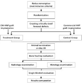
Implantation of the porous EW-HAP graft in critically sized femoral defect of animal
Twenty healthy male Wistar rats (Rattus norvegicus) aged 12 to 16 weeks with 250 to 300 g body weight were used in the study. Critically sized femoral defects were surgically created in the right thigh of all rats. At the time of surgery, rats were randomly assigned to a treatment group (n = 10), in which the femoral defect was filled with EW-HAP graft and control group (n = 10), in which the femoral defect was filled with commercial HAP graft JCPDS 09-432 (Bongros®). Clinical and physical examination was performed during 28 days experimental. Rats were sacrificed at 28 days after implantation.
Anterior-posterior and lateral radiological examination using X-ray diffractometer (XRD) of bone femour of the right thigh were performed at 28 days after implantation. Radiographs were
obtained and evaluated for bone union and bone resorption according to the International Society of Limb Salvage (ISOLS) radiological implants evaluation system after modification.
The operated bone femur sample was decalcified in formic acid working solution for seven days. Sample was then embedded in paraffin and cut longitudinally into 5 µm thick sections. Sections were stained with hemato-xyline-and-eosin (H&E). Histological examination was performed according to Lane and Shandu scoring system as modified by Heiple et al.11
commercial HAP graft is presented in FIGURE 2. All precedures have been approved by the Health Research Ethics Committee of the Faculty
of Medicine, Universitas Gadjah Mada, Yogyakarta.
FIGURE 2. The animal application flow chart of the porous EW-HAP graft
RESULTS
Fabrication of porous eggshell waste-derivet hydroxyapatite (EW-HAP) graft
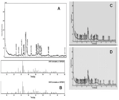
FIGURE 3 illustrates the X-ray diffraction pattern for EW-HAP (A), JCPDS (09-432) HAP control/Bongros® (B); human HAP (C) and bovine HAP (D). The X-ray diffaction pattern for EW-HAP was likely similar with the HAP standard, human HAP as well as bovine HAP.
FIGURE 3. X-R Diffraction pattern of EW-HAP (A); HAP control/Bongros®
(B) human HAP (C) and bovine HAP (D)
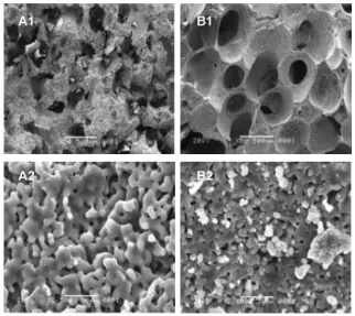
The SEM examination result of EW-HAP comparet with HAP control are presented in FIGURE 4. In magnitude of 50 times, the morphology of EW-HAP surface was likely more rough, irregular, random with irregular pore size of 150 – 500 µm. Whereas the HAP control (Bangros®) suface appeared more fine with regular pore size of 300-500 µm. In magnitude of 2500 times, both of HAP appeared
Implantation of the porous EW-HAP graft in critically sized femoral defect of animal
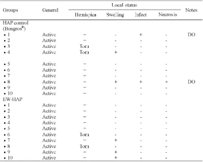
The physical and clinical conditions of rats after implantation of the porous EW-HAP graft or HAP control graft during 28 days observation are presented in TABLE 1. During 28 days observation periode, two rats on the HAP control group were excluded due to wound infection. In general the others rats exhibited
good physical and clinical conditions. On day 14, hemispica removal and operation wound care were conducted. Fixation of hemispica on four rats i.e. two in each group was torn. Post operative swelling was observed on five rats i.e. two in EW-HAP graft group and three in HAP control group. Tissue necrosis on distal hemispica was observed only in one rat of HAP control group.
TABLE 1. Physical and clinical conditions of rats on EW-HAP treatment graft group and HAP control graft group during 28 days observation
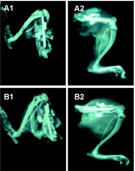
Representative of X-Ray of bone femour on 28 days after implantation of EW-HAP graft and HAP graft control are presented in TABLE. Whereas, the representative of histological
FIGURE 5. Representative of X-Ray of bone femour on 28 days after implantation. A1 (anterio-posteriol) and A2 (lateral) views of EW-HAP graft. B1 (anterio-posteriol) and B2 (lateral) views of HAP graft control
FIGURE 6. Representative histological section of bone femour on 28 days after implantation EW-HAP graft. A and B (EW-HAP graft), C (osteochondral tissue), D (osteoid and new bone cells).
The results of radiological and histological examination of of bone femour of the right thigh of rats of both groups are presented in TABLE 2. No signifantly difference in radiological and
TABLE 2. Radiological and histological examination of of bone femour of the right thigh of rats on EW-HAP graft and HAP control graft groups
Note: Min= minimum; Max=maximum; *Mann Whitney U Test with p<0.05
DISCUSSION
The synthesis HAP graft from eggshell as raw material of calcium source has been conducted by some authors.9,12,13Eggshell waste is available in huge quantity from food processing, egg breaking and hatching industries. About 250,000 tons of eggshell waste is produced annually worldwide by food processing industry only. The process involved a simple hydrothermal reaction of calcium oxide obtained from the burning of eggshell. The hydroxyapatite crystal was formed through a chemical reactions as follows:
The XRD pattern for EW-HAP was likely similar with the HAP standard, human HAP as well as bovine HAP. The EW-HAP has similar peaks with HAP standard (JCPDS 09-432). The highest peak of HAP standard was observed at 31.73oin the 2¸ with intensity of 100 Io, while the highest peak of EW-HAP was observed at 32.14o in the 2¸ with intensity of 90 Io. The similarity of the both graft was also indicated with major peaks observed between 30 and 40 oC. In addition, EW-HAP was likely similar
with the human dan bovine HAP that produced by Herliansyah.14
The morphoplogy of EW-HAP surface was likely more rough, irregular and random in the SEM examination. In this study sugar was used
to obtain a porous EW-HAP graft. Symsudin reported that the use of sugar as porogen provided HAP graft with rough, irregular and random surface.15Morphologically, it seems a sugar crystale which have cube-shaped or irregular square in the form of three-dimensio-nal mixed.16 However, the SEM examination showed the EW-HAP graft had similarity with previous studies.15,17-19The EW-HAP had ideal porous diamter to help for better osteogenic cells attachment and bone healing process.
In general, all rats exhibited good physical and clinical conditions after implantation of the porous EW-HAP or HAP control grafts during 28 days observation. The results of radiological and histological examination of bone femour of the right thigh of rats after EW-HAP graft implantation were not significantly compare to those HAP graft control. It was indicated that EW-HAP exhibited similar outcome as bone substituion compare to HAP control graft (Bangros®).
Studies the use of EW-HAP as bone graft substitution have been reported by some authors. Parket al.20evaluated of bone healing with hen eggshell-derived bone graft substitutes in rat calvaria and showed the potential efficacy of hen eggshell-derived bone graft as an osteoconductive bone substitute in a rat calvarial defect model. Lee et al.21 compared hydro-xuapatite from eggshell and synthetic hydroxyapatite for bone regeneration and reported that both eggshell and synthetic
CaCO3CaO CO2
hydroxyapatite showed highe bone formation than unfilled control. However, eggshell hydroxyapatite had significantly higher bone formation then the unfilled control at eight weels after operation. Durmus et al.22evaluated the biocompatibility and osteoproductive activity of ostrich eggshell powder in experimentally induced calvarial defect in rabits. The result showed that ostrich eggshell powder was a worth-while bone substitute because it is a safe, cheap, and easily available material.
AKNOWLEDGEMENTS
We would like to thank all technicians who gived valuable assistence druring the study.
CONCLUSION
The synthesis of hydroxyapatite from eggshell waste and sugar as raw materials has been successed performed providing porous EW-HAP graft. The structure and morphology of EW-HAP graft iss likely similar with the HAP standard (Bangros®). Furthermore, the implantation of EW-HAP graft of bone femour of the right thigh of rats provide similar outcome outcome as bone substituion compare to HAP control graft (Bangros®).
REFFERENCES
1. World Health Organization. Global burden of disease: global programme on evidence for health policy discussion paper 50. Geneva : World Health Organization, 2000.
2. Phelps JB, Hubbard GB, Wang X, Agrawal CM. Microstructural heterogeneity and the fracture toughness of bone. J Biomed Mat Res 2000; 51:735-41.
3. Lewandrowski KU, Gresser JD, Wise DL, Trantol DJ. Bioresorbable bone graft substitutes of different osteoconductivities: a histologic evaluation of osteointegration of poly(propylene glycol-co-fumaric acid)-based cement implants in rats. Biomaterials 2000; 21(8):757-64.
4. Thomas C, Burg KJL. Tissue engineered bone replacements systems. Eur Cells Mat 2007; 13(2): 1-2.
5. Camillo FX. Arthrodesis of the spine. In: Canale ST & Beaty JH editors. Campbell’s operative orthopaedics, 11th eds. Philadelphia: Mosby
Elsevier, 2007; 1851-2.
6. De Long WG, Einhorn TA, Koval K, McKee M, Smith W, Sanders, Ret al. Bone graft and bone graft substitutes in orthopaedic trauma surgery: a critical analysis. J Bone Joint Surg Am 2007;89:649-58. Doi:10.2106/JBJS.F.00465. 7. Jamali A, Hilpert A, Debes J, Afshar P, Rahban S,
Holmes R. Hydroxyapatit/calcium carbonate versus plester of Paris: a histomorphometric and radiographic study in a rabbit tibial defect model. Calcif Tissue Int 2002;71:172-78.
8. Orsini G, Ricci J, Scarano A, Pecora G, Petrone G, Iezzi G. Bone-defect healing with calcium-sulfate particles and cement: an experimental study in rabbit. J Biomed Mater Res Part B 2004; 199–208.
9. Raihanan MF, Sopyan I, Hamdi M, Ramesh S. Novel chemical conversion of eggshell to dydroxyapatite powder. In: Osman NAA, Ibrahim F, Wan WABA, Rahman HSA, Ting HN editors. Proceedings of the 4th Kuala Lumpur International Conference of Biomedical Engineering; 2008 June 25-28, Kuala Lumpur, Malaysia.
10. Sasikumar S and Vijayaraghavan. Low temperature synthesis of nanocrystalline hydroxyapatite from eggshells by Combustion method. Trens Biomater Artif Organs 2006; 19(2):70-3.
11. Heiple KG, Goldberg VM, Powell AE, Bos GD, Zika JM. Biology of cancellous bone grafts. Orthop Clin N Am 1987;18: 179–85.
12. Saeed AM, Hassan RA, Thajeel. Synthesis of calcium hydroxyapatite powder from hen’s eggshell. Iraqi J Phys 2011; 9(16): 24-8. 13. Kumar S. Eggshell derived nanophas e
bio-ceramics. Archives of BioCeramics Research Volume 9. Proceeding of the 9th
Asian Bio-ceramics Symposium; 2009 December 8-11, Nagoya, Japan.
14. Herliansyah MK. Development and fabrication of bovine hydroxyapatie bone graft for biomedical application [Ph.D thesis]. Kualalumpur: University of Malaya, Malaysia, 2009.
substitusi tulang [Tesis S–2]. Yogyakarta: Bidang Rekayasa Biomedis, Universitas Gadjah Mada. 16. Hynes RC, Le Page Y. Sucrose, a convenient test
crystal for absolute structures. J Appl Crystallograp 1991; 24 (4): 352. Doi:10.1107/ S0021889891002492.
17. Lee JH, Hwang CJ, Song BW, Koo KH, Chang BS, Lee CK. A prospective consecutive study of instrumented posterolateral lumbar fusion using synthetic hydroxyapatite (Bongros1-HA) as a bone graft extender. J Biomed Mat Res 2009; 90A(3): 804-10. DOI: 10.1002/jbm.a.32113
18. Abdurrahim T and Sofyan I. Recent progress on the development of porous bioactive calcium phosphate for biomedical applications. Recent Patents on Biomedical Engineering 2008;1: 213-29.
19. Damien E and Revell PA. Coralline hydroxyapatite bone graft substitute: a review of experimental
studies and biomedical application. J Appl Biomat Biomech 2004; 2: 65-73.
20. Pa JW, Bae SR, Suh JY, Lee DH, Kim SH, Kim H,
et al. Evaluation of bone healing with eggshell-derived bone graft substitutes in rat calvaria: a pilot study. J Biomed Mater Res A 2008; 87(1):203-14.
21. Lee SW, Kim SG, Balázsi C, Chae WS, Lee HO. Comparative study of hydroxyapatite from eggshells and synthetic hydroxyapatite for bone regeneration. Oral Sur Oral Med Oral Phathol Oral Radiol 2012;113(3):348-55.