ORIGINAL PAPER
A novel concept of Fe-mineral-based magnetoreception:
histological and physicochemical data from the upper beak
of homing pigeons
Gerta Fleissner&Branko Stahl&Peter Thalau& Gerald Falkenberg&Günther Fleissner
Received: 23 December 2006 / Revised: 21 February 2007 / Accepted: 21 February 2007 #Springer-Verlag 2007
Abstract Animals make use of the Earth’s magnetic field for navigation and regulation of vegetative functions; however, the anatomical and physiological basis for the magnetic sense has not been elucidated yet. Our recent results from histology and X-ray analyses support the hypothesis that delicate iron-containing structures in the skin of the upper beak of homing pigeons might serve as a biological magnetometer. Histology has revealed various iron sites within dendrites of the trigeminal nerve, their arrangement along strands of axons, the existence of three dendritic fields in each side of the beak with specific 3D-orientations, and the bilateral symmetry of the whole system. Element mapping by micro-synchrotron X-ray fluorescence analysis has shown the distribution of iron and its quantities. Micro-synchrotron X-ray absorption near-edge-structure spectroscopy has allowed us to unam-biguously identify maghemite as the predominating iron mineral (90 vs 10% magnetite). In this paper, we show that iron-based magnetoreception needs the presence of both of these iron minerals, their specific dimensions, shapes, and arrangements in three different subcellular compartments. We suggest that an inherent magnetic enhancement process via an iron-crusted vesicle and the attached chains of iron platelets might be sufficient to account for the sensitivity and specificity required by such a magnetoreceptor. The
appropriate alignment between the Earth’s magnetic field and the maghemite bands would induce a multiple attraction of the magnetite bullets perpendicular to the membrane, thus, triggering strain-sensitive membrane channels and a primary receptor potential. Due to its 3D architecture and physicochemical nature, the dendritic system should be able to separately sense the three vector components of the Earth’s local field, simultaneously—
allowing birds to detect their geographic position by the magnetic vector, i.e., amplitude and direction of the local magnetic field, irrespective of the animal’s posture or movement and photoreception.
Keywords Biological magnetometer . Maghemite . Magnetite . XRFS . XANES
Introduction
The magnetic sense of animals is one of their least explored sensory systems, although a great variety of animals display behavior that is modified or controlled by the Earth’s magnetic field (for review: Wiltschko and Wiltschko1995). Magnetic field parameters might serve various different functions, including compass and map orientation in true navigation or as a time cue and trigger for developmental and physiological programs (e.g., Fransson et al. 2001). Despite this importance to animals, magnetosensory organs have not been identified unambiguously (for review: Beason 2005; Johnsen and Lohmann 2005; Mouritsen and Ritz 2005). Compared to the older paradigm of a universal magnetite-based mechanism for magnetoreception (Kirschvink and Gould 1981; Kirschvink et al. 2001), an alternative mechanism or even a combination of several mechanisms is most likely: For example, the unidirectional
DOI 10.1007/s00114-007-0236-0
G. Fleissner (*)
:
B. Stahl:
P. Thalau:
G. Fleissner AG NCR, FB Biowissenschaften, J. W. Goethe-Universität, Siesmayerstr. 70,D-60054 Frankfurt a. M., Germany e-mail: [email protected]
G. Falkenberg
Hamburger Synchrotronstrahlungslabor HASYLAB, Deutsches Elektronen-Synchrotron DESY,
behavior of mole rats in the magnetic field corresponds to the horizontal vector component (like a polarity compass; Marhold et al. 1997), whereas the magnetic response of migrating birds exhibits a higher twofold rotational sym-metry and reveals their blindness for the field polarity (e.g., birds only sense the axis of the magnetic field lines in space (inclination compass; Wiltschko and Wiltschko 1997). Additionally, compass orientation and map navigation might require multimodal sensory input and appropriate processing of the central nervous information (for review: Phillips and Borland1992; Johnsen and Lohmann2005).
Several papers have reviewed possible underlying receptor principles. Three theories currently dominate the scientific discussion: electrical induction processes (Kalmijn 1978), photopigment-based magnetoreception (Ritz et al. 2000), and iron-mineral-based magnetoreception (Kirschvink et al.
2001). Evidence for these issues, as derived from theoretical and experimental data, have been recently reviewed several times in great detail (e.g., Beason 2005; Johnsen and Lohmann 2005; Mouritsen and Ritz 2005). Most of the reviewed experiments have focused only on the detection of compass mechanisms, although a combination of a compass with various types of map factors has been recognized as essential for true navigation and as timing cue, e.g., developmental processes. Sensory systems, which might function as magnetic field detectors, have not been identified with certainty, mainly due to the focus on behavioral experiments. Neurobiological studies that associate general physical principles to a detailed functional anatomy and histology are rare (for review: Beason 2005; Johnsen and Lohmann2005; Mouritsen and Ritz2005). But in the end, only such investigations will be able to elucidate the various sensory systems that serve magnetoreception.
Our interdisciplinary analysis of iron-mineral-containing dendrites in the beak of homing pigeons offers a promising receptor and neurophysiological approach to this complex problem. In this paper, we report novel essential insights based on a thorough analysis of histological details—in addition to previously published data (Fleissner et al.2003; Fleissner and Stahl2005)—in conjunction with quantitative physico-chemical measurements on iron minerals in the beak skin by spatially resolved synchrotron X-ray fluores-cence analysis (μ-SXRF) and micro-synchrotron
X-ray-absorption-near-edge-structure spectroscopy (μ-XANES).
We propose a mechanism consisting of a magneto-mechanical transducer that relies on the magnetic interac-tion of two iron minerals, magnetite and maghemite, incorporating signal amplification of the magnetic input stimulus. Such an improvement of the signal-to-noise ratio might provide the necessary sensitivity and specificity for sensing both intensity and inclination of the magnetic field, abilities seen in behavioral (e.g., Becker2000; Keeton et al.
1974) and electrophysiological experiments (e.g., Beason
and Semm 1996; also, Wang et al.2003). The aim of the present study of the sensory dendrites in the pigeon’s beak was to introduce a novel concept of a highly sensitive three-axis magnetometer based on ferri- and ferromagnetic materials and to describe their micromagnetic features.
Materials and methods
The standard histological methods for light and electron microscopic investigations used here have been described in Fleissner et al. (2003). To analyze the detailed architec-ture of the iron-mineral-containing strucarchitec-tures in the inner dermal lining of the upper beak of adult homing pigeons (raised in the loft of the Zoological Institute, University of Frankfurt a. M.) and to verify the suitability of these structures to function as a biological magnetometer, we took care to mount the beak skin naturally during fixation and subsequent histological processing. Sections were characterized under a Reichert Polyvar microscope (Vienna, Austria); then, they were documented by a digital camera (Spot, Diagnostic Instruments, Sterling Heights, IL, USA) and stack-reconstructed by Metaview Software (version 3.6 Universal Imaging, West Chester, PA, USA).
We used histological and physico-chemical methods to visualize the nature and distribution of different types of iron minerals. Only perfusion-fixed animals were used. The beak skin was then post-fixed in 4% glutaraldehyde. To evaluate possible artifacts induced by sample preparation, we tested the effects of two different buffers (Sörensen phosphate buffer and sodium cacodylate buffer) on the element distribution. The paraffin-embedded tissue was cut into 10-μm section
series. Each fourth section was mounted on glass slides and stained either with Prussian blue (PB) for Fe+3 ions or Turnbull’s blue (TB) for Fe+2ions. These sections served as a microscopic control. The three sections in between were not stained and were mounted on ultralene foil for analysis byμ-SXRF andμ-XANES. These X-ray experiments were
conducted at beamline L at HASYLAB (DESY, Hamburg, Germany). Because, in vitro, magnetite Feþ2Feþ3
2 O4
might easily be oxidized to maghemite Fe3þ
2 O3
, we demonstrated the long-term preservation of both minerals during tissue processing: Serial measurements first applied to fresh tissue were repeated with the mounted samples after 2 and 6 months. The results remained the same.
μ-SXRF is based on the analysis of the X-ray
synchrotron beam, in our case 15μm (present status at the
HASYLAB beamline L). The synchrotron beam is scanned over the sample. Data analysis is based on the X-ray spectra that are measured for all individual points of the scan. After scanning a specific region of the sample, a more detailed chemical analysis is done byμ-XANES at points with high
iron concentration. Close to the characteristic K shell absorption edge of iron (around 7,120 eV), the local chemical environment of the Fe atom will influence the details of the absorption as a function of the energy of the synchrotron radiation. By comparing the spectroscopic signal of the sample with reference samples of known composition, one can deduce the nature of chemical state of the iron (Wilke et al.2001). The iron minerals, magnetite, maghemite, and hematite are clearly distinguishable. The physical procedures at synchrotron facilities, including sample preparation, evaluation of data and statistical methods, are described in detail by Janssens et al. (2000).
Results
μ-SXRF andμ-XANES
So far, a spatially resolved physico-chemical analysis of the putative magnetoreceptive tissue in birds is lacking. We report data from X-ray analysis of the skin of the upper beak of homing pigeons, reliably showing the distribution of chemical elements and revealing characteristics of the iron minerals present.
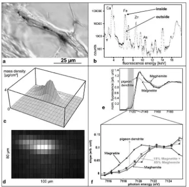
Theμ-SXRF experiment on paraffin section series (control
section, Fig.1a) taken from the beak skin of homing pigeons showed that the element spectra inside the Fe-containing dendrite and outside in the surrounding nervous tissue are clearly distinct (Fig.1b). Nervous tissue gives a strong Ca fluorescence signal that disappears in connective tissue. Fe is found within the nervous dendrite as a sharp local representation (Fig. 1c,d). The width of the elongated Fe peak in Fig. 1c,d is mainly due to the resolution limit of 15 μm of the synchrotron beam. The amount of Fe was
determined by a comparison of the absolute count rate with that of a known germanium film. The Fe within one dendrite (Fig. 1c,d) amounts to 4×10¹¹ Fe atoms, which would be equivalent to a mass of 35 pg of pure Fe.
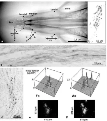
Methodological pitfallsThe chemistry during fixation and sectioning has a strong influence on the preservation of the Fe elements. This sensitivity to fixation is evident in a comparison of a fixative containing Soerensen phosphate buffer with a second fixative in natrium cacodylate buffer (Fig. 3d–f). Comparing the shape of the Fe-containing dendritic field (Fig. 3d, control section after PB staining), the Fe (Fig.3e) and As (Fig.3f) signals, in theμ-XRF data,
a perfect co-localization is evident. The As is provided as component of the cacodylate buffer. This, again, proves that it is essential to carefully survey the tissue processing (see Fleissner et al. 2003). Despite this reaction of every hundredth Fe atom with As (Fig. 3f), the dendritic field is clearly visible as an increased iron content in μ-SXRF
(Fig. 3e). The Fe signal outside this field lies below the detection threshold.
The iron content of the entire dendritic field can be estimated by the extrapolation of theμ-SXRF data taken at
the center of the field in the analyzed section. The two-dimensional Fe distribution of the section has a mean square radius of approximately 115 μm. From this, the
corresponding sphere of a three-dimensional Fe distribution (see Fig.3b) is extrapolated, leading to an estimation of the Fe content of the entire dendritic field of about 1.5×10¹⁴ atoms (equivalent to 14 ng of pure Fe). Taking the Fe content of a single dendrite, a value of 400 terminals is estimated for the whole dendritic field. All six dendritic fields should contain similar amounts of iron, giving a total of 85 ng of Fe in the skin of the upper beak. This amount of Fe would fill a cube of 22μm3.
μ-XANES data for the pigeon sample measured in the
maximum of the iron signal of Fig. 1c,d are plotted in Fig. 1e,f. Due to the presence of Fe2+ in magnetite
Fe3þ
2 Fe2
þ
O4, the characteristic absorption edge is slightly
shifted to lower photon energies compared to the signal for maghemite Fe3þ
2 O3
. It is striking how closely the fluorescence signal from the pigeon sample follows that of the reference sample of pure maghemite nanoparticles (diameter, 4 nm). The hematite signal above 7,122 eV (not shown here) would lie closer to the curve for magnetite (for an overview of XANES spectra of iron minerals: Wilke et al. 2001). The XANES signal for the pigeon is identical even after storing the sample several months. A detailed analysis of the K shell edge by differentiating the XANES signal and superimposing the magnetite and maghemite reference signal with various weights suggests a mixture of iron minerals in the pigeon sample in the range of 85% maghemite and 15% magnetite (grey line in Fig.1f). These values coincide with the estimates for the occurrence of platelets and vesicle versus clusters as gained from transmission electron microscope (TEM) images (Fig.2).
Histology
(a) Loose assembly of iron minerals: The skin lining the inner surface of the upper beak contains six patches with loose concentrations of iron minerals (Fig. 3b) seen in the light microscope after Prussian blue and Turnbull staining. The iron concentrations below the epidermis inside the stratum laxum consist of linear or lightly curved assemblies of “iron bullets” (diameter 1 μm; Figs. 1a and 2a after PB staining; Fig. 3b
camera lucida drawing from a PB-stained section series through an entire dendritic field). After Turn-bull’s reaction (for FeII ions), the bullets are pale blue, compared to bright blue after Prussian blue staining (for FeIII ions). This correlates to the presence of both Fe+2and Fe+3in magnetite. In PB-stained sections, the
red blood cells give a distinct different iron signal (Fig. 2b) that cannot be mixed up with that from the iron-mineral-containing nervous endings.
(b) New type of dermal receptor systems: The iron bullets exclusively occur in “naked” (i.e., unmyelinated) nervous endings (on average 25 μm long), as could
be shown by immunohistological investigations with various antibodies against nervous tissue (anti-neuro-filament; see also Fleissner et al.2003). As they do not contain nuclei, according to double staining with nuclear red and PB (Fig. 2a) and crystallographic search for DNA diffraction patterns (G. Miehe, unpublished), they are not nerve cell somata. As they do not stain with antibodies specific for synaptic
Fig. 1 The iron minerals in dendrites of the homing pigeon’s beak. aHistological staining methods (here, Prussian blue) reveal iron deposits in dendrite-like processes of the skin of the upper beak (10-μm paraffin section). b–d Calibrated μ-SXRF measurements quantify Fe content. bHigh-resolution element scanning within a dendrite (dotted line, high concentration) and in its vicinity (gray line, low concentration; X-ray fluorescence spectrum in logarithmic scale). Independent of its chemical state, each element has its specific emission lines; for instance, Ca at 3.69 and 4.01 keV, Fe at 6.40 and 7.06 keV, Zn at 8.64 and 9.57 keV, As at 10.53 keV.cMesh plot andddensity plot of the Fe mass distribution in
transduction (synaptophysin, synapsin, anti-CGRP: Fleissner and Klauer, unpublished), they are neither receptor cells, which propagate their excitation onto afferent nervous connections, nor do they receive efferent input.
(c) An orderly macroscopic distribution of Fe minerals: The six patches, each more than about 350μm long
and about 200 μm in diameter, vary only slightly in
shape and size (Fig.3b); they always occur at specific sites near the lateral margin of the skin of the upper
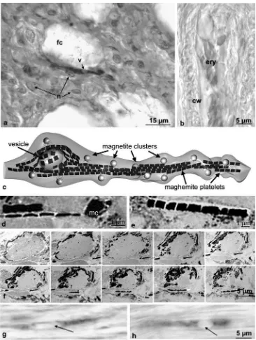
Fig. 2 Characterization and subcellular localization of iron minerals. a,bLight microscopic view of an iron-containing dendrite compared to a blood capillary with clotted erythrocytes after Prussian blue staining (10-μm paraffin section).aThe“clear zone”of the dendrites, the vesicle (arrowatv), where no iron bullets are visible, is not a site of a nucleus. This is shown by double staining with PB and nuclear red, which marks the nuclei (n) of neighboring connective tissue cells (fc, fat cell of the stratum laxum).bErythrocytes (ery) in a capillary vessel (cw, capillary wall). Here, the PB reactivity is much weaker; the nuclei additionally provide a clear distinction of these structures from
beak (Fig. 3a) and are always in bilateral symmetry, namely at the base of the cere, at the height of the distal end of the palatal“slit”, and directly proximal to the bill tip organ.
(d) Orientation of axons: Prominent is the specialized orientation of the axon bundles supporting iron-containing dendrites (Fig. 3c). Serial sections taken in various planes show that, for each of the six areas of the skin, one spatial direction prevails (Fig. 3a): The two most caudal patches have their iron-containing
endings aligned in a caudal-to-rostral direction, the middle patches mostly in a median-to-lateral orienta-tion, and the most frontal patches in a dorsal-to-ventral direction. Thinner fascicles branch into the intermedi-ate directions within the fields.
(e) Regularity: The PB-stained dendrites themselves also form a regular pattern. Several of them may align side by side but, longitudinally, they have a nearlyuniform minimum distance of about 100μm from each other.
This was confirmed in μ-SXRF-measurements of Fig. 3 Spatial distribution and quantity of iron minerals within the
putative magnetoreceptor system. a X-ray image of the upper beak showing its six iron-containing areas and the prevailing orientation of their dendritic fields (arrows:ccaudal,ddorsal,ffrontal,llateral,m median,rrostral,vventral).bCamera lucida drawing of all Prussian blue reactive‘bullets’(black dots) of one dendritic field as visible in the light microscope. (10-μm-thick paraffin sections through the right median field; Prussian blue staining).cAxon bundle, which carries multiple iron-containing dendrites. The dendrites are aligned in parallel and in a minimal distance. (10-μm paraffin section, PB-stained).eFe
mass distribution in a dendritic field seen by μ-SXRF without histological staining (dparallel control section of a ramification, PB-stained). The Fe content of the approximately 25 dendrites (pictured here) amounts to 900 pg. Outside the field, Fe is not detectable. The Fe content of the entire dendritic field can be estimated at 14 ng.f μ-SXRF also helps to identify artifacts based on “standard methods”
histologically undisturbed and unstained material (Stahl et al.2006a).
(f) Two iron minerals: Each of these dendrites contains two types of iron minerals (see Fig.1e,f ) in three different subcellular compartments (Fig. 2c–g), which can be only partly observed in the light microscope: (1) about 10–15 of iron bullets adhering to the cell membrane (Figs. 1a, 2c and3b); (2) one vesicle (diameter about 3–5 μm), which is either spherical or elongated
(Fig. 2g, h) and shows in the light microscope as an iron-void compartment of the dendrite (Fig. 2a). The vesicle is covered by a crust of a non-magnetite iron mineral and contacts the cell membrane (Fig. 2c,f ); and (3) several bands of maghemite platelets (each platelet 1 μm wide and long, less than 0.1 μm thick;
Fig. 2c–e) extend through the dendritic terminal (Fleissner et al. 2003). In the light microscope, after Prussian blue staining, they sometimes may appear as veils or blue shades in semi-thin sections.
The iron-containing structures, although hard and brittle during sectioning, immediately dissolve after contact to weak acids. They may also partly wash out or change their chemistry when fixatives or components of buffers (e.g., the arsenic acid of cacodylate buffer) bind to the unsatu-rated Fe ions (Fig.3e,f), properties that must be considered when analyzing putative structures and key particles of magnetoreceptors.
Discussion
The physico-chemical nature of the two iron oxide particles
Two iron minerals have been found in the putative magneto-sensory dendrites of the pigeon’s upper beak. The general magnetic properties of these iron-based minerals are known. In detail, they may depend, for instance, on their crystallinity and defect concentrations, size and shape, formation, tissue embedding and arrangement (see Stahl et al.2006a,2007b). Therefore, the histological architecture, the subcellular shape, and size of the components are essential to derive sound hypotheses on their respective function:
– In the pigeon’s beak, magnetite occurs as superpara-magnetic nanoparticles (2–4 nm) accumulated in clusters (diameter, about 1μm) within an organic matrix of yet
unknown nature keeping them at a distance. This was shown by TEM imaging in bright- and dark-field mode and by small area electron diffraction (SAED) (Hanzlik et al.2000; Fleissner et al.2003).
– Additionally, a non-magnetite iron material, namely maghemite, constitutes the two other
iron-mineral-containing subcellular components: Maghemite occurs as irregular crust around the vesicle (diameter, 5 μm)
and several bands (each about 10μm long) of square
platelets (1×1×0.1μm3;Fleissner et al. 2003; Stahl et
al.2006b). Three pieces of evidence suggest that these platelets are single crystalline: their (1) chemical stabilityagainst Turnbull’s reaction and especially their (2) precise and uniform shape that fits perfectly with the (3)magnetocrystalline anisotropycharacteristics of maghemite (Stahl et al.2006b; for further background information of the shape-related transition between hard and soft magnetic features in the nanoscale range, see Arcas et al.1998).
The magnetite-containing clusters can be marked as dark blue bullets by histological procedures for the light microscope (with Prussian blue as well as Turnbull’s reaction) due to their FeII and FeIII content and their high surface-to-volume-ratio as nanocrystals. Maghemite as an FeIII mineral should stain with Turnbull’s reaction. The weakness of this reaction, however, seen in the light microscope, may have to do with the crystallinity of the micron-sized platelets and, thus, it does not easily provide free ions. Consequently, the bands of platelets stay
“invisible” in the light microscope and can be seen only in the electron microscope. Reproducible quantitative measurements of the iron distribution (see Fig. 1b–d: element scanning by μ-XRF) in dendrites from different
animal preparations contradict an assumption that these maghemite platelets are only random waste or stock material for the biomineralization of magnetite. Iron is concentrated within the dendrites, only, not outside, where waste products of iron metabolism should be stored (see Fig. 1c,d).
lowest energy (for further details of micromagnetic simu-lations, see Stahl et al. 2007b). The flat quadratic shape perfectly fits the magnetocrystalline anisotropy of maghe-mite that coincides with the direction of the cubic axis (in-plane magnetization with soft magnetic features). These monocrystals have, according to their shape, a vanishing coercive force and can be easily demagnetized (for review: Herzer1997). Perhaps, the prior focus on magnetite crystals as a unifying key for iron-based magnetoreception has occluded the consideration of a possible role of additional non-magnetite minerals, although they have been found in several cases (for review: Kirschvink and Hagadorn2000).
The subcellular components of the sensory unit suggest a specific primary transduction process
The two iron minerals have clearly distinct functions in a three-step transduction process: The iron-crusted vesicle concentrates the magnetic flux, and the magnetic behavior of the maghemite platelets yields the specific input stimulus, whereas the opening of membrane channels results from the strain by magnetite bullets. The complex elongated entity of iron minerals in the sensory dendrites of the pigeon’s beak is proposed to reversibly acquire a net magnetization, when in parallel to the magnetic field, and to relax, when the field is rotated. On the ground of their presumed soft magnetic behavior, the bands of
maghemite platelets may become reversibly magnetized (see also Arcas et al. 1998) and then would be able to locally amplify the Earth’s magnetic field. The magnetite
clusters may react to this local field enhancement by the stabilization of their net magnetic moment; that is, a preferred direction of the nanomagnets inside the clusters is established. This could lead to a displacement or, in higher order, a deformation of the clusters. Thus, a magneto-mechanical transduction in the dendrite mem-brane may be initiated by these displacing forces. In the amplified Earth’s magnetic field, the induced net magnetic moment of the centrally located bands of maghemite platelets rather exerts an isometric pull to the clusters vertical to the membrane, than yielding a real displace-ment or deformation, which clearly would need more energy. This model matches the actual concept of a ligand-bound mechanoreception (for review: Kung2005).
In earlier papers, two other principles have been proposed for the transduction process in iron-based mag-netoreceptors, which are clearly distinct from our concept of the pigeon-type magnetoreceptor:
(1) A solid permanent magnet of single-domain magnetite: Such an iron bar could rotate like a compass needle with the changing magnetic field and induce the primary mechanosensory processes by this torque
(Diebel et al.2000; Kirschvink and Gould1981). So far, this hypothetic structure could not be described down to the subcellular level of a sensory system, although magnetite has been localized in the nervous system of various organisms (for review: Kirschvink et al. 2001). Another aspect is that such a sensor could only serve as magnetic compass and would not help to understand the sensory principles of a magnetometer.
(2) Ferrofluid-like behavior: The magnetite clusters in the pigeon’s beak might react to changes of a magnetic field like ferrofluid droplets (Bacri et al.1982). Based on model experiments with technical ferrofluids (typical particle size 15–20 nm), a distortion (Winklhofer et al.2001) or dislocation (Davila et al.
2003) is assumed to simulate the behavior of magne-tite clusters in the pigeon’s beak, despite the lack of data on their visco-elasticity. These ferrofluid-like processes should yield early receptor potentials via excitation of strain-sensitive membrane channels. The ferrofluid concept, with proposed forces onto the cell membrane, however, does not fit to the arrangement and morphology of the iron mineral compartments found in the pigeon dendrites. Additionally, all model calculations with these artificial ferrofluids (Shcherbakov and Winklhofer 1999) contradict the assumption that such a process can explain the behav-iorally and electrophysiologically observed high sensi-tivity of the magnetic sense in pigeons and other birds with the given size and shape of subcellular compart-ment in the pigeon’s iron-mineral-containing dendrites.
Therefore, a completely new receptor hypothesis, in-cluding enhancement and filter mechanisms, is necessary.
The sensory system in the pigeon’s beak provides evidence for signal filtering, receptor adaptation, and reset
mechanisms
Tracking down magnetic sensing requires more than the occurrence of magnetic iron minerals—which are reported to be distributed in, e.g., diseased nervous tissue (Quintana et al. 2006)—and various body parts (Abracado et al.
functional context. An essential prerequisite for addressing a structure as a magnetoreceptor candidate is that magnetic entities occur in a specific well-ordered assembly. Further-more, the iron-based entities have to be identified not only as “iron” but also by their crystalline structure, magnetic features, oxidation states, and correlation to other chemical elements to predict their response to physiologically relevant changes of the Earth’s magnetic field.
The putative magnetoreceptor system in the pigeon’s beak may function like a sensitive biological magnetometer, with special structural features standing for the encoding field intensity and inclination as well.
Specificity of the receptor The specificity of the receptor system is achieved, again, by a two-step process. Each dendrite is preferably excited by the magnetic field in one axis only:
(1) The iron-mineral-containing dendrites of the beak skin are of similar size, shape, and ultrastructure. In principle, they will become strongly magnetized when the field lines are exactly in parallel to their long axis and will be transformed to an about-20-μm-long
magnetic dipole. This assumption matches the finding that those dendrites, which are attached to the same axon bundle, obviously have to keep a minimum distance from each other, as could be seen inμ-SXRF
measurements (Stahl et al.2006a).
(2) If not disturbed by sectioning or other histological procedures, the dendrites appear in parallel to guiding axon bundles, aligned in series, ready to cooperatively register information on the strength of the magnetic vector in a specific direction. These dendritic arrange-ments along axon bundles in the three different spatial orientations seem to be adapted to analyze separately the three vector components of the magnetic field: The bundles are arranged about perpendicular to each other in a bilateral symmetry.
Selectivity and filter processes Whether the main axes of the dendritic areas are strictly in parallel to the body axes or tilted by a certain angle is a still open question and will be analyzed in unsectioned samples with a high resolution, e.g., by μ-SXRF with microfocus capillaries. The
bilater-ally symmetrical arrangement is a general phenomenon observed in all sensory systems and often recognized as essential for spatial localization of stimuli. This kind of selectivity would also constitute a way to reduce the influence of noise or interfering signals. It has to be analyzed whether the tiny bundles with dendrites deviating from the preferred direction serve this filtering function or establish a neuronal bias (P. Schlegel, personal communication).
Amplification The sensitivity of the sensory system has not yet been investigated, but several peripheral and also central nervous amplification processes may be assumed: The magnetite clusters inside the dendrite would be attracted by the magnetized maghemite bands and exert a simultaneous multiple mechanical strain perpendicular to the membrane, which is depending on both magnetic field intensity (amplified by maghemite) as well as the distance between magnetite bullets and maghemite bands (maximal-ly 1μm due to the diameter of the dendrite), resulting in a
composite receptor potential. Additionally, all dendrites, as the smallest sensory units, with the same directionality, are similarly affected and might, by summation, serve to amplify the magnetic signal encoding field strength parallel to their axis. A deviating field direction will yield a decreased receptor signal in these dendrites, which explains the results found in the earlier electrophysiological experi-ments (e.g., Beason and Semm 1996). Pilot calculations based on the histological data have already shown that the magnetic field effects inside the dendrites might well be strong enough to satisfy these assumptions (Solov'jov and Greiner personal communication). More precise predictions concerning dynamics and working range are not yet possible with the actual state of the mathematical codes.
Reset and adaptation The role of the iron-crusted vesicle is still unclear in this context. It expands through the entire diameter of the dendrite and may react to magnetic field changes, e.g., flatten, when the field lines are in parallel to the long axis of the dendrite. Whether this observed deformation may contribute to the threshold of the primary receptor processes or whether it is part of a complex magneto-mechanical feedback loop, serving as a driving or reset mechanism, must be analyzed in the future. It is an interesting idea to assume that, by such a mechanism, the dendrite may support an adaptation of the system to various background field intensities, which differ depending, e.g., on the geographical position of the homeloft.
(for review: Dubbeldam 1998), its multimodal sensory endings only in the skin of the upper beak. Hence, we could not detect similar iron-containing structures in the lower beak (Fleissner, unpublished).
So far, it is unknown which trigeminal ganglia may represent the primary targets of these putatively magneto-receptive afferents of the pigeon. In a pilot study with mole rats, Nemec et al. (2001) showed layered accumulation of c-fos
expression in the superior colliculus after magnetic stimuli. In any case, the proposed magnetoreceptor is a complex system. This means that it can fulfil its function only when it is intact. Neurophysiological experiments in the periph-ery, for example electrophysiological recordings in one of the dendritic fields or at the afferent nervous connections of the ROM, must take into account the specificity of the respective sensory endings. The stimuli must be scanned to determine the optimal direction of field lines; otherwise, the single receptor responses might be small or non-detectable. Beason and Semm (1987) assumed that only about 10 to 20% of ROM fibers respond to magnetic field stimulation with action potentials or changes of the spontaneous discharge rate. Therefore, this is no indication for the real number of magnetoresponsive fibers. Further lesion and also tracing experiments might contribute to solve the multidimensional puzzle of sensory afferents in the future.
The magnetoreceptor model of homing pigeons has now several consequences:
– If the iron-containing structures are sensory endings, information processing does not occur along the axonal pathway from beak to brain. The separate peripheral
“channels” encoding the three spatial components of the magnetic field have to be composed to the resultant vector in the central nervous neuropils—an important fact for the (re)evaluation of all electrophysiological recordings and lesion experiments.
– As soon as the bird’s head turns relative to the Earth’s magnetic field, the generated receptor signals would decrease or increase depending on the receptor subunits, whereas the resulting total field vector as a composition of these three components will stay the same.
– As soon as the magnetic field changes either its intensity or inclination, the complex receptor system will signal this change as well.
The sensory units are dendrites of distinct axonal connections with an established relevance for magnetic orientation
It is still an open question how the described dendritic system in the beak might contribute to magnetic orientation and how information from this system has to interact with
the compass mechanism possibly located in the retina (for review: Wiltschko and Wiltschko 2005) or other sensory systems used in navigation. Our new model for a magnetic field receptor in the beak offers a reliable new approach to further elucidate these complex problems.
Our paper does not aim to solve the problem of magnetic field guided behavior; rather, it tries to describe a complex structure that is not an accidental feature but based on general physiological and physical principles and that, thus, provides a most promising candidate for a magnetic field sense. Indirect evidence for its function may be derived from behavioral experiments. The sophisticated architecture of the iron-containing dendrites in the pigeon’s beak, which we have described above, lead us to assume that this system might be an indicator of the magnetic field strength which implies directional information as well. This constitutes then a new compass mechanism independent of photore-ception. Several observations on the loss of an inclination compass by migratory birds during experiments with increased light intensity still reveal a so-called “fixed direction” (Wiltschko et al. 2003) of orientation. This phenomenon is interpreted as a polar compass, which is independent of light but follows changing magnetic field conditions. The features of the iron-based system in the beak would match such an additional compass mechanism. Magnetic pulse experiments with adult animals (e.g., birds: Wiltschko et al. 2002; turtles: Irwin and Lohmann 2003,
2005) result in a changed heading for home, possibly due to a partial “blinding”of the direction finding system. If the pulse is applied under a certain angle, direction finding changes accordingly. Recovery from this directional effect is reported to take several days and to pass through a period of misorientation. Our model of a separate recording of the vector components of the magnetic field matches these results. We still do not know whether the dendritic structure or entire information processing is affected negatively by the pulse experiments. Replications of similar experiments under well-controlled conditions need to be followed by histological and physical controls. Lesions to the median branch of the trigeminal nerve in pigeons destroy their ability to recognize a magnetic anomaly. This was shown in conditioning experiments by Mora et al. (2004) and even in field experiments with local anesthesia of the upper beak skin (Wiltschko and Wiltschko, personal communication).
Based on our ongoing histological studies, we expect that the pigeon-type receptor system in the skin of the upper beak might turn out to be a universal feature of all birds, as we have found similar iron-containing dendrites in robins, garden warblers, and chicken (Stahl et al.2007a), although these birds have to solve quite distinct navigational tasks.
expected. However, factors such as motivation (for exam-ple, hunger or search for the companion), physiological state (for example, migratory restlessness), multimodal stimulation (for example, simultaneous activation by flight control systems), and also the age-related maturing of the sensory structures must be consequently considered in all kinds of neurobiological experiments, also in those analyz-ing the nature and meananalyz-ing of magnetoreceptive systems.
Acknowledgments W. and R. Wiltschko (University Frankfurt a. M.) and P. Schlegel (University Munich) gave helpful comments on data presented in this paper and discussed the theories supporting our model. P. Brownell (University Corvallis, Oregon) critically com-mented on the receptor physiological part of the model. We acknowledge the support by M. Wilke (University Potsdam) during the μ-XANES experiments and C. Taylor-Dorenkamp (University Boston) who helped us improve the English version of the text. We also thank the reviewers of an earlier version of the manuscript who gave helpful and encouraging comments. We thank E. Thielen, M. Stöhr (both at University Frankfurt a. M.) and W. Hofer (MPI Hirnforschung, Frankfurt a. M.) for their expert technical help in the histology labs. This project is supported by grants from the Deutsche Forschungsgemeinschaft to G. F. (Fl 177/15-1) and HASYLAB at DESY Hamburg to B.S. (I-04-012, I-05-095). All experimental procedures followed the legal requirements of the German law for the protection of animals.
References
Abracado LG, Esquivel DM, Alves OC, Wajnberg E (2005) Magnetic material in head, thorax, and abdomen ofSolenopsis substitutaants: a ferromagnetic resonance study. J Magn Reson 175:309–316 Arcas J, Hernando A, Barandiaran JM, Prados C, Vazquez M, Marin
P, Neuweiler A (1998) Soft to hard magnetic anisotropy in nanostructured magnets. Phys Rev B 58:5193–5196
Bacri JC, Salin D, Massart R (1982) Study of the deformation of ferrofluid droplets in a magnetic field. J Physique Lett 43:L179–L183 Bazylinski DA (1999) Synthesis of the bacterial magnetosome: the
making of a magnetic personality. Int Microbiol 2:71–80 Beason RC (2005) Mechanisms of magnetic orientation in birds.
Integr Comp Biol 45:565–573
Beason RC, Semm P (1987) Magnetic responses of the trigeminal nerve system of the bobolink (Dolichonyx oryzivorus). Neurosci Lett 80:229–234
Beason RC, Semm P (1996) Does the avian ophthalmic nerve carry magnetic navigational information? J Exp Biol 199:1241–1244 Becker M (2000) Die Nutzung des Erdmagnetfeldes innerhalb der
Navigation von Brieftauben (Columba livia). PhD thesis, Universität Frankfurt a. M
Davila A, Fleissner G, Winklhofer M, Petersen N (2003) A new model for a magnetoreceptor in homing pigeons based on interacting cluster s of superparamagnetic magnetite. Phys Chem Earth 28:647–652
Diebel CE, Proksch R, Green CR, Neilson P, Walker MM (2000) Mag-netite defines a vertebrate magnetoreceptor. Nature 406:299–302 Dubbeldam JL (1998) The sensory trigeminal system in birds: input,
organization and effects of peripheral damage. A review. Arch Physiol Biochem 106:338–345
Fleissner G, Stahl B (2005) Magnetrezeption bei Brieftauben. In: Rossmann T, Tropea C (eds) Bionik. Springer, Berlin, pp 501–515 Fleissner G, Holtkamp-Rötzler E, Hanzlik M, Winklhofer M, Fleissner G, Petersen N, Wiltschko W (2003) Ultrastructural analysis of a
putative magnetoreceptor in the beak of homing pigeons. J Comp Neurol 458:350–360
Fransson T, Jakobsson S, Johansson P, Kullberg C, Lind J, Vallin A (2001) Bird migration: magnetic cues trigger extensive refuelling. Nature 414:35–36
Hanzlik M, Heunemann C, Holtkamp-Rötzler E, Winklhofer M, Petersen N, Fleissner G (2000) Superparamagnetic magnetite in the upper beak tissue of homing pigeons. BioMetals 13:325–331 Herzer G (1997) Nanocrystalline soft magnetic alloys. In: Buschow KHJ (ed) Handbook of magnetic materials, vol 10. Elsevier, Amsterdam, pp 415–462
Irwin WP, Lohmann KJ (2003) Magnet-induced disorientation in hatchling sea turtles. J Exp Biol 206:497–501
Irwin WP, Lohmann KJ (2005) Disruption of magnetic orientation in hatchling loggerhead sea turtles by pulsed magnetic fields. J Comp Physiol A 191:475–480
Janssens KHA, Adams FCV, Rindby A (2000) Microscopic X-ray fluorescence analysis. Wiley, Chichester
Johnsen S, Lohmann KJ (2005) The physics and neurobiology of magnetoreception. Nat Rev Neurosci 6:703–712
Kalmijn AJ (1978) Experimental evidence of geomagnetic orientation in elasmobranch fishes. In: Schmitt-Koenig K, Keeton WT (eds) Animal migration, navigation and homing. Springer, Berlin, Heidelberg New York, pp 347–353
Keeton WT, Larkin TS, Windsor DM (1974) Normal fluctuations of the earth’s magnetic field influence pigeon orientation. J Comp Physiol 95:95–103
Kirschvink JL, Gould JL (1981) Biogenic magnetite as a basis for magnetite-based magnetic field detection in animals. Biosystems 13:181–201
Kirschvink JL, Hagadorn JW (2000) A grand unified theory of bio-mineralisation. In: Bäuerlein E (ed) The biomineralisation of nano-and microstructures. Wiley-VCH Verlag, Weinheim, pp 139–150 Kirschvink JL, Walker MM, Diebel CE (2001) Magnetite-based
magnetoreception. Curr Opin Neurobiol 11:462–467
Kung C (2005) A possible unifying principle for mechanosensation. Nature 436:647–654
Mann S, Sparks NH, Walker MM, Kirschvink JL (1988) Ultrastruc-ture, morphology and organization of biogenic magnetite from sockeye salmon,Oncorhynchus nerka: implications for magneto-reception. J Exp Biol 140:35–49
Marhold S, Burda H, Wiltschko W (1997) A magnetic polarity compass for direction finding in a subterranean mammal. Naturwissenschaften 84:421–423
Mora CV, Davison M, Wild JM, Walker MM (2004) Magneto-reception and its trigeminal mediation in the homing pigeon. Nature 432:508–511
Mouritsen H, Ritz T (2005) Magnetoreception and its use in bird navigation. Curr Opin Neurobiol 15:406–414
Nemec PJ, Altmann J, Marhold S, Burda H, Oelschläger HHA (2001) Neuroanatomy of magnetoreception: the superior colliculus involved in magnetic orientation in a mammal. Science 294: 366–368
Phillips JB, Borland SC (1992) Behavioural evidence for use of a light-dependent magnetoreception mechanism by a vertebrate. Nature 359:142–144
Quintana C, Bellefqih S, Laval JY, Guerquin-Kern JL, Wu TD, Avila J, Ferrer I, Arranz R, Patino C (2006) Study of the localization of iron, ferritin, and hemosiderin in Alzheimer’s disease hippocam-pus by analytical microscopy at the subcellular level. J Struct Biol 153(1):42–54
Ritz T, Adem S, Schulten K (2000) A model for photoreceptor-based magnetoreception in birds. Biophys J 78:707–718
Shcherbakov VP, Winklhofer M (1999) The osmotic magnetometer: a new model for a magnetite-based magnetoreceptor in animals. Eur Biophys J 28:380–392
Stahl B, Fleissner G, Barnert E, Falkenberg G (2006a) Element scanning by μXRF in putative magnetic field receptors in the upper beak skin of homing pigeons. HASYLAB Annual Report 2005. DESY, Hamburg, pp 1029–1030
Stahl B, Fleissner G, Falkenberg G, Fleissner G (2006b) Magnetite nanoparticles alone are not able to explain iron mineral-based magnetoreception in homing pigeons. In: Kyriakopoulos A, Michalke B, Graebert A, Behne D (eds) Proceedings of the 4th fall conference on metalloproteins and metalloidproteins. Herbert Utz Verlag, München, pp 63–68
Stahl B, Fleissner G, Fleissner G, Falkenberg G (2007a) Cross-species unveiling of a putative avian magnetoreceptor. HASYLAB Annual Report 2006. DESY, Hamburg, 1269–1270
Stahl B, Fleissner G, Fleissner G, Holub-Krappe E (2007b) Micromagnetic aspects of magnetoreception of homing pigeons based on iron mi-nerals. XAFS 13 Proceedings, American Inst Physics 882:755–757 Walker M, Diebel CE, Haugh CV, Pankhurst PM, Montgomery JC,
Green CR (1997) Structure and function of the vertebrate magnetic sense. Nature 390:371–376
Wang JH, Shaun SD, Cain D, Lohmann KJ (2003) Identification of magnetically responsive neurons in the marine molluscTritonia diomedea. J Exp Biol 206:381–388
Wilke M, Farges F, Petit PE, Brown GE Jr, Martin F (2001) Oxidation state and coordination of Fe in minerals: an Fe K XANES spectroscopic study. Am Mineral 86:714–730
Wiltschko R, Wiltschko W (1995) Magnetic orientation in animals. Springer, Berlin, Heidelberg New York
Wiltschko W, Wiltschko R (1997) Magnetic orientation in birds. J Exp Biol 199:29–38
Wiltschko W, Munro U, Ford H, Wiltschko R (2003) Magnetic orientation in birds: non-compass responses under monochromatic light of increased intensity. Proc R Soc Lond B 270:2133–2140
Wiltschko W, Wiltschko R (2005) Magnetic orientation and magneto-reception in birds and other animals. J Comp Physiol A 191:675–693 Wiltschko W, Munro U, Wiltschko R, Kirschvink JL (2002) Magnetite-based magnetoreception in birds: the effect of a biasing field and a pulse on migratory behavior. J Exp Biol 205: 3031–3037 Winklhofer M, Holtkamp-Rötzler E, Hanzlik M, Fleissner G, Petersen