Document heading doi: 10.1016/S2222-1808(13)60071-7 襃 2013 by the Asian Pacific Journal of Tropical Disease. All rights reserved.
A
ntibacterial and brine shrimp lethality effect of marine actinobacterium
S
treptomyces
sp.
CAS72
against human pathogenic bacteria
Palaniappan Sivasankar*,Panchanathan Manivasagan, Packiyaraj Vijayanand, Kannan Sivakumar, Shanmugam Sugesh, Subramaniam Poongodi, Viswanathan Maharani, Shanmugam Vijayalakshmi, Thangavel Balasubramanian
Faculty of Marine Sciences, Centre of Advanced Study in Marine Biology, Annamalai University, Parangipettai -608 502, Tamil Nadu, India
Asian Pacific Journal of Tropical Disease
journal homepage: www.elsevier.com/locate/apjtd
*Corresponding author: Sivasankar Palaniappan, Faculty of Marine Sciences, Centre of Advanced Study in Marine Biology, Annamalai University, Parangipettai -608502,
Tamil Nadu, India.
Tel: +914144243223235 /+919944376973
Fax: +914144243223
E-mail: [email protected]
Foundation Project: Faculty of Marine Science - MoES-COMAPS (XIIth Plan) for providing financial assistance through fellowships with No. G4(1)/11742/2012.
1. Introduction
Microbial bioactive molecules are the source for the development of new medicines. Bio-resources of the ocean has exploited over many decades for microbial bioactive molecules to safeguard our lives. The bioactive molecules
have wide applications in agriculture, veterinary and pharmaceutical industry. As for as microbes concern, a group of actinobacteria comes within the order of
Actinomycetales, have the potential to produce many different biologically active secondary metabolites such as antibiotics, herbicides, pesticides, anti-parasitic
PEER REVIEW ABSTRACT
KEYWORDS
Actinobacteria, Brine shrimp lethality, Uppanar estuary
Objective:To investigate the in vitro antibacterial activity against human pathogenic bacteria and brine shrimp lethality bioassay of the marine actinobacterium.
Methods: Forty six marine actinobacterial strains were isolated from sediment samples of
Uppanar estuary, Cuddalore, India. Preliminary screening was done by cross-streak method and the potential strain was identified by morphological, chemotaxonomical and molecular methods. Fermentation was done and the metabolite was obtained by liquid-liquid extraction using ethyl acetate and purified by silica gel (100-200 mesh) column chromatography. The purified metabolite was tested for antibacterial activity, minimal inhibitory concentration and brine shrimp lethality bioassay.
Results: Among the forty six strains, CAS72 was found effective against human pathogenic
bacteria. The strain CAS72 was identified as Streptomyces sp. The purified metabolite exhibited a significant in vitro antibacterial activity. The MIC value was also determined against human pathogenic bacteria and a strong cytotoxic activity in brine shrimp lethality assay was observed and the LC50value was 23.5µg/mL.
Conclusions: The present investigation reveals that the marine actinobacteria are well obtainable in Uppanar estuary environment and it can provide a definite source for novel bioactive metabolites.
Contents lists available at ScienceDirect
Peer reviewer
Dr. K. Senthilkumar. Ph.D, Post
Doctoral Fellow, Marine Biochemistry
Laboratory, Department of Chemistry,
Marine Bioprocess Research Center,
Pukyong National University, 599-1
Daeyeon 3-dong, Nam-gu, Busan,
608-737, South Korea.
E-mail: [email protected]
Comments
This is a good study in which authors focused on specific area.
Marine actinobacteria was isolated from estuary. From this 46 marine
actinobacterial strains 23 isolates
was isolated and screened. Then secondary metabolites were isolated, characterized, purified and checked its different antimicrobial activites compared with standard antibiotic nalidixic acid.
Details on Page 292
Article history:
Received 10May 2013
Received in revised form 15May, 2nd revised form 25May, 3rd revised form 15Jun 2013
Accepted 1Jul 2013
compounds and enzymes like cellulose, xylanase[1]. Among these compounds, antibiotics are commercial important and therapeutically valued products[2]. Hence, the actinogenic molecules are the threshold for novel drug discovery.
Among the filamentous actinobacteria group,
Streptomycetes have been realized as new opportunity for novel drug discovery[3]. These organisms are prominent in producing new secondary metabolites than other common actinobacteria[4]. Streptomyces can produce biologically active molecules of pharmaceutical use, as well as the development of other valuable products like enzymes, nutraceuticals and cosmetics[5]. Of these, 75% of the molecules derived from the genus of Streptomyces and more than 5000 known bioactive compounds documented
in this genus[6]. In the present study, marine actinobacteria were isolated from the sediment of Uppanar estuary,
Cuddalore, India, and tested for its brine shrimp lethality and antibacterial potential against human pathogenic bacteria. In addition, an attempt made to identify the strain up to species level with morphological, physiological and chemo-taxonomical analysis.
2. Materials and methods
2.1. Chemicals and reagents
All the solvents used in the experiment and silica gel for thin layer chromatography (TLC) were obtained from Sigma-Aldrich, USA. All bacteriological media components were purchased from Hi-Media (Mumbai, India). All other chemicals and reagents were extra pure grade.
2.2. Pathogenic strains
The human pathogenic bacteria viz., Escherichia coli
-MTCC 1610, Klebsiella pneumoniae-MTCC 4030, Listeria
monocytogenes-MTCC 839, Salmonella enterica-MTCC
9844, Shigella flexneri-MTCC1457, Staphylococcus aureus
-MTCC 3160, Staphylococcus haemolyticus-MTCC 3383,
Streptococcus pneumoniae-MTCC 1935, Vibrio cholerae
-MTCC3904 and Vibrio vulnificus-MTCC1145 were procured
from Microbial Type Culture Collection, Institute of
Microbial Technology, Chandigarh, India during March
2008 for evaluating the antagonistic potential of the marine
actinobacteria. The lyophilized cells were taken from the vials, inoculated in nutrient broth, and incubated at 37°C
for 24 h. After incubation, the human pathogenic bacteria were sub cultured in nutrient agar slants and stored under refrigeration till the next use.
2.3. Sample collection and enrichment
Sediment samples were collected from five different stations of Uppanar estuary, Cuddalore, India during
2008 in summer. The collected sediment samples were transferred in sterile polyurethane bags tightly covered and stored in the refrigerator until use. One gram of each sediment sample was added in 10 mL of sterile distilled water and stirred for 15 min[7]. The suspension had been allowed to stand for 30 min and the supernatant was
separated. Prior to isolation, the samples were enriched by a modified method proposed by Shirling[8]. The supernatant was transferred to conical flasks containing
100 mL of sterile starch casein broth. Samples incubated at 28依2 °C for 7 d in static condition with intermittent
shaking.
2.4. Isolation, media and culture conditions
Isolation, 0.1 mL of the enriched media was spread plated onto a starch casein agar medium (g/L): starch 10,
casein 0.3, KNO32, NaCl2, K2HPO42, MgSO4.7H2O0.05, and CaCO30.02, FeSO4.7H2O 0.01 and agar 18supplemented
with Nystatin 10 µg/mL and Nalidixic acid 25 µg/mL[9,10].
The plates were incubated at 28依2 °C for seven to ten d
and colonies appeared on the plate were purified and maintained in SCA slants for further studies. The isolated marine actinobacterial strains were segregated based on their morphological differences, labeled with numbers for individualization and used for preliminary antibacterial screening.
2.5. Preliminary screening for antibacterial activity
Antibacterial activity of the isolated marine actinobacterial strains were tested against the above mentioned human pathogenic bacteria by a modified cross streak method[11]. Single streak of actinobacteria was made on the surface of the Modified Nutrient agar
(50%NA+50%SCA) as a straight line in the left side corner
of the petriplate and incubated at room temperature (28
依2) °C. After observing a good ribbon-like growth of
the actinobacteria on the petriplates, the pathogenic bacteria were streaked at right angles to the original streak of the actinobacterial isolates and incubated at
(28依2) °C. The zone of inhibition was measured after 24
2.6. Identification of potential marine actinobacteria
2.6.1. Morphological and physiological characteristics
The potential strain CAS72 was taken for further
identification. Cultural characteristics of the strain CAS72
was studied by growing the strain on various International
Streptomyces Project media[8]. The morphology of the spore bearing hyphae with entire spore chain was examined under light microscope by using inclined cover slip technique[12]. Spore chain morphology and ornamentation of the spore surface was examined by using scanning electron microscope[13]. The spore mass colour was visually examined and estimated. Analysis of carbon source utilization was done by following the protocol of Pridham and Gottlieb[14].
2.6.2. Chemo-taxonomical analysis
Whole cell hydrolysate was used to analyze the cell wall amino acids and sugars. The diaminopimelic acid isomers and whole cell sugars were analysis and was done by the method of Kämpfer[15]. Based on these analysis the strain CAS72 was tentatively grouped in Streptomyces genus. To precise the strain in genus and species level, molecular identification was performed.
2.6.3. Molecular identification of strain CAS72
2.6.3.1. DNA extraction
Genomic DNA extraction was done by modifying the method of Everest et al[16]. Actinobacteria strain was grown in 20 mL of TSB media for 7 d. Cells were harvested by centrifugation (8000 r/min for 2 min), washed with 500 µL
of 10mmol/LTris-HCl/1 mmol/LEDTA buffer (pH7.7) and
resuspended in 500µLTE buffer (pH 7.7). The sample was kept in boiling water bath for 10 min, cooled for 5 min and
centrifuged (8000 r/min for 3 min). The supernatant (300µL)
was transferred to a clean tube and stored at 4°C.
2.6.3.2. PCR amplification
The isolated DNA was purified and amplified according to Karuppiah et al[17]. A 50 µL amplification reaction
contained 1 µL template DNA (50-200 ng), 5 µL 10×PCR buffer, 1µL each PCR primer (20 mmol/L)(27F, 1492R), 1µL
dNTP mix (10 mmol/L), 6 µL MgCl2(25 mmol/L), 2.5 U Taq
DNA polymerase, 5 µLDMSO(5%) and 29µL sterile MilliQ
water. The reaction conditions were initial denaturation at 95 °C for 5 min, followed by 30 cycles of denaturation
at 95°C for 30 seconds, annealing at 55°C for 30 seconds
and extension at 72 °C for 90 seconds. A final extension
was performed at 72°C for 10 min. Reaction products were
electrophoresed on a 1% agarose gel and the bands were
visualized with ethidium bromide under UV light.
2.6.3.3. Sequencing and analysis of 16S rRNA gene
The amplified PCR product was purified. Both strands of
16S rRNA gene were sequenced using a Terminator Cycle
Sequencing kit-ABI Prism 310 Genetic Analyzer (Applied
Biosystems, USA). To assess the similarity of the sequence
BLAST (http: //www. ncbi.nlm.nih.gov/blst) program was
used and the sequence was submitted in GeneBank.
2.7. Fermentation and extraction of bioactive metabolites
The potential strain CAS72 was grown at (28依2) °C in
starch-casein agar for one week. Spores were collected from the slant culture with 10 mL of the same medium broth. 1 mL of the spore suspension was transferred to 100
mL of SGG production medium (Glucose 1.0 g, glycerol 1.0 g, corn steep powder 0.25 g, peptone 0.5 g, soluble
starch 1.0 g, yeast extract 0.2 g, CaCO30.3 g, NaCl 0.1 g
and pH 7.3) advised by Goodfellow and Fiedlerfor novel
drug synthesis[4]. The media was incubated at (28依2) °C
for 6 d on a rotary shaker (120 r/min). After incubation, the culture broth was centrifuged at 10000伊g for 30 min
at 4 °C by maintaining all the physicochemical factors at
optimum levels for the culture. Growth and production were estimated at the temperature, 30 °C, pH, 7.5 and sodium
chloride concentration, 2.0%. Extracellular metabolites
extracted from the supernatant by liquid-liquid extraction, the filtrate was transferred aseptically into the conical
flask. An equal volume of ethyl acetate was added separately to the cell-free culture filtrate and shaken for 2
h[18]. Metabolite extraction was carried out using the ethyl acetate solvent on the basis of best solubility and maximum antibacterial activity. The metabolite in the solvent was evaporated in a rotary evaporator, lyophilized and stored for further use.
2.8. Purification
The crude metabolite (5 mg) was chromatographed on
silica gel (Merck, particle size 0.100-0.200 mm) column (22
伊5 cm) and eluted with chloroform-ethyl acetate (70:30) to
give 10 major fractions (Fr SMI-X). The dried residues of all the 10 major fractions were dissolved, each in a specified
volume of ethyl acetate, to give 1 mg/mL concentration and tested for their antibacterial activities against the test organisms by well-diffusion method. Among the 10
fractions, SMV fraction exhibited good activity. The active fraction SMV was purified by chromatography on Sephadex
LH20(Ethyl acetate) to obtain fractions SMVa to SMVe. The
fraction SM-Vd was found to possess good antibacterial activity. This active fraction SM-Vd was further purified by chromatography on Sephadex LH20 (Ethyl acetate)
were scanned in a spectrophotometer (UV-1800-Shimadzu scientific instruments, USA) to determine their absorption spectra of UV-visible light at wavelengths ranging from
200 to 900 nm. All the extracts and fractions were subjected to Thin Layer Chromatography (TLC) on silica gel (60F254
MERCK; 25TLC aluminium sheets 20伊20 cm MERCK) and
run in chloroform-ethyl acetate (70:30) to visualize the
efficiency of separation of UV fluorescent substances. Each batch was repeated several times to confirm the results.
2.9. Antibacterial activity
The purified active fraction SM-Vd was dissolved in
DMSO at different concentrations (25-100µg/mL) and used to
determine antibacterial activity by a modified well diffusion assay[19], against the above listed human pathogenic bacteria. For comparison, Nalidixic acid (100 µg/mL) was
used as standard antibiotic and a well filled with DMSO as negative control. Mueller-Hinton Agar (Difco) was used for
this study. The plates were incubated at 37°C for 18 h. The
antibacterial activity of the purified fraction was determined by measuring the respective zones of inhibition (mm).
2.10. Determination of minimal inhibitory concentration
The antibiotic susceptibility testing of the broth micro dilution was carried out by modifying the method of Al
Momani et al[20].A96 well micro dilution tray was used; the active fraction was dissolved in DMSO in the concentration of 50µg/mL and thoroughly mixed. Five different (volume)
concentrations viz. 1 µg/mL, 2 µg/mL, 4 µg/mL, 8 µg/
mL and 16 µg/mL were used to evaluate the efficiency
of antibacterial activity against the clinical isolates. The wells are then inoculated with a standardized bacterial suspension 5×10⁵ CFU/mL using disposable device that transfers 0.01-0.05 mL into each well of the micro dilution tray. Wells containing broth alone was used as control. After
16–20 h of incubation, MICs are determined by examining the tubes macroscopically for visible evidence of bacterial growth in the form of turbidity. Wells containing the least concentration of metabolite showing no visible sign of growth was taken as the minimum inhibitory concentration.
2.11. Brine shrimp lethality bioassay
Cytotoxic activity of the purified metabolite was evaluated by using brine shrimp (Artemia salina)[21]. The eggs were hatched in seawater. A constant oxygen supply was maintained throughout the process. Mature nauplii were used for this study. The metabolite was dissolved in
DMSO(10 mg/mL) was applied to the nauplii in each vial.
However, not more than 50µL of DMSO was added to the
vials containing the brine shrimp. For each concentration, a vial containing the same volume of DMSO plus seawater and brine shrimp was used as control. The number of survivors was counted after 24 h. Based on these data, percentage of lethality of the brine shrimp nauplii was calculated. From this value, the LC50 of the sample was determined.
3. Result
In the course of our screening program for antagonistic actinobacteria, we isolated 46 marine actinobacterial
strains. The strains were screened for antibacterial activity.
From the preliminary screenings, 23 isolates showed
antagonistic activity against human pathogenic bacteria.
Of the 23 isolates, strain CAS72 showed higher inhibition
potential against human pathogenic bacteria. The potential antagonistic actinobacteria was identified further. It was studied for physiological and morphological characteristics
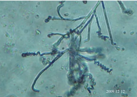
(Table 1). In general, aerial mycelium was red; Sporophores were spirales (Figure 1). Conidia were oblong, the surface
of which was smooth (Figure 2). The chemotaxonomy of the strain CAS72 showed LL-diaminopimelic acid with glycine in their cell wall and no characteristic sugar pattern was observed. Hence, the strain belongs to the cell wall type-I.
Based on these observations, the strain was grouped in to
Streptomyces genus. Further, the strain was analyzed for its
16S rRNA gene, sequenced and identified as Streptomyces sp. CAS72(Accession number: JQ425073).
Table 1
Phenotypic characteristics of the Streptomyces sp.CAS72.
Characteristics Streptomyces sp.CAS72
Growth on ISP2 Good
Growth on ISP4 Good
Growth on ISP7 Poor
Aerial spore mass colour Red
Spore chain Spirales
Spore surface Smooth
Melaniod pigment +
Reverse side pigment Reddish orange
Soluble pigment Violet
Growth on sole carbon sources
D-Glucose +
L-Arabinose +
D-Xylose +
Inositol +
D-Mannitol +
D-Fructose +
Rhamnose +
Sucrose +
Raffinose +
20091212 Figure 1. Spirales spore chains of the actinobacteria Streptomyces sp.
CAS72 using light microscopy at 100伊.
Figure 2. Smooth surface of spirales spores of the Streptomyces sp.
CAS72 viewed under Scanning Electron Microscopy at 5000伊. 20kv 伊5 000 5µm 0000 19 24 SEI
The Streptomyces sp. was further studied for fermentation and extraction of bioactive metabolites. The SGG medium was found to be appropriate for production of novel bioactive metabolites. Post fermentation, the metabolites were extracted by liquid-liquid extraction and purified by silica gel column chromatography. A23.8 mg of metabolite
was obtained from 100 mL of the culture filtrate. The purified antibacterial metabolite with Rf value 0.74(silica
gel GF254, solvent system, chloroform: ethyl acetate (5:1)
was identified from ethyl acetate extract of the metabolites as reddish crystals with a melting point of 110-120 °C.
The metabolite was odorless and appeared as a pinkish brown spot on the TLC plate. The metabolite showed the maximum absorption peak at the wavelength, 249 nm.
The purified metabolite was tested for antibacterial activity with different concentrations in well diffusion assay. The concentration with antibacterial activity was compared with a standard antibiotic Nalidixic acid (Table
2). The maximum zone of inhibition was found to be 8
依0.2 (100 µg/mL) for Klebsiella pneumoniae-MTCC 4030
(Figure 3), 5依0.1 (100µg/mL) for Staphylococcus aureus
-MTCC 3160 (Figure 4), 4.5依0.1 (100 µg/mL) for Listeria
monocytogenes-MTCC 839 (Figure 5). Whereas, the
ampicillin showed 11依0.1 as maximum inhibition. When compared to standard antibiotic our purified metabolite produced relatively higher inhibition (Figure 3).Minimum inhibitory concentration of the purified metabolite was also analyzed (Table 3). The minimum inhibitory concentration (MIC) values indicated that the purified metabolite has shown less antibacterial potency, when compared to the standard antibiotic (ampicillin) tested, as revealed by
minimum inhibitory concentration. For purified metabolite, the MIC values were 19, 10, 12, 10, 8, 13, 11, 13, 10, 12, 16, 11, 13 and 10 µg/mL, respectively for Escherichia
coli-MTCC 1610, Klebsiella pneumoniae-MTCC 4030, Listeria
monocytogenes-MTCC 839, Salmonella enterica-MTCC
9844, Shigella flexneri-MTCC1457, Staphylococcus aureus
-MTCC 3160, Staphylococcus haemolyticus-MTCC 3383,
Streptococcus pneumoniae-MTCC 1935, Vibrio cholerae
-MTCC3904 and Vibrio vulnificus-MTCC1145. Whereas the standard antibiotic showed MIC values between 15-22µg/
mL for different human pathogenic bacteria tested. Table 2
Antibacterial activity of the purified metabolite and nalidixic acid against human pathogenic bacteria (Zone of inhibition mm).
Commercial antibiotic
(Nalidixic acid) Concentration of purified metabolite (µg/mL)
Pathogens N P(100µg/mL) 25 50 75 100
MTCC1610 - 5依0.1 Nil Nil Nil 1依0.1
MTCC4030 - 10依0.1 1.5依0.1 3.5依0.1 4依0.1 8依0.2*
MTCC839 - 10依0.1 2依0.1 2依0.1 3依0.1 4.5依0.1*
MTCC9844 - 6依0.1 Nil Nil Nil 1.5依0.1
MTCC1457 - 5依0.2 Nil Nil 1.5依0.1 2依0.1
MTCC3160 - 11依0.1 1依0.1 2.5依0.1 4依0.1 5依0.1*
MTCC3383 - 10依0.1 1依0.1 1.5依0.1 2依0.1 3.5依0.1
MTCC1935 - 8依0.1 Nil Nil Nil Nil
MTCC3904 - 10依0.1 Nil Nil 1依0.1 2依0.1
MTCC1145 - 10依0.1 Nil Nil 1.5依0.1 2依0.1
*The results of Klebsiella pneumoniae, Staphylococcus aureus is significant at 100(µg/mL) purified metabolite vs nalidixic acid (P<0.05)
One-way ANOVA by Dunnett’s test. N: Nalidixic acid, P: purified metabolite.
Table 3
Minimum inhibitory concentration of the purified metabolite and standard antibiotic (nalidixic acid) against selected human pathogenic
bacteria.
Pathogens Purified metabolite (µg/mL) Nalidixic acid (µg/mL)
MTCC1610 19 22
MTCC4030 10 15
MTCC839 12 16
MTCC9844 10 18
MTCC1457 8 15
MTCC3160 13 18
MTCC3383 11 16
MTCC1935 13 17
MTCC3904 10 19
Figure 5. Antibacterial activity of the purified metabolite and
Nalidixic acid against MTCC4030. (ZOI 4.5依0.1 at 100µg/mL)P-
Positive control, N- Negative control, 25-25µg/mL, 50-50µg/mL,
75-75µg/mL, 100-100µg/mL
栟
P
25
N
75 100
50
MTCC 1457
In brine shrimp lethality bioassay, mortality rate of the brine shrimp nauplii was found to increase with the increasing concentration of the sample and a plot concentration versus percent morality on graph showed an almost linear correlation. The metabolite, also subjected to the brine shrimp lethality bioassay for probable cytotoxic activity. The metabolite demonstrated a strong cytotoxic activity with a LC50 value of 23.5µg/mL(Table 4).
Table 4
Cytotoxic effect of the purified metabolite on brine shrimp nauplii.
Concentration of the
metabolite (µg/mL) Log concentration %Mortality
LC50 value (µg/
mL)
0 0 0
10 1.0 34
25 1.4 55 23.5
50 1.7 85
100 2.0 100
200 2.3 100
4. Discussion
Marine environmental conditions are extremely different from the terrestrial environment. It infers that the marine microorganisms are different in their characteristics from those that of their terrestrial counterparts. Therefore, the marine actinobacteria are producing novel bioactive metabolites. Based on this preliminary evaluation and screening the antibacterial activity of the purified metabolite showed that it had some degree of antibacterial activity against human pathogenic bacteria. In an earlier study, Oskay et al.reported 32 mm (50µg/disc) inhibition
zone against Klebsiella pneumoniae[22]. Singh et al. reports that the extract of Streptomyces tanashiensis strain A2D
showed antibacterial activity against Bacillus subtilis
(15 mm), Staphylococcus aureus (25 mm), Escherichia
coli (21 mm) and Klebsiella pneumoniae (23 mm)[23]. The observed minimal inhibitory concentration of the purified metabolite against the human pathogenic bacteria showed that it has less antibacterial and antifungal potency. Figure 3.Antibacterial activity of the purified metabolite and
Nalidixic acid against MTCC4030. (↑ZOI 8依0.2 at 100µg/mL)P-
Positive control, N- Negative control, 25-25µg/mL, 50-50µg/mL,
75-75µg/mL, 100-100µg/mL.
栚
P
N
100
15
50
MTCC 4030
25
Figure 4. Antibacterial activity of the purified metabolite and
Nalidixic acid against MTCC1457. (ZOI5依0.1 at 100µg/mL)P-
Positive control, N- Negative control, 25-25µg/mL, 50-50µg/mL,
75-75µg/mL, 100-100µg/mL.
栛
P
N
100
15
50 25
However, performance of the present purified metabolite is better than that of some other sources: 0.375-3 mg/mL in
Streptomyces sp.[24] and 16-64 mg/mL of chromium based chemical complex[25]. The brine shrimp lethality bioassay for probable cytotoxic activity demonstrates that a strong cytotoxic activity with a LC50 value of 23.5 µg/mL. Ruhul
Amin et al.have reported 17.78 µg/mL as LC50 value of
penicillin extract, against the brine shrimp mortality, which is quite lower[26]. Ullah et al. also reported LC50 value of a plant extract as 8.447 to 60.323µg/mL, which is very lower
than our result[21]. Therefore, the purified metabolite can be as antitumor compound.
The search for novel metabolites especially from the marine actinobacteria requires a large number of isolates
(over thousands) in order to discover a novel compound of
pharmaceutical interest. The present investigation reveals the efficacy of the metabolite produced by Streptomyces sp. CAS72 as a bioactive compound against a variety of
opportunistic human pathogenic bacteria. The strong cytotoxic activity also reveals the potentiality of the marine actinobacteria towards antitumor drug development.
Streptomyces sp. CAS72, However, needs to be improved in their genetic level to synthesize/produce more bioactive potentials. Actinobacteria are metabolic cell factories of the marine environment. In this context, current study focused on biologically active molecules of marine actinobacteria and its availability in Uppanar estuary. Moreover, further studies need to explore such untapped resources.
Conflict of interest statement
The authors hereby declare that we have no conflict of interest whatsoever.
Acknowledgements
Authors are obliged to our Annamalai University authorities for providing permission and necessary support to carry out this study. Corresponding author is thankful to the Project Co-ordinator, Faculty of Marine Science - MoES-COMAPS (XIIth Plan) for providing financial
assistance through fellowships with No. G4(1)/11742/2012.
Comments
Background
Marine microbiology is developing strongly in several countries with a distinct focus on bioactive compounds.
Actinobacteria are well known as secondary metabolite producers and hence of high pharmacological and commercial interest. Naturally occurring about 23,000
antibiotics have been discovered from microorganisms.
It has been estimated that approximately 10000 of them
were isolated from actinomycetes. Actinomycetes, mainly the genus Streptomyces, have the ability to produce a wide variety of secondary metabolites as bioactive compounds, including antibiotics. The genus Streptomyces is represented in nature by the largest number of species among all the genera of actinomycetes and figures over 500 species.
Actinomycetes comprise about 9% of the bacteria colonising
marine aggregates and can be isolated from various marine sources. Many actinomycete isolates from the depths of the oceans contain non-ribosomal polyketide synthase and polyketide synthase pathways, the hallmarks of secondary metabolite production.
Research frontiers
Studies are performed to determine the significant anti microbial effects from the isolated and characterized actinobacteria of the genus Streptomyces. The samples were collected from Cuddalore estuary in India.
Related reports
Abdelmohsen et al. (2010) isolated 90 actinomycetes
from 11 different species of marine sponges that had
been collected from offshore Ras Mohamed (Egypt)
and anti-infective activities was performed against clinically relevant, Gram-positive (Enterococcus faecalis,
Staphylococcus aureus) and Gram-negative (Escherichia
coli, Pseudomonas aeruginosa) bacteria, fungi (Candida
albicans) and human parasites (Leishmania major,
Trypanosoma brucei). Uthiraselvam et al. (2011) isolated
17 actinomycetes from leaves of 5 different halophytic
plants such as Avicennia marina, Bruguiera cylindrica,
Rhizophora mucronata, Salicornia brachiata and Suaeda monoica and tested for antibacterial activity against some bacterial pathogens. Valli et al. (2012) isolated 21
Actinomycetes species from marine environment and tested from antibacterial activity against pathogenic bacteria.
Innovations & breakthroughs
Data regarding this study Actinobacteria was isolated from the sediment of Uppanar estuary, Cuddalore, India. The isolated actinobacterial strains were analyzed antimicrobial screening. Marine actinobacteria are the most economically as well as biotechnologically valuable prokaryotes. This paper focused on specific topics and can be of high interest to the readers.
Applications
Peer review
This is a good study in which authors focused on specific area. Marine actinobacteria was isolated from estuary.
From this 46 marine actinobacterial strains was isolated
and screened 23 isolates. Then secondary metabolites were isolated, characterized, purified and checked the different antimicrobial activity compared with standard antibiotic nalidixic acid.
References
[1] Subramani R, Aalbersberg W. Marine actinomycetes: An ongoing source of novel bioactive metabolites. Microbiol Res 2012; 167(10): 571-580.
[2] Djinni I, Defant A, Kecha M, Mancini I. Antibacterial polyketides from the marine alga-derived endophitic Streptomyces sundarbansensis: A study on hydroxypyrone tautomerism. Mar Drugs2013; 11: 124-135.
[3] Pimentel-Elardo S, Kozytska S, Bugni T, Ireland C, Moll H,
Hentschel U. Anti-parasitic compounds from Streptomyces sp. strains isolated from Mediterranean sponges. Mar Drugs2010; 8:
373-380.
[4] Goodfellow M, Fiedler HP. A guide to successful bioprospecting: informed by actinobacterial systematics. Antonie Van Leeuwenhoek2010; 98(2): 119-142.
[5] Imhoff JF, Labe A, Wiese J. Bio-mining the microbial treasures of the ocean: New natural products. Biotechnol Adv 2011; 29: 468 -482.
[6] Anderson AS, Wellington EM. The taxonomy of Streptomyces and related genera. Int J Syst Evol Microbiol2001; 51: 797-814.
[7] El-Nakeeb MA, Lechevalier HA. Selective isolation of aerobic actinomycetes. Appl Microbiol1963; 11(2): 75-77.
[8] Shirling EB, Gottlieb D. Methods for characterization of
Streptomyces species. Int J Syst Bacteriol1966; 16(3): 313-340. [9] Kuster E, Williams ST. Selection of media for isolation of
streptomycetes. Nature1964; 202: 928-929.
[10] Williams ST, Davies FL. Use of antibiotics for selective isolation and enumeration of actinomycetes in soil. J Gen Microbiol 1965; 38(2): 251-261.
[11] Ramesh S, Mathivanan N. Screening of marine actinomycetes isolated from the Bay of Bengal, India for antimicrobial activity and industrial enzymes. World J Microbiol Biotechnol2009; 25(12): 2103-2111.
[12] Whitman WB, Goodfellow M, Kämpfer P, Busse H-J, Trujillo
ME, Ludwig W, et al, editors. Bergey’s manual of systematic
bacteriology. Vol 5. New York: Springer, 2012.
[13] Kumar V, Bharti A, Gupta VK, Gusain O, Bisht GS. Actinomycetes from solitary wasp mud nest and swallow bird mud nest: isolation and screening for their antibacterial activity. World J Microbiol Biotechnol 2012; 28(3): 871-880.
[14] Pridham TG, Gottlieb D. The utilization of carbon compounds
by some Actinomycetales as an aid for species determination. J Bacteriol1948; 56(1): 107-114.
[15] Kämpfer P. Genus I. Streptomyces Waksman and Henrici
1943, 339AL emend. Witt and Stackebrandt 1990, 370, emend.
Wellington, Stackebrandt, Sanders, Wolstrup and Jorgensen
1992, 159. In: Goodfellow M, Kämpfer P, Busse H-J, Trujillo ME,
Suzuki K-I, Ludwig W, editors. Bergey’s manual of systematic
bacteriology. Part B, Vol 5, 2nd ed. New York: Springer; 2012, p. 1455-1767.
[16] Everest GJ, le Roes-Hill M, Omorogie C, Cheung SK, Cook AE,
Goodwin CM, et al. Amycolatopsis umgeniensis sp. nov., isolated from soil from the banks of the Umgeni River in South Africa.
Antonie Van Leeuwenhoek2013; 103(3): 673-681.
[17] Karuppiah V, Aarthi C, Sivakumar K. Enhancement of PCR
amplification of actinobacterial 16S rRNA gene using an adjuvant, dimethyl sulphoxide. Curr Sci 2011; 101(1): 22-23.
[18] Vijayakumar R, Panneerselvam K, Muthukumar C, Thajuddin
N, Panneerselvam A, Saravanamuthu R. Optimization of antimicrobial production by a marine actinomycete Streptomyces afghaniensisVPTS3-1 isolated from Palk Strait, East cost of
India. Indian J Microbiol2011; 52(2): 230-239.
[19] Saengmee-anupharb S, Srikhirin T, Thaweboon B, Thaweboon
S, Amornsakchai T, Dechkunakorn S, et al. Antimicrobial effects of silver zeolite, silver zirconium phosphate silicate and silver zirconium phosphate against oral microorganisms. Asian Pac J Trop Biomed2013; 3(1): 47-52.
[20] Momani WMA, Taha ZA, Ajlouni AM, Shaqra QMA, Zouby MA. A
study of in vitro antibacterial activity of lanthanides complexes with a tetradentate Schiff base ligand. Asian Pac J Trop Biomed 2013; 3(5): 367-370.
[21] Ullah MO, Haque M, Urmi KF, Md Zulfiker AH, Anita ES, Begum
M, et al. Anti-bacterial activity and brine shrimp lethality bioassay of methanolic extracts of fourteen different edible vegetables from Bangladesh. Asian Pac J Trop Biomed 2013; 3(1): 1-7.
[22] Oskay M, Tamer AU, Azeri C. Antibacterial activity of some actinomycetes isolated from farming soils of Turkey. Afr J Biotechnol2004; 3(9): 441-446.
[23] Singh LS, Mazumder S, Bora TC. Optimisation of process parameters for growth and bioactive metabolite produced by a salt-tolerant and alkaliphilic actinomycete, Streptomyces tanashiensis strain A2D. J Mycol Med2009; 19(4): 225-223.
[24] Arasu MV, Duraipandiyan V, Agastian P, Ignacimuthu S.
Antimicrobial ativity of Streptomyces spp. ERI-26 recovered from
Western Ghats of Tamil Nadu. J Mycol Med2008; 18(3): 147-153.
[25] Al-Bari MAA, Khan A, Rahman BM, Kudrat-E-Zahan M,
Mossadik MA, Islam MAU. In vitro antimicrobial properties and cytotoxic activities of chromium complexes. Res J Agric Biol Sci 2007; 3(6): 599-604.
[26] Ruhul Amin ARM, Jabbar A, Rashid MA. Antibacterial and cytotoxic activities of metabolites isolated from a Penicillium