EXPERIMENTAL STUDY OF THE BEHAVIOR OF BIPHASIC MATERIALS
TENG KUAN HUI
I
SUPERVISOR DECLARATION
“I hereby declare that I have read this thesis and in my opinion this report is sufficient in terms of scope and quality for the award of the degree of Bachelor of
Mechanical Engineering (Structure & Materials)”
Signature : ………
Supervisor : DR MOHD JUZAILA BIN ABDUL LATIF
II
EXPERIMENTAL STUDY OF THE BEHAVIOR OF BIPHASIC MATERIALS
TENG KUAN HUI
This report was submitted in fulfillment of the requirement for the award of Bachelor of Degree of Mechanical Engineering with Honours (Structure &
Materials)
Faculty of Mechanical Engineering Universiti Teknikal Malaysia Melaka
III
DECLARATION
“I hereby declare that the work in this report was my own except for summaries and quotations which have been duly acknowledged.”
Signature : ………
Author : TENG KUAN HUI
IV
ACKNOWLEDGEMENT
In preparing this report, there were many people had involved helping me to complete the report, so, I would like to express my grateful and thankful to them.
I am very appreciate the help from my final year project supervisor, Dr. Mohd Juzaila Bin Abdul Latif who has gave a lot of useful idea, suggestions and knowledge to me. He has spent his precious time in guiding me with his expertise and giving his moral support. Furthermore, I also want to express my thankful and acknowledgement to the panel of this project, Prof. Dr. Md. Radzai Bin Said for this
excitement and willingness to provide feedback regarding to this project.
V
ABSTRAK
Osteoarthritis (OA) merupakan salah satu jenis penyakit sendi yang bergejala dan sering dihidapi oleh kumpulan umur sederhana dan kumpulan warga umur emas yang berlaku di bahagian tangan, lutut, pinggang dan tulang belakang. Ia dicirikan oleh sakit sendi dan disfungsi, dan seterusnya mengakibatkan sendi dan otot atrofi dan kecacatan bahagian tulang belakang. OA yang diakibatkan oleh kemerosotan fungsi sendi sinovia akan semakin mewujudkan kehausan progresif rawan artikular dalam banyak contoh pembentukan tulang subchondral dan osteophytes marginal. Dalam eksperimen yang dilakukan sebelum ini, articular cartilage telah diandaikan secara rata. Walau bagaimanapun, articular cartilage yang sebenarnya berada di dalam sendi sinovia manusia manghasilkan permukaan lengkung dan kejadian ini akan menjadikan ketidaktentuan untuk pencirian cirri-ciri. Oleh itu, projek ini bertujuan untuk mengkaji tindak balas biomekanik bahan bifasa yang berkemukaan rata dan lengkung dengan mengaplikasikan ujian kasturi jalar. Tiga jenis span yang berlainan dipilih untuk mangganti articular cartilage kerana mereka merupakan jenis bahan yang sama. Ujian kasturi jalar dijalankan kerana ia lebih senang untuk
VI
ABSTRACT
Osteoarthritis (OA) is the most common joint disease and symptomatic health problem for middle aged and senior citizen group which frequently occurs at the hands, knees, hips and spine. It was characterised by joint pain and dysfunction, and in its advanced stages, joint contractures, muscles atrophy and limb deformity spine. OA results from degeneration of a synovial joint which generally progressive loss of articular cartilage in many instances the formation of subchondral bone cysts and marginal osteophytes. In previous experimental studies, the cartilage has been assumed to be flat. However, the actual cartilage surface in human synovial joint possesses curvature and this could contribute to inaccuracy of characterised properties. Hence, this project is aims to study and investigate the biomechanical behavior of flat and curved surfaces of biphasic material by perform creep indentation test. Three different types of sponge were taken as model to replace the actual articular cartilage since it were also biphasic material. The creep indentation test was performed because it was much easier to set up compared to confined compression test and unconfined compression test. The relationship between dry to
VII
TABLE OF CONTENT
CHAPTER CONTENT PAGE
SUPERVISOR DECLARATION I
DECLARATION III
ACKNOWLEDGEMENT IV
ABSTRAK V
ABSTRACT VI
TABLE OF CONTENT VII
LIST OF FIGURES IX
LIST OF TABLES XI
LIST OF SYMBOLS XII
LIST OF ABBREVIATION XIII
CHAPTER 1 INTRODUCTION 1
1.0 Introduction 1
1.1 Problem Statement 2
1.2 Objective 2
1.3 Scope 2
CHAPTER 2 LITERATURE REVIEW 3
2.0 Introduction 3
2.1 Articular Cartilage 3
2.2 Confined Compression Test 8 2.3 Unconfined Compression Test 11 2.4 Creep Indentation Test 13 2.5 Instantaneous Deformation 16
CHAPTER 3 DEVELOPMENT OF INDENTATION TEST
APPARATUS 21
VIII
3.1 Development of the Experimental Apparatus 22
CHAPTER CONTENT PAGE
3.1.1 Design of Indentation Test Rig 23 3.1.1.1 Design Approaches 24 3.1.1.2 Design of Specimen Fixture 24
3.1.2 LVDT Sensor 25
3.1.3 Data Collection 28
CHAPTER 4 CREEP INDENTATION TEST 30
4.0 Introduction 30
4.1 Materials 30
4.2 Indentation Test 32
4.2.1 Indentation Calibration 33
4.2.2 Reliability Test 34
4.2.3 Effects of Dry to Wet Ratio 35 4.2.4 Curvature Sensitivity Study 35
CHAPTER 5 RESULTS AND DISCUSSION 36
5.0 Introduction 36
5.1 Reliability Test 37
5.2 Effect of Dry to Wet Ratio 39 5.3 Curvature Sensitivity Study 40
5.4 Discussion 43
CHAPTER 6 CONCLUSION AND RECOMMENDATION 45
6.0 Conclusion 45
6.1 Recommendation 46
REFERENCES 47
IX
LIST OF FIGURES
FIGURE TITLE PAGE
2.1 The schematic diagram of the synovial joint. 4 2.2 The schematic diagram of the articular cartilage. 5 2.3 The schematic drawing of an apparatus used to perform a
confined compression test of cartilage.
9
2.4 The schematic diagram of the apparatus for the confined compression test.
10
2.5 The schematic diagram of the apparatus for the unconfined compression test
11
2.6 Graph of stress against time of the unconfined compression test.
12
2.7 The schematic diagram of the indentation test. 14 2.8 The schematic representation of an apparatus used to perform
an indentation test on articular cartilage.
15
2.9 Graph obtained for the deformation of cartilage against the
time.
16
2.10 Graph obtained for the applied load against the deformation for the indentation test
17
2.11 The schematic diagram of the articular cartilage been tested in the indentation test.
18
3.1 The schematic diagram of the indentation test rig. 21 3.2 The schematic diagram of the indentation test rig designed by
using software of Solidwork 2010.
23
3.3 The isometric view of the indentation test rig after development.
X
3.4 The schematic diagram of the LVDT sensor. 25 3.5 The effect of the movement of the core to the change of the
secondary signal.
25
3.6 The effect of the movement of the core to the change of the signal.
26
3.7 The sign of the movement direction from the central zero point.
26
3.8 The examples of the LVDT sensor used. 27
3.9 Indentation Test Rig 28
3.10 E309 Transducer Indicator 29
3.11 DLS Dracton Photon Analyser 29
3.12 Laptop with installation of software RT PRO SIGNAL ANALYSIS
29
4.1 Flat surface specimens used in the test. 31 4.2 Concave surface specimens used in the test 31 4.3 Convex surface specimens used in the test 31 4.4 Graph represents the mean (±SD) of the measurements taken
in the indentation apparatus displacement calibration.
34
5.1 Sponge deformation (mean ± SD) of three repeated
indentation tests undertaken on the same specimens (dry to
wet ratio: 2%).
37
5.2 Sponge deformation (mean ± SD) of three repeated
indentation tests undertaken on the same specimens (dry to wet ratio: 9%).
38
5.3 Sponge deformation (mean ± SD) of three repeated
indentation tests undertaken on the same specimens (dry to wet ratio: 10%).
38
5.4 Comparison between the three different dry to wet ratio of each type of materials to the displacement.
39
5.5 Comparison of the curved surface and flat surface to the displacement of specimen by 2% dry to wet ratio.
41
5.6 Comparison of the curved surface and flat surface to the displacement of specimen by 9% dry to wet ratio.
XI
5.7 Comparison of the curved surface and flat surface to the displacement of specimen by 10% dry to wet ratio.
XII
LIST OF TABLES
TABLE TITLE PAGE
2.1 Linear biphasic biomechanical properties of articular cartilage in human synovial joints.
8
2.2 Analytical solutions by two different mathematical equations.
19
XIII
LIST OF SYMBOLS
E – Young’s Modulus
k – Permeability
v – Poisson’s ratio
r – Indenter radius
H – Aggregate modulus
Vave – Average fluid velocity
ΔP – Pressure gradient
HA – Equilibrium moduli (constraining parameter)
EZ– Equilibrium moduli (constraining parameter)
λ– Equilibrium moduli (constraining parameter)
s – Second (time)
P – Load
h – Cartilage thickness
w – Depth of penetration
– Radius of the contact region between cartilage and indenter pin
V – Voltage
XIV
LIST OF ABBREVIATION
AC – Alternating current
BOM – Bill of material
CT – Computed Tomography
DC – Direct current
EDTA – ethylenediaminetetraacetic
LVDT – Linear variable differential transformer
MRI – Magnetic resonance imaging
OA – Osteoarthritis
1
CHAPTER 1
INTRODUCTION
1.0 INTRODUCTION
2
1.1 PROBLEM STATEMENT
In previous experimental studies, the cartilage has been assumed to be flat. However, the actual cartilage surface in human synovial joint possesses curvature and this could contribute to inaccuracy of characterised properties.
1.2 OBJECTIVE
Therefore, the objective of this project is to study the biomechanical behavior of flat and curved surfaces of biphasic material by developing an indentation test apparatus.
1.3 SCOPE
The scopes of this project were:
1. To develop an indentation test apparatus.
2. To perform creep indentation test on biphasic material.
3
CHAPTER 2
LITERATURE REVIEW
2.0 INTRODUCTION
There are mainly three experimental methods to characterise the biomechanical properties of the articular cartilage, which are confined compression test, unconfined compression test and creep indentation test respectively. The background of the articular cartilage and all this methods shall be studied before conducting the real testing method.
2.1 ARTICULAR CARTILAGE
4
composed by solid matrix phase which consist of collagen and proteoglycans where it were about 20 percent of the total tissue mass by weight and an interstitial fluid
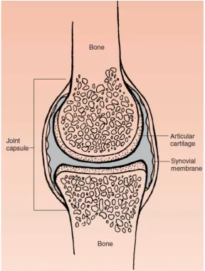
phase which was water by containing about 80 percent of the total tissue mass by weight. The intrinsic mechanical properties of each phase as well as the mechanical interaction between these phases afford the tissue its interesting rheological interaction (Mow et al., 1980, Mak et al., 1987, Mow et al., 1989). The location of the articular cartilage in the synovial joint was shown in Figure 2.1.
Figure 2.1 The schematic diagram of the synovial joint. Adapted from Mansour (1975).
5
bind or aggregated to a backbone of hyaluronic acid in order to form a macromolecule with a weight up to two hundred million. It was about 30 percent of
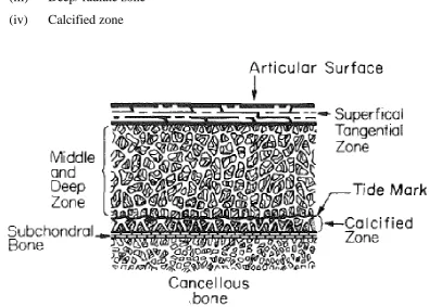
the dry weight of the articular cartilage when composed by the Proteoglycans. The concentration of this Proteoglycans and water content was varied through the depth of the tissue. The concentration of Proteoglycan was relatively low but the water content was highest at near the articular cartilage surface. Otherwise, the Proteoglycan concentration was greatest in the deeper regions of the cartilage which near the subchrondral bone while the water content was the lowest. Furthermore, the collagen was a fibrous protein that makes up sixty to seventy percent of the fry weight of the tissue.
The structure of the articular cartilage as shown in Figure 2.2 was often described in terms of fours zones between the articular surface and the subchrondral bone:
(i) Surface/ superficial tangential zone (ii) Intermediate/ middle zone
(iii) Deep/ radiate zone (iv) Calcified zone
6
This composition makes the articular cartilage structure inhomogeneous, and possesses anisotropic and nonlinear properties both in tension and compression. The
calcified zone was the boundary layer between the cartilage and the underlying subchrondral bone. In the calcified and deep zones, the collagen fibers with radial orientation were arranged in tightly packed bundles where those bundles were linked by numerous fibrils. The radial orientation becomes less distinct while the collagen fibrils form a network that which the chondrocytes were surrounded from the upper deep zone into the middle zone. The interface between the deep zone and calcified cartilage was known as tidemark. The structure of the articular cartilage has been revealed by using several of microscopy methods such as optical microscopy, scanning electron microscopy and transmission electron microscopy. The fibers in the superficial zone were finer than in the deeper zones and the collagen structure was organised into several layers (Abdul Latif et al., 2012, Mansour, 1975).
Articular cartilage plays a role as the bearing surface which permits the smooth motion between the adjoining bony segments in the freely moveable synovial joints (diarthroses) (Abdul Latif, 2011). Knee and elbow are examples of the synovial joints. In a typical synovial joint, the ends of opposing bones are covered with a thin layer of articular cartilage where it is normally white colour and its surface was smooth and glistening. The articular cartilage does not have a blood supply in normal mature animals since it was aneural (Mansour, 1975). A fluid which was known as synovial fluid was secreted by the inner surface of which was lined with the synovial membrane whereby, the entire joint was enclosed in a fibrous tissue capsule. This synovial fluid was clear to yellowish and was stringy where it was resembles egg white, and this resemblance was giving these joints name, synovia by which means “with egg” (Mansour, 1975).
7
interaction of its predominant components which including proteoglycans, collagens and interstitial fluid (Mansour, 1975).
The articular cartilage was distributed inhomogeneously and yields a variable thickness within the major synovial joints of human (Adam et al., 1998). There are two types of methods which are compression testing and imaging methods are used to determine the cartilage thickness. Magnetic Resonance Imaging (MRI) use a strong magnetic field and high frequency of radio waves to produce an image of
organs inside the body (Eckstein et al., 1995, Millington, 2007, Vanwanseele, 2004). On the other hand, Computed Tomography (CT) uses the ioning radiation (X-ray) to generate detailed images of structures inside the body (Yogananda et al., 2003). The indenter tip needle was used for compression testing to penetrate the cartilage until a significant increase in measured load was obtained (Swann and Seedhom, 1989, Athanasiou et al., 1998, Schenck et al., 1994).
The biomechanical properties of the articular cartilage such as Young
Modulus (E), Permeability (k) and Poisson’s ratio (v) were commonly determined by tensile test and compression methods. The tensile tests have been utilised to obtain single phase cartilage properties such as elastic modulus ultimate tensile stress, fracture stress and tensile fatigue properties (Kempson et al., 1968, Weightman et al., 1978, Akizuki et al., 1986, Kempson, 1991).
8
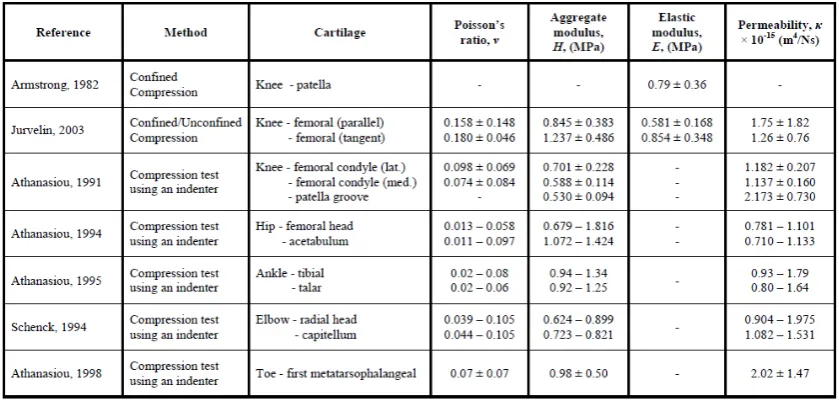
Table 2.1 Linear biphasic biomechanical properties of articular cartilage in human synovial joints. Adapted from Abd Latif (2011).
2.2 CONFINED COMPRESSION TEST
9
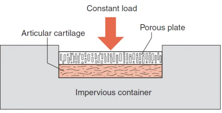
Figure 2.3 The schematic drawing of an apparatus used to perform a confined compression test of cartilage. Adapted from Mansour (1975).
The cartilage was deformed under a constant load in the creep mode as shown in Figure 2.4, but the deformation was not in the instantaneous response, as it would be in a single-phase elastic material such as spring. The fluid cannot escape from the matrix instantaneously caused this displacement of the articular cartilage results that as a function of time. The displacement was rapidly in initially and then this corresponds to a relatively large of the flow of fluid out of the cartilage. The flow of fluid was likewise to slow as the rate of the displacement slows and it was