IDENTIFICATION OF POTENTIAL ENTOMOPATHOGENIC FUNGI
OF Tetranychus kanzawai Kishida
(Tetranychidae: Acarina)
USING ITS-5.8s rDNA
REGION AS MOLECULAR MARKER
Yayan Sanjaya 1*), Virginia R. Ocampo 2) and Barbara L. Caoili 2)
1) Biology Program, Universitas Pendidikan Indonesia Jl Setiabudi No. 229, Bandung 40154 West Java Indonesia
2) Crop Protection Cluster, College of Agriculture, University of the Philippines Los Baños, College, Laguna 4031, Philippines
*) Corresponding author E-mail: [email protected]
Received: April 19, 2015/ Accepted: March 19, 2016
ABSTRACT
Fungi has been tested as one of the potential control agents for insect pests, which raises hopes for developing fungi as good biopesticides. The high variation within fungi species made taxonomic identification procedures more complex, thus molecular identification techniques are needed in addition to traditional morphological characteristics currently used as primary methods to classify fungi species. The objective of this research was to identify the species of the most pathogenic fungi to Tetranychus kanzawai Kishida using RAPD-PCR. The internal transcribed spacer of 5.8s rDNA (ITS-5.8s rDNA) sequence of these fungal isolates were amplified using two sets of universal primers for ITS and then analyzed. Molecular identification showed that these isolates had a higher of simi-larity to Metarhizium anisopliae than Metarhizium flavoviride.
Keywords: entomopathogenic fungi, ITS, mole-cular characterization, Tetranychus kanzawai
INTRODUCTION
The use of fungi as biopesticide covers a wide range of applications, that includes their use as control agents for insect pests - One of fungi classes recently used for that purpose is Hyphomycetes (Deuteromycetes), which is distinguished from other fungal groups by the morphology of its conidia, conidiogenous cells, and by hosts. However, it is almost impossible to distinguish individual isolates using only morphological characters because of limited distinctive characteristics (Samson, 1974).
Moreover, neither standard laboratory bioassays nor interactions with their natural hosts offer sufficient information to identify fungi on the subspecies level (Osborne and Landa 1992). Once selected for development as pest control agent, fungi isolates must be identified at the subspecies level (Jenkins and Grzywacz, 2000). This will place the fungi isolate within a taxonomic rank, preferably at species level. The ensuing problem is that fungi species are widely distributed in the environment worldwide and thus have an enormous number of strains. For instance, more than 700 species of fungi from about 90 genera are known as entomopathogens, composed of various subspecies, pathotypes, strains, and isolates (Charnley, 1989). Individual isolates of a particular entomopathogenic and mycoparasitic fungi may display considerable specialization in host range, and it is becoming increasingly apparent that identification at the species level is no longer adequate (Humber, 1997).
Several methods have been used to describe the variation within a species of entomo-pathogenic and mycoparasitic fungi. These include morphological characteristics of spores and colonies, extracellular protein profiles, patho-genicity, and growth or nutrient requirements (Samson, 1981). Obviously, taxonomic proce-dures are becoming more complex, and it is generally accepted that some forms of molecular identification techniques are needed in addition to the traditional morphological characteristics for-mally used to classify fungi species (Bridge and Arora, 1998). Different molecular techniques are used for various applications and on different entomopathogenic and mycoparasitic fungi, in-cluding identification of fungi isolates based on DNA polymorphism using RAPD-PCR technique
Cite this as: Sanjaya, Y., V.R. Ocampo and B.L. Caoili. 2016. Identification of potential entomopathogenic fungi of
Tetranychus kanzawai Kishida (Tetranychidae: Acarina) using ITS-5.8s rDNA region as molecular marker. AGRIVITA Journal of Agricultural Science. 38(2): 186-192. Doi: 10.17503/agrivita.v38i2.560
Accredited : SK No. 81/DIKTI/Kep/2011
Yayan Sanjaya et al.: Identification of Potential Entomopathogenic Fungi……….
(Joshi and St Leger, 1999). The RAPD (Random Amplified Polymorphic DNA) technique was intro-duced in 1990 (Samšiňáková et al., 1983). This technique could reveal polymorphism within completely unknown samples without the need of probe hybridization or DNA sequencing. Only one short oligonucleotide primer (6–12 bases) is used for the reaction, and primers sequence is fully arbitrary. The product is a spectrum of DNA fragments differing from each other in length and nucleotide sequence. The total number of pro-ducts and the length of each DNA sequence depend on DNA and primer templates used and are specific for a particular combination. The application of RAPD markers is similar to those of other DNA polymorphism detection methods. It can be used also for characterization of a fungi isolate by constructing a specific finger print or for genetic stability testing of an individual isolate. RAPD has already been used to estimate the diversity of a population, for genotype charac-terization or constructing the molecular phylogeny of closely related taxons (Tigano-Milani et al., 1995). Previous study showed that among the seven entomopathogenic fungi isolates tested against T. kanzawai, isolates Ma4, Ma5 and Ma6 were the most pathogenic (Sanjaya et al., 2013). The objective of this research was to identify the most pathogenic fungi using RAPD technique.
MATERIALS AND METHODS
Molecular Characterization
DNA extraction. Mycelia and conidia from each of the three most pathogenic isolates previously identified (Sanjaya et al., 2013) were inoculated on potato dextrose agar (PDA) and single spore colony were grown on potato dextrose broth (PDB), incubated on shaker (150 rpm) at 20˚C for 5-7 days. DNA extraction was done following the manufacturer’s protocol using Animal and Fungi DNA Preparation Kit (Jena Bioscience, Germany).
RAPD-PCR Amplification. Internal transcribe spacer (ITS) region for 5.8s ribosomal DNA (rDNA) used for the classification of the unknown fungi were amplified using three different types of ITS primers: ITS 1, ITS 2 and ITS 4 (Table 1). The primer-pairing were set based on reported ITS region for ITS-5.8s rDNA of the common fungi associated with insect. Amplification reactions were performed using 2x Taq Master Mix (Vivantis Technologies, Malaysia) with a total
volume of 50 μl, consisting of 20μl nuclease free sterile water, 25 μl 2xTaq Master mix, 1 μl of 2.5 mM MgCl2, 1 μl 5 μMol each primer, and 2μl of DNA template (20-25 ng). Controlled reactions were also run containing all components except genomic DNA. Thermal conditions was as follows: one cycle of initial denaturation at 95°C for five minutes, follow by 35 cycles with denaturation at 94°C for one minute and 30 sec, annealing at 55°C for two minutes, and extension at 72°C for three minutes, and a final extension at 72°C for five minutes.
Table 1. Internal Transcribe Spacer (ITS) primers used in the amplification of the target ITS-5.8s rDNA sequence of Metarhizium anisopliae Ma4, Ma5 and Ma6 isolates fungal isolates
ITS Primer
Code Sequence
ITS 1 5’-TCCGTAGGTGAACCTGCGG-3’
ITS 2 5’-GCTGCGTTCTTCATCGATGC-3’
ITS 4 5’-TCCTCCGCTTATTGATATGC-3’
PCR products were separated by electrophoresis in 1% agarose gel, containing 0.01% SYBR safe DNA gel stain (Invitrogen molecular probe), run with TBE (Tris-Boric EDTA) buffer by comparison with 1 kbp DNA Ladder were placed in a new and sterilized 1.5 ml micro centrifuge tube and then brought to MACROGEN Inc. in Seoul, South Korea (www.macrogen.com; email: [email protected]) through FedEx® Express (www.fedex.com/ph) for sequencing.
Data Analysis
Duncan multiple range test (P<0.05) using the SAS software package.
RESULTS AND DISCUSSION
Molecular Characterization
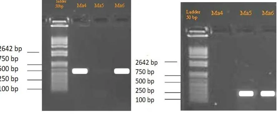
Polymerase Chain Reaction (PCR). The deve-lopment of PCR and the design of primers for the amplification of the various rDNA regions have considerably facilitated taxonomic studies of fungi. ITS sequences are generally constant, or show little variation within species but vary between species in a particular genus. The results of gel electrophoresis are shown in Figure 1.
A PCR product of approximately 200 bp was obtained from the three entomopathogenic isolates using the ITS1-ITS2 primer pair. This 200 bp PCR product, however, was not observed in Ma4. All of PCR products sizes can be amplified with the primer pairs ITS1, ITS2, and the 5.8s rDNA gene and also with 50 bp of the 3' end of the 18s rDNA and 50 bp of the 5' end of the 28s rDNA. A PCR product of approximately 560 bp was amplified from all the isolates, Ma4, Ma5,
Ma6, using the ITS1-ITS4 primer pair. Fungal universal primers of ITS1 and ITS4 have been commonly used to amplify ITS regions. Three Metarhizium isolates can be detected with ITS1 and ITS4 primers.
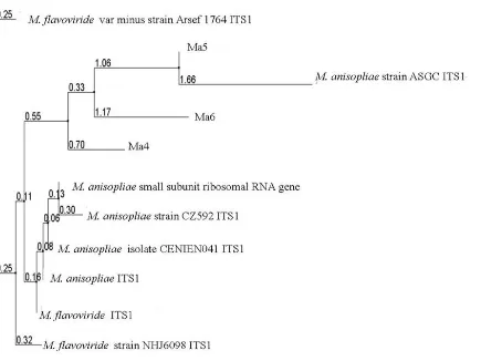
TheITS-5.8s rDNA sequence of the three fungi isolates were also observed using ClustalW multiple sequence alignment (Figure 2). Differences in the nucleotide sequence imply that Ma4, Ma5 and Ma6 were three different species. The BLAST similarity search sequences were obtained from samples of commonly known as entomopathogenic fungi, namely from Beauveria, Metarhizium, and Paecilomyces genus, from the GenBank. The identity of Ma4, Ma5 and Ma6 isolates as a species under the genus Metarhizium was verified using multiple sequence alignment of the ITS and 5.8s region sequence shown in (Table 2). Further, phylogenetic sequence analysis using different Metarhizium species showed a close relationship of Ma4, Ma5 and Ma6 with Metarhizium anisopliae (Strain CZ592) (Figure 2).
Yayan Sanjaya et al.: Identification of Potential Entomopathogenic Fungi……….
Figure 2. Rooted phylogenetic tree of ITS-5.8s rDNA region of Metarhizium anisopliae isolates Ma4, Ma5, and Ma6 compared with M. flavoviride and M. anisopliae.
Phylogram
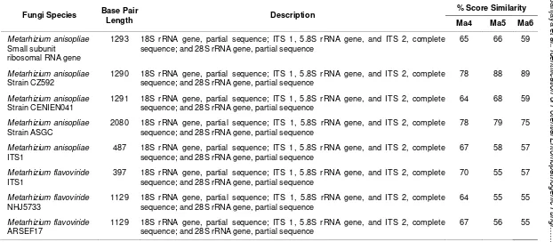
The use of ITS 5.8s rDNA sequence has been found to be a powerful tool for Metarhizium anisopliae. Observations showed that it can distinguish not only between isolates from different geographical origins, but also between isolates from the same country (Figure 1). The Blast of Ma4, Ma5 and Ma6 with isolates of Metarhizium from GenBank was obtained to identify the percent score of similarity (Table 2).
n
5.8s rDNA sequences in the Gen Bank
Fungi Species Base Pair
Length Description
sequence; and 28S rRNA gene, partial sequence
65 66 59
Metarhizium anisopliae Strain CZ592
1290 18S rRNA gene, partial sequence; ITS 1, 5.8S rRNA gene, and ITS 2, complete
sequence; and 28S rRNA gene, partial sequence
78 88 89
Metarhizium anisopliae Strain CENIEN041
1291 18S rRNA gene, partial sequence; ITS 1, 5.8S rRNA gene, and ITS 2, complete
sequence; and 28S rRNA gene, partial sequence
64 68 59
Metarhizium anisopliae Strain ASGC
2080 18S rRNA gene, partial sequence; ITS 1, 5.8S rRNA gene, and ITS 2, complete
sequence; and 28S rRNA gene, partial sequence
78 79 75
Metarhizium anisopliae ITS1
487 18S rRNA gene, partial sequence; ITS 1, 5.8S rRNA gene, and ITS 2, complete
sequence; and 28S rRNA gene, partial sequence
67 58 57
Metarhizium flavoviride ITS1
397 18S rRNA gene, partial sequence; ITS 1, 5.8S rRNA gene, and ITS 2, complete
sequence; and 28S rRNA gene, partial sequence
70 55 57
Metarhizium flavoviride NHJ5733
1129 18S rRNA gene, partial sequence; ITS 1, 5.8S rRNA gene, and ITS 2, complete
sequence; and 28S rRNA gene, partial sequence
64 55 55
Metarhizium flavoviride ARSEF17
1129 18S rRNA gene, partial sequence; ITS 1, 5.8S rRNA gene, and ITS 2, complete
sequence; and 28S rRNA gene, partial sequence
Yayan Sanjaya et al.: Identification of Potential Entomopathogenic Fungi………. Phylogenetic sequence analysis showed a close relationship of Ma4, Ma5 and Ma6 with Metarhizium anisopliae (Strain CZ592) (Figure 2). In regards to the performance variability of the different isolates, Sosa-Gómez and Alves (1983) reported a high enzymatic activity in more pathogenic isolates of M. anisopliae from several Brazilian regions, and suggested that they are probably associated with the presence of enzymes that influence the penetration process of the fungus (St Leger et al., 1988; De La Rosa et al., 1997), as well as with toxins such as destruxins and beauvericin, present in M. anisopliae and B. bassiana respectively, which vary in different isolates (Roberts and St. Leger, 2004). However, unlike insecticides, fungal infection takes 4-6 days after application to kill a mite. During this time the infected mite can cause serious damage to the crops (St. Leger et al., Oligonychus yothersi (McGregor), recorded 77 to 98% mortality. On the other hand isolates of M. anisopliae, causes 12.0 to 45.0%, and LT50 of 8.6 to 18.4 days.
CONCLUSION
The internal transcribed spacer of 5.8s rDNA (ITS-5.8s rDNA) characteristics confirmed that Ma4, Ma5 and Ma6 were Metarhizium anisopliae.
ACKNOWLEDGEMENTS
The authors wish to express their sincerest gratitude and profound appreciation to the following persons and organizations: SEAMEO-SEARCA for the scholarship grant.
REFERENCES
Bridge, P.D. and D.K. Arora. 1998. Interpretation of PCR methods for species definition. In: Applications of PCR in mycology. P.D. Bridge, D.K. Arora, C.A. Reddy and R.P.
Elander (eds.). Wallingford: CAB Inter-national. pp. 63-84.
Charnley, A.K. 1989. Mycoinsecticides: present use and future prospects. In: Progress and prospects in insect control. N.R. Macfarlane (ed.). Proceedings of an International Conference. England: British Crop Protection Council. pp. 131-144. De La Rosa, W., R. Alatorre, J. Trujillo and J.F.
Barrera. 1997. Virulence of Beauveria bassiana (Deuteromycetes) strains a-gainst the coffee berry borer (Coleoptera: Scolytidae). Journal of Economic Ento-mology 90 (6): 1534-1538. doi: 10.1093/ jee/90.6.1534
Humber, R.A. 1997. Fungi: identification. In: Manual of techniques in insect pathology. L.A. Lacey (ed.). London: Academic Press. pp. 153-185.
Jenkins, N.E. and D. Grzywacz. 2000. Quality control of fungal and viral biocontrol agents - assurance of product perform-ance. Biocontrol Science and Technology 10 (6): 753-777. doi: 10.1080/095831500 20011717
Joshi, L. and R.J. St. Leger. 1999. Cloning, expression, and substrate specificity of MeCPA, a zinc carboxypeptidase that is secreted into infected tissues by the fungal entomopathogen Metarhizium anisopliae. The Journal of Biological Chemistry 274 (14): 9803-9811. doi: 10.1074/jbc.274.14. 9803
Oliveira, I., J.A. Pereira, T. Lino-Neto, A. Bento and P. Baptista. 2012. Fungal diversity asso-ciated to the olive moth, Prays Oleae bernard: a survey for potential entomo-pathogenic fungi. Microbial Ecology 63 (4): 964-974. doi: 10.1007/s00248-011-9955-z Osborne, L.S. and Z. Landa. 1992. Biological control of whiteflies with entomopath-ogenic fungi. Florida Entomologist 75 (4): 456-471. doi: 10.2307/3496127
Tolypocladium, and Culicinomyces (in Czech). Ochrana Rostlin 19 (3): 195-204. Samson, R.A. 1974. Paecilomyces and some allied Hyphomycetes. In: Studies in mycology, no. 6. Baarn: Centraalbureau voor Schimmelcultures. p. 119.
Samson, R.A. 1981. Identification: entomopath-ogenic Deuteromycetes. In: Microbial control of pests and plant diseases 1970-80. H.D. Burges (ed.). London: Academic Press. pp. 93-106.
Sanjaya, Y., V.R. Ocampo and B.L. Caoili. 2013. Infection process of the entomopath-ogenic fungi, Metarhizium anisopliae in the Tetranychus kanzawai (Kishida) (Tetranychidae: Acarina). Agrivita Journal of Agricultural Science 35 (1) : 64-71. Sosa-Gómez, D.R. and S.B. Alves. 1983.
Characterization than eleven isolation of Metarhizium anisopliae (Metsch.) Sorok. Standardization, virulence and enzyme activity (in Spain). Revista de Investi-gacíon Educativa 1: 83-101.
St. Leger, R.J., L. Joshi and D. Roberts. 1998. Ambient pH is a major determinant in the
expression of cuticle-degrading enzymes and hydrophobin by Metarhizium aniso-pliae. Applied and Environmental Microbiology 64 (2): 709-713.
St. Leger, R.J., L. Joshi, M.J. Bidochka, N.W. Rizzo and D.W. Roberts. 1996. Biochem-ical characterization and ultra-structural localization of two extracellular trypsins produced by Metarhizium anisopliae in infected insect cuticles. Applied and Environmental Microbiology 62 (4): 1257-1264.
Tamai, M.A., S.B. Alves, R.B. Lopes and P.S. Neves. 1998. Evaluation of entomo-pathogenic fungi for control of Tetrany-chus urticae Koch (in Portugis). Abstracts 17th Congress of Entomology. Rio de Janeiro. p. 1066.