VOL. 5, NO. 2, pp. 60-64, May, 2015
Intraperitoneal Injection of High Tumor Necrosis Factor (TNF-α) Serum Increase Soluble Fms-like
Tyrosine Kinase 1 (sFlt-1) and Blood Pressure of Pregnant Mice
Bramantyo Aji Wicaksono1*
, Siti Candra Windu Baktiyani2
, Loeki Enggar Fitri3
1
Master Program in Biomedical Science, Faculty of Medicine, University of Brawijaya, Malang, Indonesia.
2Department of Obstetrics and Gynecology RSSA/Faculty of Medicine, University of Brawijaya, Malang, Indonesia. 3Department of Parasitology Faculty of Medicine, University of Brawijaya, Malang, Indonesia.
ABSTRACT
Preeclampsia has major symptoms of hypertension and proteinuria and is a cause of significant maternal and infant mortality in the world. The slow development of preeclampsia research possibility created by the diffi -culty in acquiring animal preeclampsia. Many existing animal model have been developed, but most of them are expensive to do. The purpose of this study was to determine the effects of intraperitoneal injection of pregnant pa -tients serum with high TNF-α levels toward sFlt-1 serum concentration and blood pressure of pregnant mice. Pregnant patients serum with high TNF-α levels (>20 pg/mL) was injected intraperitoneally to pregnant mice at gestational age 13 and 14 days. At 18 days of gestation, the blood pressure was measured, then the mice were dis -sected and the serum was taken to measure serum sFlt-1 concentration using ELISA kit (Bioassay Technology Laboratory, E0611Mo). The results showed there was a significant increase in blood pressure (p= 0.000) and the sFlt-1 levels (p= 0.002) of injected pregnant mice group compare to control group. These finding demonstrated that intraperitoneal injection of pregnant patients serum with high TNF-α levels to pregnant mice can increase blood pressure and sFlt-1 serum concentration of mice.
Keywords: blood pressure, preeclampsia, sFlt-1, TNF-α
Preeclampsia is a syndrome in pregnancy that oc-curs in over 20 weeks of gestational age, with hyperten-sion and proteinuria as the main symptoms [1]. Preeclampsia is a cause of maternal and infant mortal-ity in the world and also can lead 12-25% occurrence of fetal growth restriction. The incidence of preeclamp-sia ranges from 3% to 10% of pregnancies worldwide, and the impact of preeclampsia is more prevalent in developing countries [2].
Preeclampsia is preceded by the disruption of the placental invasion of the endometrium [3]. These con-ditions will cause placental hypoxia that can lead the placental damage, followed by high inflammatory and immune responses in the placenta. Activated immune responses will result in producing the angiotensin II type 1 receptor autoantibodies (AT1-AA) which then synthesize TNF-α [4]. Tumor necrosis factor α
(TNF-α) induces the formation of high antiangiogenic factor levels in serum, one of which is soluble fms-like tyro-sine kinase-1 (sFlt-1).
The sFlt-1 is a vascular endothelial growth factor receptor 1 (VEGFR-1) or Flt-1 that lack of transmem-brane and a cytoplasmic domain. The sFlt-1 is not bind on the plasma membrane of the endothelial cell but soluble in the serum. This lack of transmembrane and cytoplasmic domain make the sFlt-1 can not continue the second messenger of this receptor. [5]. The increas-ing of sFlt-1 serum levels resultincreas-ing in decreasincreas-ing serum levels of it is a ligand, the vascular endothelial growth factor (VEGF), and placental growth factor (PlGF). The decreasing of VEGF and PlGF will cause clinical symptoms of preeclampsia like hypertension and pro-teinuria [6].
One of the preeclampsia treatments is by giving magnesium sulfate to prevent seizures. However, this treatment does not treat or decrease the progression of preeclampsia [1, 7]. The slow progress of research in finding therapies preeclampsia likely influenced by the difficulty of obtaining experimental preeclampsia ani-mal models. A previous study revealed that injecting INTRODUCTION
*Corresponding author: Bramantyo Aji Wicaksono
Master Program in Biomedical Science, Faculty of Medicine, University of Brawijaya, Malang, Indonesia
TNF-α in pregnant mice will lead to hypertension and proteinuria [8].
Hence in this study we want to know the effects of intraperitoneal injection of pregnant patients serum with high TNF-α levels toward sFlt-1 serum levels and blood pressure of pregnant mice. We also want to know whether this study method possibility can be used as preeclampsia mice model.
Experimental Design
This research is a purely experimental (true experi-mental) laboratory study using a posttest only con-trolled group design. This study was performed in vivo to determine the effects of intraperitoneal injection of pregnant patients serum with high levels of TNF-α (>20 pg/mL) toward sFlt-1 serum concentration and blood pressure in pregnant mice.
Experimental Subjects
This study used pregnant patients as serum sources and pregnant mice as an experimental subject. Preg-nant patients were patients who control to Obstetrics Department of dr. Saiful Anwar Hospital at age 28-35 years and 30-40 weeks gestation with an agreement to participate in this study. Serum was collected from pregnant patients serum with TNF-α levels more than 20 pg/mL. Patients with infectious diseases and other chronic diseases are excluded from this study. Pregnant mice were used as many as 18 mice and were divided into two groups (each group consists of nine mice). One group was not injected and included as a control
group, and the other group was injected with the serum of pregnant patients with high TNF-α levels (TNF-α group). This study was approved by the Ethics Committee of the Faculty of Medicine, University of Brawijaya.
Pregnant Patients TNF-α Serum Levels Measurement
Blood was drawn from a pregnant patients vein us-ing five cc syrus-inge and put in plain vacutainer. Blood stored in a refrigerator at 4°C for 12 hours and then centrifuged at 6000 rpm for 10 minutes. Serum was taken and measured for the TNF-α levels using ELISA kit (BioLegend, USA, catalog number 430207). Serum with high levels of TNF-α then grouped and homoge-nized by the strong mixture. After measuring TNF-α levels in the serum with ELISA, we found that the mean of TNF-α serum levels from six pregnant pa-tients were 27.59 ± 5.39 pg/mL.
Serum Intraperitoneal Injection
Pregnant patients serum with high TNF-α levels (0.1 cc) was injected to pregnant mice at the age of 13 and 14 days of pregnancy [4, 9]. The Serum was in-jected on intraperitoneal of the upper right abdomen with a slope of 45° and 0.5 cm deep with one cc sy-ringe and 27G needle.
Blood Pressure Measurement
Mice blood pressure was measured at the age of 18 days of pregnancy before dissected. Mice were placed on mice stabilizer then mounted by the cuff and pulse recorded on the tail. Mice non-invasive blood pressure measuring machine (UgoBasile, Italy, product number 58500) was stand run to measure systolic and diastolic MATERIALS AND METHODS
blood pressure of mice. The measurement results were analyzed using independent t-test analysis.
Mice sFlt-1 Serum Levels Measurement
Mice were dissected at the age of 18 days of preg-nancy using chloroform. Mice blood was taken directly from the right heart using one cc syringe and 27G nee-dle. Blood was stored in a refrigerator at 4°C for 12 hours and then centrifuged at 6000 rpm for 10 min-utes. Serum was taken and measured the sFlt-1levels using ELISA kit (Bioassay Technology Laboratory, China, catalog number E0611Mo). The measurement results were analyzed using independent t-test analysis.
Mice Blood Pressure
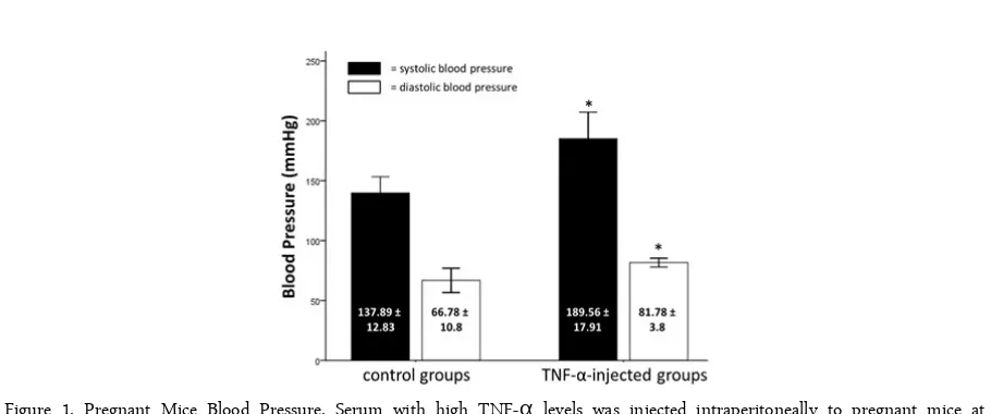
We injected serum with high TNF-α levels to the injected mice group as described above and measured their systolic and diastolic blood pressure. Systolic and diastolic blood pressure increase in the group of mice injected with serum with high TNF-α levels. The re-sults of measurements of systolic and diastolic blood pressure can be seen in Figure 1.
This study used independent t-test analysis (p <0.05) on systolic and diastolic blood pressure. There was a significant increase in the systolic blood pressure in mice group injected with serum with high TNF-α levels compare to control group (189.56 ± 17.91 vs. 137.89 ± 12.83; p=0.00). The diastolic blood pressure also significantly increase, and there was a significant difference between mice group injected with serum with high TNF-α levels and control group (81.78 ± 3.8 vs. 66.78 ± 10.8; p=0.01).
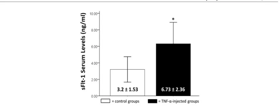
Mice sFlt-1 Serum Levels
We injected serum with high TNF-α levels to the injected mice group as described above and measured their sFlt-1 serum levels using ELISA kit. Mice sFlt-1 serum levels increase in the group of mice injected with serum with high TNF-α levels. The results of measurements of serum levels of sFlt-1 mice can be seen in Figure 2. This study used independent t-test analysis (p <0.05) on sFlt-1 serum levels. There was a significant increase of sFlt-1 serum levels in mice group injected with serum with high TNF-α levels compare to control group (6.73 ± 2.36 vs. 3.2 ± 1.53; p=0.00).
This study showed that intraperitoneal injection of serum with high TNF-α levels may increase sFlt-1 serum levels of pregnant mice. The TNF-α in the serum may bind with mice TNF-α receptor. This bind-ing stimulate the formation of hypoxia-inducible factor 1 (HIF-1) by increase the transcription of HIF-1 sub-unit, the HIF-1α [10]. The HIF-1 acts as a transcrip-tion activator protein of sFlt-1 [11]. The HIF-1 is con-sisted of HIF-1α and HIF-1β subunits. The HIF-1β is an 1 subunit expressed in constant, whereas HIF-1α is an HIF-1 subunit that normally expressed stably in hypoxic conditions by blocking its breakdown mechanism. To perform its functions as activating transcription of a gene, HIF-1α and HIF-1β should form dimers into HIF-1 [12]. In a normal oxygen con-dition (normoxia) HIF-1α is hydroxylated, acetylated, polyubiquitinated and braked down by the proteasome. All of that mechanism is oxygen dependent. Therefore, the amount of HIF-1α is low and can not bound with HIF-1βto become HIF-1 dimer [13]. In conditions of high inflammatory factors, such in high levels of TNF-RESULTS AND DISCUSSION
α, HIF-1α can be synthesized with higher numbers de-spite normoxia. The TNF-α will activate NFκB, a transcription factor that has a downstream signal to HIF-1α gene transcription so that HIF-1α will be syn-thesized more and expressed stably [14]. The high number of HIF-1, sFlt-1synthesis will be increased.
Tumor necrosis factor α (TNF-α) will also increase IL-6, which is the product of TNF-α downstream sig-nal. The previous data showed that there is higher lev-els of TNF-α and IL-6 in patients with preeclampsia compare to normal pregnant patients [15, 16]. The high levels of IL-6 will induce the differentiation of T cells into Th17 cells and followed by decreasing in the regulatory function of Treg cells that would reduce the ratio of Treg: Th17 [17]. This condition will activate response and differentiation of B cells into B cells CD19+ CD5+. These type of B cells are autoreactive cells that can secrete high angiotensin II type 1 recep-tor agonistic autoantibodies (AT1-AA) in patients with preeclampsia [18]. The AT1-AA that is formed will bind and activate angiotensin 1 receptor (AT1). The activated AT1 receptor will further increase the produc-tion of TNF-α and forming more sFlt-1 [4].
This study also showed that intraperitoneal injec-tion of serum with high TNF-α levels will increase blood pressure pregnant mice. Increased blood pres-sure is caused by high levels of sFlt-1 that will decrease angiogenic factors, such as VEGF and PlGF [6]. De-creased VEGF and PlGF will reduce the bond between VEGF to VEGFR-1 and VEGFR-2, and the bond be-tween PlGF with VEGFR-1. Reduction of VEGFR-2 ac-tivation will reduce acac-tivation of eNOS that have an ef-fect on the reduction of NO synthesis [19]. Nitric Ox-ide (NO) is a natural vasodilator blood vessels and serves to maintain the permeability of blood vessels [20]. Reduced NO in blood vessels will increase en-dothelin 1 (ET-1) which is a blood vessels vasoconstric-tor, thus causing hypertension [21, 22].
Vasoconstriction that occurs in blood vessels due to the presence of ET-1 will cause endothelial dysfunction or damage to the endothelium [22]. Endothelial dys-function that occurs in the kidneys will cause damage to the kidney glomerulus, thus causing proteinuria [23]. Endothelial dysfunction that occurs in the pla-centa will cause disruption of plapla-cental vascularization [24]. Disruption of placental vascularization will aggra-vate the condition of preeclampsia because it will in-crease the damage to the placenta and inin-crease the pla-centa inflammatory condition [25].
This research has shown the possibility of pregnant patients serum with high TNF-α levels injected into
pregnant mice can mimic preeclampsia condition such hypertension and increase sFlt-1 serum concentration. However, some limitations are also found in this study. In our present study, we did not evaluate urine protein levels of mice that indicate proteinuria which also the main symptom of preeclampsia. Moreover, we did not evaluate the mechanism of how pregnant patients serum with high TNF-α levels can cause hypertension and increase of sFlt-1 serum concentration. Measure-ment of VEGF and PlGF serum concentration may be useful to explain that mechanism. Therefore, this does not rule out the possibility of opening further research in the future to cover the limitations of this study.
The results of this present study indicate that preg-nant patients serum with high TNF-α levels that injected into pregnant mice can increase blood pressure and sFlt-1 serum concentration of mice. Thus, our present study may be developed as an experimental mice model for preeclampsia.
We thank Mirza Zaka pratama, MD and Syahroni Yudistian Agmar, MD for helping this experiment. We thank to Bunga Prihardina, S.Si dan Wahyudha Ngatiril Lady, S.Si at Laboratory of Biomedic Brawijaya University for guiding us on ELISA analysis at the laboratory.
1. ACOG Committee on Obstetric Practice (2002) ACOG practice bulletin No. 33: diagnosis and management of preeclampsia and eclampsia. Obstet Gynecol. 99: 159-167. 2. Duley L (2009) The global impact of preeclampsia and
eclampsia. Semin Perinatol. 33: 130-137.
3. Smith RA, Kenny LC (2006) Current thoughts on the pathogenesis of preeclampsia. The Obstetrician & Gynae-cologist. 8: 7-13.
4. Zhou CC, Roxanna AI, Zhang Y, Blackwell SC, Mi T, Wen J, Shelat H, Geng YJ, Ramin SM, Kellems RE, Xia Y (2010) Angiotensin receptor autoantibody-mediated tumor necrosis factor-αinduction contributes to increased solu-ble endoglin production in preeclampsia. Circulation. 121: 436-444.
Libermann TA, Morgan JP, Sellke FW, Stillman IE, Ep-stein FH, Sukhatme VP, Karumanchi SA (2003) Excess placental soluble fms-like tyrosine kinase 1 (sFlt-1) may contribute to endothelial dysfunction, hypertension, and proteinuria in preeclampsia. J Clin Invest. 111: 649-658. 7. Coetzee EJ, Dommisse J, Anthony J (1998) A randomized
controlled trial of intravenous magnesium sulphate versus placebo in the management of women with severe preeclampsia. Br J Obstet Gynaecol. 105: 300-303. 8. Parrish MR, Murphy SR, Rutland S, Wallace K, Wenzel
K, Wallukat G, Keiser S, Ray LF, Dechend R, Martin JN, Granger JP, LaMarca B (2010) The effect of immune fac-tors, tumor necrosis factor-alpha, and agonistic autoanti-bodies to the angiotensin II type I receptor on soluble fms-like tyrosine-1 and soluble endoglin production in re-sponse to hypertension during pregnancy. Am J Hyper-tens. 23: 911-916.
9. Kalkunte S, Boij R, Norris W, Friedman J, Lai Z, Kurtis J, Lim KH, Padbury JF, Matthiesen L, Sharma S (2010) Sera from preeclampsia patients elicit symptoms of human dis-ease in mice and provide a basis for an in vitro predictive assay. Am J Pathol. 177(5): 2387-2398.
10. Sandau KB, Zhou J, Kietzmann T, Brune B (2001) Regu-lation of the hypoxia-inducible factor-1alpha by the in-flammatory mediators nitric oxide and tumor necrosis fac-tor-alpha in contrast to desferroxamine and phenylarsine oxide. J Biol Chem. 276: 39805-39811.
11. Kendall RL, Thomas KA (1993) Inhibition of vaskular en-dothelial cell growth factor activity by an endogenously encoded soluble receptor. Proc Natl Acad Sci USA. 90: 10705-10709.
12. Ke Q, Costa M (2006) Hypoxia-inducible factor-1 (HIF-1). Mol Pharmacol. 70(5): 1469-1480.
13. rahimi-Horn C, Mazure N, Pouyssegur J (2005) Signaling via the hypoxiainducible factor-1alpha requires multiple posttranslational modifications. Cell Signal. 17: 1-9. 14. Jung YJ, Isaacs JS, Lee S, Trepel J, Liu ZG, Neckers L
(2003) Hypoxia-inducible factor induction by tumour necrosis factor in normoxic cells requires receptor inter-acting protein dependent nuclear factor kappa B activa-tion. Biochem J. 370: 1011-1017.
15. Benyo DF, Smarason A, Redman CW, Sims C, Conrad
KP (2001) Expression of inflammatory cytokines in pla-centas from women with preeclampsia. J Clin Endocrinol Metab. 86: 2505-2512.
16. Vince GS, Starkey PM, Austgulen R, Kwiatkowski D, Redman CW (1995) Interleukin-6, tumour necrosis factor and soluble tumour necrosis factor receptors in women with preeclampsia. Br J Obstet Gynaecol. 102: 20-25. 17. Laresgoiti-Servitje E (2013) A leading role for the immune
system in the pathophysiology of preeclampsia. J Leukoc Biol. 94: 247-257.
18. Jensen F, Wallukat G, Herse F, Budner O, El-Mousleh T, Costa SD, Dechend R, Zenclussen AC (2012) CD19 CD5 cells as indicators of preeclampsia. Hypertension. 59: 861-868.
19. Facemire CS, Nixon AB, Griffiths R, Hurtwitz H, Coff-man TM (2009) Vascular endothelial growth factor recep-tor 2 controls blood pressure by regulating nitric oxide synthase expression. Hypertension. 54: 652-658.
20. Olsson A, Dimberg A, Kreuger J, Claesson-Welsh L (2006) VEGF receptor signaling in control of vascular function. Nature. 7: 359-371.
21. Murphy SR, LaMarca BB, Cockrell K, Granger JP (2010) Role of endothelin in mediating soluble fms-like tyrosine kinase-1 (sFlt-1)-induced hypertension in pregnant rats. Hypertension. 55: 394-398.
22. George EM, Granger JP (2011) Endothelin: key mediator of hypertension in preeclampsia. Am J Hypertens. 24(9): 964-969.
23. Collino F, Bussolati B, Gerbaudo E, Marozio L, Pelissetto S, Benedetto C, Camussi G (2008) Preeclamptic sera in-duce nephrin shedding from podocytes through endothe-lin-1 release by endothelial glomerular cells. Am J Physiol Renal Physiol. 294: 1185-1194.
24. Fiore G, Florio P, Micheli L, Nencini C, Rossi M, Cerre-tani D, Ambrosini G, Giorgi G, Petraglia F (2005) En-dothelin-1 triggers placental oxidative stress pathways: pu-tative role in preeclampsia. J Clin Endocrinol Metab. 90: 4205-4210.