566
Darsono et al., Int J Med Res Health Sci. 2015;4(3):566-571
International Journal of Medical Research
&
Health Sciences
www.ijmrhs.com Volume 4 Issue 3 Coden: IJMRHS Copyright @2015 ISSN: 2319-5886 Received: 1stApr 2015 Revised: 25thApr 2015 Accepted: 26thMay 2015
Research article
EX VIVO STUDY OF GARCINIA MANGOSTANA L. (MANGOSTEEN) PEEL EXTRACT AND XANTHONES AS ANTI-ADIPOGENESIS IN HEPG2 CELL MODEL
Lusiana Darsono1, Meilinah Hidayat1, Maesaroh Maesaroh2, Nurul Fauziah2, *Wahyu Widowati1 1
Medical Research Center, Faculty of Medicine, Maranatha Christian University, Jl. Prof drg. Suria Sumantri No.65, Bandung 40164, West Java, Indonesia
2
Biomolecular and Biomedical Research Center, Aretha Medika Utama, Jl. Babakan Jeruk II. No. 9 Bandung 40163, West Java, Indonesia
*Corresponding author email: [email protected] ABSTRACT
Background: Anti-adipogenesis is one of proposed mechanism for anti-obesity. Adipogenesis regulation of obesity is important, so identification of anti-adipogenic activity is a potential strategy to find anti-obesity agent. Aim: The aim of this study is to evaluate the anti-adipogenesis potential of Garcinia mangostana L. peel extract (GMPE) compared to xanthones in HepG2 cells line as model. Material and Methods: GMPE was performed based on maceration method using distilated ethanol 70% as the solvent. The level of triglyceride and cholesterol and the inhibitory activity of triglyceride (TG) and cholesterol (CHOL) in HepG2 cells were assayed and determined as the anti-adipogenesis parameter. Results: The most active subtance to lower the triglyceride level was showed by GMPE in every concentration followed by the garcinone-C, γ -mangostin, garcinone-D and α -mangostin respectivelly. The highest activity to decrease the cholesterol level was showed by GMPE and followed by γ -mangostin, α -mangostin, garcinone-c, garcinone-d respectively. Conclusion: GMPE posses the anti-adipogenesis potential in inhibiting TG and CHOL synthesis was better than any other xanthone (α -mangostin, γ -mangostin, garcinone-C and garcinone-D).
Keywords: Obesity, Adipogenesis, Garcinia mangostana L., HepG2, Triglyceride, Cholesterol.
INTRODUCTION
Obesity is one of the most common global metabolic disorders defined as an excessive body weight in the shape of fat accumulation.[1] Recently, the metabolic syndrome including obesity represents one of the most serious problem worldwide.[2] Obesity has a strong association with the chronic disease such as diabetes, cardiovascular diseases, hypertension, osteoarthritis, some cancer and inflammation-based pathologies.[3] At the cellular level, obesity is characterized by an excess accumulation of adipose tissue is largely comprised of fat cells.[3-5] Obesity including excessive differentiation and growth of adipocytes which leads to increase fat cell mass and number, adipogenesis, lipid accumulation and lipogenic enzyme expression and surplus energy accumulation stored as triglyceride (TG) in
adipocytes.[1] Adipose tissue growth involves in formation of new adipocytes from precursor cells, further leading to an increase in adipocyte size.[6] Adipogenesis is a multi step process involving a cascade of transcription factors and cell cycle protein regulating gene expression and leading to adipocyte development.[7]The number of studies to prevent and treat obesity continues to rise.[8] Developing anti-obesity drugs that are efficacious and have minimal side effects become a pressing need.[1] Anti-adipogenesis is one of the proposed mechanisms of anti-obesity.[9,10] Adipose tissues are specialized for high capacity to accumulate TG.[11]Human hepatoma HepG2 cells is the most suitable and accessible human-derived cells that retain many of the biochemical functions of human liver parenchymal
567
Darsono et al., Int J Med Res Health Sci. 2015;4(3):566-571
cells for the ex vivo study including anti-adipogenesis screening.[12] Subjects with established obesity have an increased lipogenesis in hepatocytes (not in adipocyte) that might contribute to develop and/or retain the excessive fat mass.[13] Excess hepatic lipid accumulation is associated with nutritional factors, drugs, and multiple genetic defects in energy metabolism.[14] Many phytonutrients were investigated for their potential therapeutic properties. Some phytochemical bioactive have been shown to inhibit adipocyte differentiation and induce adypocyte apoptosis.[15,16] Garcina mangostana L. (mangosteen) is a tropical fruit originated from Southeast Asia, has been used in traditional therapy in the treatment of great variety of medical conditions for decades.[17] The pericarps of this fruit have been used for many years as traditional medicine in treating sicknesses such as trauma, skin infection, abdominal pain, dysentery, and wounds.[18]The major bioactive secondary metabolites of G.mangostana are xanthone derivates.[19] Xanthones could be isolated from peel, whole fruit, bark, and leaves of mangosteen.[20] Xanthones were repoted to have a great variety of pharmacological activities including antioxidant, antifungal, antibacteria, cytotoxic, anti-inflammation, anti-histamine, anti-HIV, and other activities.[19] The previous study confirm that mangosteen peel extract contained α -mangosteen (105 ppm), γ -mangosteen (7.20 ppm), garcinone C (3.50 ppm), and garcinone D (9.92 ppm) based on high performance liquid chromatography (HPLC).[21] Adipogenesis regulation of obesity is important, so identification of anti-adipogenic activity is a potential strategy to find anti-obesity agent. Therefore, the aim of this study is to evaluate the anti-adipogenesis potential of G. mangostana peel extract (GMPE) compared to xanthones including γ -mangostin, α -mangostin, garcinone-C, garcinone-D in HepG2 cells by using the inhibitory activity to lower triglyceride and cholesterol synthesis as the parameters.
MATERIAL AND METHODS
The present study was carried out in Biomolecular and Biomedical Research Center, Aretha Medika Utama, Bandung, West Java, Indonesia in collaboration with Medical Research Center, Faculty of Medicine, Maranatha Christian University. HepG2 cells (human liver hepatocellular carcinoma cell line) were used as cells model after cells were induced to
differentiate as adipocyte. The laboratory experiment was performed 4 months.
Plant material preparation and extraction: Garcina mangostana L. was collected from Cisalak-Subang, West Java, Indonesia. The plant was identified by herbarium staff, Department of Biology, School of Life Sciences and Technology, Bandung Institute of Technology, Bandung, West Java, Indonesia. The peel were collected, chopped, and kept in drier tunnel service. Extraction was performed based on the maceration method with distilated ethanol 70% as solvent.[21-23]
HepG2 cell culture and adipocyte differentiation induction: HepG2 cells (human liver hepatocellular carcinoma cell line) was cultured in DMEM (Dulbecco’s Modified Eagle Medium, Biowest) supplemented with 10% FBS (Fetal Bovine Serum, Biowest) and 100 U/mL penicillin-streptomycin (Biowest) then incubated for 24 hours at 37°C humidified atmosphere and 5% CO2.
[14]
After the cells were confluence, medium was discharged and cells were harvested after tripsin-EDTA treatment in 2500 rpm centrifuge for 4 minutes, cells then resuspended by 1 mL new medium and seeded in the 6 well plate (5 x 10⁵ cells/well) with DMEM supplemented with 10% FBS and 100 U/mL penicillin-streptomycin then incubated for 48 hours until confluent. Medium then discharged and suplemented with starving medium (DMEM + 1% antibiotic solution) then incubated for 24 hours. Starving medium then discharged and supplemented with induction medium (DMEM, 1:2 of 1mM palmitic acid: 1mM linoleic acid, BSA, and GMPE or xanthone). Cells then incubated in 37°C humidified atmosphere and 5% CO2.
Cells Lysate: Cell lysate was performed according to Biorad #163-2068 kit protocol. Confluent cells after GMPE or xanthone treatment were harvested using 500 g centrifuge for 4 minutes. Supernatan was discharged and work solution was added into the pelet cell. The solution briefly was sonicated for 30 second four times. Cells then centrifuge in 16.000 rpm for 30 minutes in room temperature. Supernatant then transfered to 2 mL ependorf tube and placed in -80°C as the sample for the TG and CHOL levels assay.[24]
568
Darsono et al., Int J Med Res Health Sci. 2015;4(3):566-571
reagent with 5µL sample was incubated in 37°C for 5 minutes. Double-distiled water (ddH2O) was used as blank and standard reagent was used as standard. Seven diferent concentration (2.180; 1.090; 0. 545; 0.273; 0.136; 0.068 and 0.034 mmol/L) were prepared by serial dilution for standard solution. The absorbance was measured in 500 nm of wave length. The TG level was calculated based on the ratio of sample absorbance to standard absorbance multiplied by the standard concentration.[25]
Cholesterol Levels and Inhibitory Activity Assay: The CHOL level assay was measured according to the Chol Kit Randox CH 200 protocol kit. Briefly 500µL mix reagent was added into 24 well plate and 5µL sample was added into the sample well. 5µL of ddH2O was used as blank. Seven diferent concentration (5.170; 2.585; 1.293; 0.646; 0.323; 0.162; and 0.081 mmol/L) were prepared by serial dilution for standard solution. 5µL standard solution was added into the well as standard. The absorbance was measured in 500 nm of wave length. The reaction then incubated at 37°C for 5 minutes. The absorbance was measured in 500 nm of wave length. The cholesterol concentration was calculated based on the ratio of sample absorbance to standard absorbance multiplied by the standard concentration.[26]
Statistical Analysis: The every treatment was done in three replication. Statistical analysis was conducted using SPSS software (version 17.0). Significant differences between the groups were determined using the Analysis of Variance (ANOVA) and Tukey Post Hoc Test. Statistical significance was set at p<0.05. The data were presented as mean ± standard deviation.
RESULTS
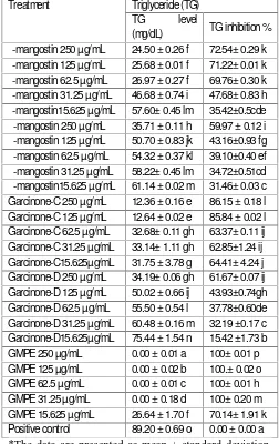
Table 1 showed that GMPE and xanthones including γ -mangostin, α -mangostin, garcinone-C and garcinone-D have lower TG levels in HepG2 cells compared to the triglyceride level in the cell lysate without GMPE and xanthones. The most active substance was showed by GMPE in every concentration followed by the garcinone-C, γ -mangostin, garcinone-D and α -mangostin respectivelly showed by the TG inhibitory activity. High plasma TG is associated with obesity.[27]
Table 1: Triglyceride (TG) Level and Inhibition Activity of GMPE and Xanthones in Various Concentration
Treatment Triglyceride (TG) TG level
(mg/dL) TG inhibition % γ -mangostin 250 µg/mL 24.50 ± 0.26 f 72.54± 0.29 k γ -mangostin 125 µg/mL 25.68 ± 0.01 f 71.22± 0.01 k γ -mangostin 62.5 µg/mL 26.97 ± 0.27 f 69.76± 0.30 k γ -mangostin 31.25 µg/mL 46.68 ± 0.74 i 47.68± 0.83 h γ -mangostin15.625 µg/mL 57.60± 0.45 lm 35.42±0.5cde α -mangostin 250 µg/mL 35.71 ± 0.11 h 59.97 ± 0.12 i α -mangostin 125 µg/mL 50.70 ± 0.83 jk 43.16±0.93 fg α -mangostin 62.5 µg/mL 54.32 ± 0.37 kl 39.10±0.40 ef α -mangostin 31.25 µg/mL 58.22± 0.45 lm 34.72±0.51cd α -mangostin15.625 µg/mL 61.14 ± 0.02 m 31.46± 0.03 c Garcinone-C 250 µg/mL 12.36 ± 0.16 e 86.15 ± 0.18 l Garcinone-C 125 µg/mL 12.64 ± 0.02 e 85.84 ± 0.02 l Garcinone-C 62.5 µg/mL 32.68± 0.11 gh 63.37± 0.11 ij Garcinone-C 31.25 µg/mL 33.14± 1.11 gh 62.85±1.24 ij Garcinone-C15.625µg/mL 31.75 ± 3.78 g 64.41± 4.24 j Garcinone-D 250 µg/mL 34.19± 0.06 gh 61.67± 0.07 ij Garcinone-D 125 µg/mL 50.02 ± 0.66 ij 43.93±0.74gh Garcinone-D 62.5 µg/mL 55.50 ± 0.54 l 37.78±0.60de Garcinone-D 31.25 µg/mL 60.48 ± 0.16 m 32.19 ±0.17 c Garcinone-D15.625µg/mL 75.44 ± 1.54 n 15.42 ±1.73 b GMPE 250 µg/mL 0.00 ± 0.01 a 100± 0.01 p GMPE 125 µg/mL 0.00 ± 0.02 b 100.± 0.02 o GMPE 62.5 µg/mL 0.00 ± 0.01 c 100± 0.01 h GMPE 31.25 µg/mL 0.00 ± 0.18 d 100± 0.20 m GMPE 15.625 µg/mL 26.64 ± 1.70 f 70.14± 1.91 k Positive control 89.20 ± 0.69 o 0.00 ± 0.00 a
*The data are presented as mean ± standard deviation. Different letters in the same column (a-o and its combination) indicate significant differences among the means of groups (GMPE and xanthones in various
concentrations) based on Tukey’s pos hoc comparison
(P<0.05).
569
Darsono et al., Int J Med Res Health Sci. 2015;4(3):566-571
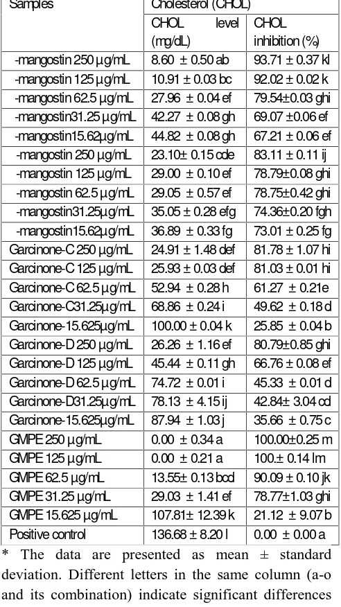
Table 2: Cholesterol (CHOL) Level and Inhibition Activity of GMPE and Xanthones in Various Concentrations
Samples Cholesterol (CHOL) CHOL level (mg/dL)
CHOL inhibition (%) γ -mangostin 250 µg/mL 8.60 ± 0.50 ab 93.71 ± 0.37 kl γ -mangostin 125 µg/mL 10.91 ± 0.03 bc 92.02 ± 0.02 k γ -mangostin 62.5 µg/mL 27.96 ± 0.04 ef 79.54±0.03 ghi γ -mangostin31.25 µg/mL 42.27 ± 0.08 gh 69.07 ±0.06 ef γ -mangostin15.62µg/mL 44.82 ± 0.08 gh 67.21 ± 0.06 ef α -mangostin 250 µg/mL 23.10± 0.15 cde 83.11 ± 0.11 ij α -mangostin 125 µg/mL 29.00 ± 0.10 ef 78.79±0.08 ghi α -mangostin 62.5 µg/mL 29.05 ± 0.57 ef 78.75±0.42 ghi α -mangostin31.25µg/mL 35.05 ± 0.28 efg 74.36±0.20 fgh α -mangostin15.62µg/mL 36.89 ± 0.33 fg 73.01 ± 0.25 fg Garcinone-C 250 µg/mL 24.91 ± 1.48 def 81.78 ± 1.07 hi Garcinone-C 125 µg/mL 25.93 ± 0.03 def 81.03 ± 0.01 hi Garcinone-C 62.5 µg/mL 52.94 ± 0.28 h 61.27 ± 0.21e Garcinone-C31.25µg/mL 68.86 ± 0.24 i 49.62 ± 0.18 d Garcinone-15.625µg/mL 100.00 ± 0.04 k 25.85 ± 0.04 b Garcinone-D 250 µg/mL 26.26 ± 1.16 ef 80.79±0.85 ghi Garcinone-D 125 µg/mL 45.44 ± 0.11 gh 66.76 ± 0.08 ef Garcinone-D 62.5 µg/mL 74.72 ± 0.01 i 45.33 ± 0.01 d Garcinone-D31.25µg/mL 78.13 ± 4.15 ij 42.84± 3.04 cd Garcinone-15.625µg/mL 87.94 ± 1.03 j 35.66 ± 0.75 c GMPE 250 µg/mL 0.00 ± 0.34 a 100.00±0.25 m GMPE 125 µg/mL 0.00 ± 0.21 a 100.± 0.14 lm GMPE 62.5 µg/mL 13.55± 0.13 bcd 90.09 ± 0.10 jk GMPE 31.25 µg/mL 29.03 ± 1.41 ef 78.77±1.03 ghi GMPE 15.625 µg/mL 107.81± 12.39 k 21.12 ± 9.07 b Positive control 136.68 ± 8.20 l 0.00 ± 0.00 a * The data are presented as mean ± standard deviation. Different letters in the same column (a-o and its combination) indicate significant differences among the means of groups (GMPE and xanthones in various concentrations) based on Tukey’s pos hoc comparison (P<0.05).
DISCUSSION
Obesity is a risk factor for severe disease such as diabetes, atherosclerosis, coronary heart disease and several cancer.[29]Primarily, obesity is a disorder of lipid metabolism and the enzyme involved in this process could be selectively targeted to develop anti-obesity drugs.[8] Different parts of medicinal plants like stem, flower, seed, root, fruit, etc. are used to obtain pharmacologically active metabolite.[30] Mangosteen peel has been used in medicinal in both Chinese and Ayurvedic. The yellow exudate from
mangosteen peel contain xanthone as the major class of compounds including α -mangostin, β -mangostin, γ -mangostin, garcinone-C and garcinone-D along with mangostinone, tanins, and flavonoid called epicatechin.[31]In this ex vivo study, we evaluated the anti-adipogenesis potential of GMPE and xanthones on HepG2 cells. Our results demonstrated that GMPE and xanthones exhibited potential to decrease the level of CHOL and TG as anti-adipogenesis parameters compared to the cell without GMPE or xanthones treatment.
In present study, GMPE could lower TG level compared to the cells without GMPE or xanthones treatment. Inhibition of TG metabolism was mediated by decreasing of gene expression of FAS (Faty Acid Synthase), ACC(Acetyl-CoA Carboxylase), and malic enzyme, among other factors.[32] The decrease of TG level may result from decreasing lipid synthesis.[33]Excess fat is stored as TG in the adipose tissue.[34] The decreased levels of TG indicated the adipogenesis inhibition. The potential for suppresing of adipogenesis and reducing lipid accumulation in cells mode were showed by some xanthones including α -mangostin and γ -mangostin.[35] α -mangostin also showed its ability to inhibit FAS correlated with intracellular lipid accumulation in differentiating adipocytes and stimulated lipolysis in mature adipocytes.[18]
Adipocyte normaly contain free CHOL and will redistributed from the plasma membran to the lipid droplet as TG storage increase. Adipocyte CHOL levels will increase in proportion to TG level.[34,35] GMPE also showed have CHOL inhibitory activity in concentration dependent maner showed by the decrease HOL level compared to the cell without GMPE or xanthones treatment as anti adipogenesis parameters. Previous study also found that G.mangostana posses the CHOL level reduction.[36] The GMPE showed anti-adipogenic activity through suppressing proliferator-activated receptor gamma (PPARγ ) expression and FAS activity.[37] Maria et al (2007) study observed that diets supplemented with mangosteen positively affect plasma lipid levels and plasma antioxidant activity in rats fed cholesterol-containing diets.[38] Based on our ex vivo study, we recommended the GMPE have beneficial effects as anti-adipogenesis agent was comparable with xanthones through the inhibition activity on CHOL and TG synthesis in HepG2 cells model.
570
Darsono et al., Int J Med Res Health Sci. 2015;4(3):566-571
CONCLUSSION
The GMPE posses the anti-adipogenesis potential on decreasing CHOL and TG levels in HepG2 cell better than xathones (α -mangostin, γ -mangostin, garcinone-C and garcinone-D). However, in vivo test in an animal model still needed to confirm the anti-adipogenesis activity of the GMPE and xanthones.
ACKNOWLEDGMENT
We gratefully acknowledge the financial support of the Research Center and Service Commuinity, Maranatha Christian University for research grant 2014. We are thankful to Pande Putu Erawijantari from Biomolecular and Biomedical Research Center, Aretha Medika Utama, Bandung, Indonesia for her valuable assistance. This research was also supported by Biomolecular and Biomedical Research Center, Aretha Medika Utama, Bandung, Indonesia for research method and laboratory facilities.
Conflict of Interest: Nil
REFERENCES
1. Kang J, Nam D, Kim K, Huh J, Lee J. Effect of Gambisan on the inhibition of adipogenesis in
3T3-L1 Adipocytes. J Evid Based
Complementary Altern Med. 2013; 10: 1-11 2. Holubkova A, Penesova A, Sturdik E, Mosovska
S, Mikusova L. Phytochemicals with the potential effects in metabolic syndrome prevention and therapy. Acta Chimica Slovaca. 2012; 5(2):186-99.
3. Williams D, Edwards D, Hamernig I, Jian L, James A. Vegetables containing phytochemicals with potential anti-obesity properties: a review. Food Res Int. 2013; 52(1):323-33.
4. Stephens J. The fat controller: adypocyte development. PLOS Biol. 2012; 10(11):1-3 5. Furuyashiki T, Nagayasu H, Aok I, Bessho H,
Hashimato T, Kanazawa K, et al. Tea ctechin suppresses adipocyte differentiation accompanied by down-regulation of PPARgamma2 and C/EBPalpha in 3T3-L1 cells. Biosci Biotechnol Biochem. 2004; 68(11): 2353-59.
6. Rayalam S, Fera M, Baile C. Phytochemicals and regulation of the adipocyte life cycle. J Nutr Biochem. 2008; 19: 717-26.
7. Rayalam S, Fera M, Baile C. Phytochemicals and
regulation of the adipocyte life cycle. J Nutr Biochem. 2008; 19: 717-26.
8. Navarrete J, Real J. Adipocyte differentiation. In: Symond M. Adipose Tissue Biology. Spain: Springer Science Business Media; 2012.vol,issue pp.17-38.
9. Deshpande M, Shengule S, Apte K, Wani M, Piprode V, Parab P. Anti-obesity activity of Ziziphus mauritiana: a potent pancreatic lipase inhibitor. Asian J Pharm Clin Res. 2013;vol,issue,page no
10. Wang Y, Jones P. Conjugated linoleic acid and obesity control: efficacy and mechanism. Int J Obes Relat Metab Disord. 2004; 28: 941-55. 11. Kodali G, Kakarla S, Seru G. Screening of crude
plants extracts for anti-diapogeneis activity in3T3-L1 cells. J Pharm Res. 2014; 8(1): 81-86. 12. Attie A, Scherer P. Adipocyte metabolism and
obesity. J Lipid Res. 2009; 50:395-99.
13. Inoue N, Nagao K, Sakata K, Yamano N, Gunawardena P, Han SY, et al. Screening of soy protein-derived hypotriglyceridemic di-peptides in vitro and in vivo. Lipid Health Dis. 2011; 10(85):1-10.
14. Gotstynki M, Gutzwiller F, Kuulasma K, Doring A, Ferrario M, Grafnetter D, et al. Analysis of the relationship betweem total cholesterol age, body mass index among males and females in the WHO MONICA project. Int J Obesity. 2004; 28:1082-90.
15. Liu Z, Li Q, Huang J, Liang Q, Yan Y, Lin H, et al. Proteomic analysis of the inhibitory effect of epigallocatechin gallate on lipid accumulation in human HepG2 cells. Proteome Sci. 2013; 11(32):1-11.
16. Lin J, Della-Fera M, Baile C. Green tea polyphenol epigallocatechin gallate inhibits adipogenesis and induces apoptosis in 3T3-L1 adipocytes. Obes Res. 2005; 13: 982-90.
17. Yang JY, Della-Fera M, Rayalam S, Ambati S, Baile C. Enhanced pro-apoptotic and anti-adipogenic effects of genistein plus guggulsterone in 3T3-L1 adipocytes. BioFactors. 2008; 30: p. 159-69.
571
Darsono et al., Int J Med Res Health Sci. 2015;4(3):566-571
Mol Nutr Food Res. 2014; 58: p. 239-3-247. 19. Ibrahim M, Hashim N, Mariod A, Mohan S,
Abdulla M, Abdelwahab S, et al. Alpha-mangostin from Garcina mangostana Linn: An update review of its pharmacological properties. Arabian J Chem. 2014; 02(11): p. 1-13.
20. Palakawong C, Sophanodora P, Pisuchpen S, Phongpaichit S. Antioxidant and antimicrobial activities of crude extracts from mangosteen (Garcinia mangostana L.) parts and some essential oils. Int Food Res J. 2010; 17:583-89 21. Widowati W, Darsono L, Suherman J, Yellianty
Y, Maesaroh M. High performance liquid chromatography (HPLC) analysis, antioxidant, antiaggregation of mangosteen peel extract (Garcinia mangostana L.). Int J Biosci Biochem Bioinforma. 2014; 4(6):458-66.
22. Widowati W, Rusmana D, Hardiman H, Tiono H, Wargasetia T, Pujimulyani D, et al. Mangosteen peel (Garcinia mangostana L.) extract for effervescent tablet. Eng Tech. 2013; 2013(82): 190-95.
23. Tjahjani S, Widowati W. The potency of xanthones as antioxidant and antimalarial, and their synergism with artemisinin in vitro. J Indon Med Assoc. 2013; 63(3): 95-99.
24. Liu Z, Li Q, Huang J, Liang Q, Yan Y, Lin H, et al. Proteomic analysis of the inhibitory effect of epigalloctechin gallate on lipid accumulation in human HepG2 cells. Proteome Sci. 2013; 11(32): 1-11.
25. Soeng S, Evacuasiany E, Widowati W, Fauziah N, Manik VT, Maesaroh M. Inhibitory potential of rambutan seeds extract and fractions on adipogenesis in 3T3-L1 cell line. J Exp Integr Med. 2015; 5(1): 55-60.
26. Hidayat M, Soeng S, Prahastuti S, Erawijantari PP, Widowati W. Inhibitory potential of ethanol extract of Detam 1 Soybean (Glycine max) seed and jati belanda (Gauzuma ulmifolia) leaves on adipogenis and obesity models in 3T3-L1 cell line. JSRR. 2015; 6(4): 304-312
27. Wang L, Li L, Ran X, Long M, Zang M, Tao Y, et al. Ellagic acid reduces adipogenesis through inhibition of differentiation-prevention of the induction of Rb Phosphorylation in3T3-L1 adipocytes. J Evid Based Complementary Altern
Med. 2013; 20113:1-11.
28. Lei F, Zhang X, Wang W, Xing D, Xie W, Su H, et al. Evidence of anti-obesity effects of promegranate leaf extract in high-fat diet induced obese mice. Int J Obesity. 2007; 31:. 1023-29. 29. Yang JY, Della-Fera M, Rayalam S, Ambati S,
Baile C. Enhanced pro-apoptotic and anti-adipogenic effects of genistein plus guggulsterone in 3T3-L1 adipocytes. BioFactors. 2008; 30: 159-69.
30. Sivasangari S, Vijayanand N, Rathinavel S. Antidiabetic activity of Cynodon dactylon (L.) Pers. extract in alloxan induced rats. Int J Pharm Pharm Sci. 2014; 6(4): 1-5.
31. Shibata MA, Matoba Y, Tosa H, Linuma M. Effects of mangosteen pericarp extracts against mammary cancer. Altern Integ Med. 2013; 2(8):1-5.
32. Kondo T, Kishi M, Fushimi T, Ugajin S, Kaga T. Vinegar intake reduces body weight, body fat mass and serum triglyceride levels in obese japanese subjects. Biosci Biotechnol Biochem. 2009; 73(8):1873-43.
33. Hsieh YH, Wang SY. Lucidone from Lindera erythrocarpa Makino fruits suppresses adipogenesis in 3T3-L1 cells and attenuates obesity and consequent metabolic disorders in high-fat diet C57BL/6 mice. Phytomedicine. 2013; 20: 394-400.
34. Chui P, Guan H, Lehrke M, Lazar M. PPAR gamma regulates adipocyte cholesterol metabolism via oxidized LDL receptor 1. J Clin Invest. 2005; 115(8): 2244-56.
35. Liu Q, Wang YT, Lin L. New insights in the anti-obese activity from Garcina mangostana. Food Funct. 2014;:2-37.
36. Karim A, Azlan A. Fruit pod extracts as a source of nutraceuticals and pharmaceuticals. Molecules. 2012; 17: 11931-46.
37. Gutierrez-Orozco F, Failla M. Biological activities and bioavailability of mangosteen and xanthones: a critical review of the current evidennce. Nutrients. 2013; 5: 3163-3183.