ISOLATION OF BIOACTIVE COMPOUNDS FROM
Aspergillus terreus
LS07
Rizna Triana Dewi
1,*, Sanro Tachibana
2, Puspa Dewi
1, L.B.S. Kardono
1, and Muhammad Ilyas
3 1Research Center for Chemistry - Indonesian Institute of Sciences, Kawasan PUSPIPTEK Serpong, Tangerang Selatan, Banten 15314, Indonesia
2
Department of Applied Biosciences, Faculty of Agriculture, Ehime University, 3-5-7 Tarumi Matsuyama, Ehime 790-8566 Japan
3
Research Center for Biology - Indonesian Institute of Sciences, Cibinong Science Center Jl. Raya Jakarta Bogor KM. 46, Cibinong 16911, Indonesia
Received December 10, 2013; Accepted July 18, 2014
ABSTRACT
This study aims to search for the active compounds from Aspergillus terreus LS07 which isolated from an
Indonesian soil. Bioassay-guided fractionations of the ethyl acetate (EtOAc) extract against α-glucosidase and
DPPH free radical to give four isolated compounds: oleic acid (1), ergosterol (2), butyrolactone I (3), and butyrolactone II (4). The structures of these metabolites were assigned on the basis of detailed spectroscopic analysis. Oleic acid (1) was showed significant activity toward α-glucosidase with IC50 value of 8.54 μM, but not for antioxidant. Butyrolactone I (3) and II (4) were showed significant activities against the α-glucosidase with their IC50
values at 52.17 and 96.01 μM, and those against DPPH free radicals at 51.39 and 17.64 μM, respectively. On the
other hand, ergosterol (2) did not show any activities.
Keywords:Aspergillus terreus LS07; α-glucosidase inhibitor; unsaturated fatty acid
ABSTRAK
Penelitian ini bertujuan untuk mencari senyawa aktif dari Aspergillus terreus LS07 yang diisolasi dari tanah
Indonesia. Pemisahan ekstrak etil asetat (EtOAc) berdasarkan pengujian bioaktivitas terhadap enzim α-glukosidase
dan radikal bebas DPPH menghasilkan empat senyawa terisolasi: asam oleat (1), ergosterol (2), butirolakton I (3), dan butirolakton II (4). Struktur metabolit tersebut dielusidasi berdasarkan metode analisis spektroskopi. Asam oleat (1) menunjukkan aktivitas yang signifikan terhadap α-glukosidase dengan nilai IC50 8,54 μM, tetapi tidak untuk antioksidan. Butirolakton I (3) dan II (4) menunjukkan aktivitas yang signifikan terhadap α-glukosidase dengan nilai IC50 pada 52,17 dan 96,01 μM, dan peredaman radikal bebas DPPH pada 51,39 dan 17,64 μM, masing-masing. Di sisi lain, ergosterol (2) tidak menunjukkan aktivitas apapun.
Kata Kunci:Aspergillus terreus LS07; penghambat α-glukosidase, asam lemak tidak jenuh
INTRODUCTION
α-Glucosidase (EC 3.2.1.20) is an enzyme that
plays a central role in carbohydrate metabolism by hydrolyzing the terminal glycosidic bonds at the non-reducing end of saccharide polymers to release
α-glucose [1]. Recently there had been widespread
interest in these enzymes because of their promising therapeutic potential in the treatment of disorders such as diabetes, human immunodeficiency virus (HIV) infection, metastatic cancer, and lysosomal storage diseases. Their potential as therapeutic targets,
especially, the inhibition of α-glucosidase had been
found to help control postprandial blood glucose levels in diabetic patients because they slow the uptake of dietary carbohydrates [2]. Clinical trials showed that the
α-glucosidase inhibitor improved long-term glycemic
control as measured by decreased hemoglobin A1c(HbA1c) in patients with type 2 diabetes and delay the development of type 2 diabetes in patients with impaired glucose tolerance [3-4].
Generally, the α-glucosidase inhibitor can be
α-glucosidase inhibitor were isolated from
microorganism such as: validamycin A was isolated from Streptomyces hygroscopicus var. limoneus, broth of Bacillus subtilis B2 also possessed strong
α-glucosidase activity [5], the new N-containing maltooligosaccharide GIB-638 was isolated from a culture filtrate of Streptomyces fradiae PWH638 [7] and Aspergillusol A was isolated from marine-derived fungus Aspergillus aculeatus [8]. Interest in the isolation of
α-glucosidase inhibitors from certain microorganisms
has increased due to fast growing characteristic of microorganisms.
The genus Aspergillus represents a diverse group of fungi that are among the most abundant fungi in the world [9].Aspergillus is a filamentous, cosmopolitan and ubiquitous fungus commonly found in soil, plant debris and indoor air environment. The genus Aspergillus includes over 185 species and is famous for the production of bioactive secondary metabolites (e.g. antibiotic, mycotoxin, antifungal compounds, etc) [10]. Aspergillus terreus is ubiquitous fungus isolated from both marine and terrestrial environments, however common in tropical or sub-tropical areas. The compounds isolated from A. terreus mostly posses’ pharmacological and commercial values. Lovastatin was one of the antihyperlipidemic drugs, which inhibits the cholesterol biosynthesis and was a major drug agent in the treatment of heart disease and atherosclerosis [11]. However, there have been relative few studies on
α-glucosidase inhibitors and antioxidants from species of
Aspergillus.
Previously study, we reported that the EtOAc extract of A. terreus showed potential inhibitory activity
toward α-glucosidase [12] and produced antioxidants
[13]. As continuous of the research on this species,
α-glucosidase inhibitors and antioxidant compounds
fromA. terreusLS07 are reported herein.
EXPERIMENTAL SECTION
Materials
α-Glucosidase [(EC 3.2.1.20)] Type I: from
Saccharomyces cereviseae, p-nitrophenyl
α-D-glucopyranoside (p-NPG), DMSO, 1,1-diphenyl-2-picrylhydrazyl (DPPH), quercetin dehydrate, linolenic acid ((CH3(CH2CH:CH3)3(CH2)7COOH), and Silica gel
(60-200 mesh Wako gel) were purchased from Wako Pure Chemical Industries, Ltd (Osaka, Japan). Stearic acid (C18H36O2) and linoleic acid (C18H32O2) were
purchased from TCI, Tokyo Chemical Industry. Co. Ltd. All the solvent used in this study were purchased from Wako Pure Chemicals and distilled prior to use.
The fungus A. terreus LS07 was isolated from a sea shore in Teluk Kodek, Pamenang area, West Nusa
Tenggara Province, Indonesia, in April 2009. This fungus was identified as A. terreus based on the sequence data of ITS rDNA. This fungus was deposited in the LIPI Microbial Culture Collection (LIPI MC), Research Center for Biology, Indonesian Institute of Sciences.
Instrumentation
Optical rotation values were measured with a Jasco P-2100 polarimeter. UV-Vis absorption spectra of the active compound in MeOH were recorded on a Hitachi U-1600 spectrophotometer. The mass spectra of the compound were measured with high-resolution FAB-MS. The nuclear magnetic resonance (NMR) spectra were recorded at 500 MHz for1H and 125 MHz for 13C on a JEOL JNM-ECA 500 with TMS as the internal standard. HMQC and HMBC techniques were used to assign correlations between 1H and 13C
signals. The chemical shift values (δ) are given in parts
per million (ppm), and coupling constant (J) in Hz. The GC-MS analysis for TMS derivatizations sample were
analyzed on the GC-MS QP-2010 gas
chromatographed equipped with a silica capillary
SPBTM-50 (30 m, id x 0.25 mm x 0.2 μm). Helium was
used as carrier gas, sample were injected an oven temperature of 60 °C then oven was heated at 10 °C/min to 280 °C where it was maintained for
10 min. Injected volume was 1 μL was split ratio 100.
TLC was run on silica gel 60 F254 pre-coated plates
(Merck 5554) and spots were detected by UV light.
Procedure
Fermentation, extraction and isolation
The stock culture of A. terreus was grown on PDA, incubated at 25 °C for seven days. Two discs (8 mm) of fungal mycelia were used to inoculate 150 mL of CzY(3% sucrose, 0.2% sodium nitrate, 0.1% K2HPO4, 0.05% magnesium sulphate, 0.05%
potassium chloride, 0.001% ferrous sulphate, and 0.5% yeast extract) in Erlenmeyer flask 500 mL and incubated at 25 °C on shaking condition (60 rpm) for seven days. After harvesting, fermentation broth (10 L) was extracted with EtOAc (10 x 2 L), followed by concentration in vacuo to afford 2.7 g as oily brown gummy. The EtOAc extract (2.7 g) was fractionated on silica gel column and eluted by n-hexane: CHCl3
gradient to give eleven fractions (F1-F11). Fraction F2 (100 mg) was further fractionated using Sephadex LH-20 column (n-hexane: CHCl3: MeOH 1:5:1) to afford
colorless oil F2.1 (1, 30 mg). Fraction F8.1 (2, 20 mg) was isolated as a colorless crystal from fraction F8 (150 mg) by recrystallization from CHCl3: MeOH.
to give F10.3 (3, 300 mg). Purification of F11 through column chromatography using stepwise gradient elution from 70% n-hexane in EtOAC to 100% EtOAC and preparative layer chromatography (PLC) with CHCl3:
Acetone (3:1) to afford F11.2 (4, 100 mg).
Compound 1: colorless oil, 1H-NMR (CDCl3, 500 MHz) δ: 5.33 (2H, m, H-9,10), 2.34 (2H, t, H-2), 2.01
(4H, m, H-8,11), 1.62 (2H, m, H-3), 1.25-1.30 (22H, m, H-4-8, 11-17), 0.88 (3H, t, H-18), MS m/z (%) = 282 ([M], 264 ([M-H2O]), 97 (64), 83 (72), 69 (80), 55 (84),
41 (13.5); deduced for C18H34O2.
Compound 2: colorless crystal, mp 166-168 °C,
1
H-NMR (CDCl3, 500 MHz) δ: 5.57 (dm, 1H, H-6), 5.38 (dm, 1H, H-7), 5.17 (m, 2H, H-22,23), 3.62 (m, 1H, H-3), 2.46 (dm, 1H, H-5), 2.35 (m, 2H, H-20,24), 2.09-1.93 (m, 3H), 1.92-1.89 (m, 4H), 1.88-1.55 (m, 4H), 1.50-1.40 (m, 3H), 1.38-1.16 (m, 3H), 1.02 (d, J=7.2, 3H, CH3-21), 0.93
Compound 3: yellowish gum. UV spectra (MeOH)
λmax 307 (log ε 4.3). 22.5 158.93 (C-4’), 154.81 (C-4”), 139.12 (C-2), 132.48 (C-9”), 132.36 (C-2”), 130.20 (C-6’ and C-3’), 129.60 (C-6”), 122.85 (C-1’), 128.27 (C-3”), 124.94 (C-1”), 123.43 (C-8”), 127.95 (C-3), 116.67 (C-5’ and C-2’), 115.05 (C-5”), 86.02 (C-4), 53.76 (OCH3),
39.32 (C-6), 28.62 (C-7”), 25.95 (C-10”), 17.82 (C-11”). HRFABMS: [M+H]+ m/z =425.1607, calcd for C24H25O7]
13 degrees of unsaturation.
Compound 4: colorless gum, UV (MeOH) λmax nm
(log ε): 307.5 (4.17). 28.7 168.83 (C-1), 158.91 (C-4’), 157.38 (C-4”), 139.28 (C-2), 132.33 (C-2’and C-6’), 130.12 (C-2” and C-6”), 128.06 (C-1”), 124.89 (C-1’), 122.78 (C-3), 115.47 (C-3’ and C-5’), 116.66 (C-3” and C-5”), 85.97 (C-4), 53.79 (OCH3), 39.18 (C-6). FABMS: [M+H]
+
m/z 357 for C19H17O7.
GC-MS analysis
The GC-MS analysis for TMS derivatizations sample were analyzed on the gas chromatography
coupled with mass spectrometry (GC-MS Shimadzu QP-2010). For MS detection, the electron ionization mode with ionization energy of 70 eV was used, with mass range at m/z 50-550. An SPB-50 column
(30 m x 0.25 mm i.d., film thickness of 0.25 μm) was
used for GC-MS. Helium was used as carrier gas and the flow rate was maintained at 1 mL/min. Samples were injected on oven temperature of 60 °C then oven was heated at 10 °C/min to 280 °C where it was
maintained for 10 min. Injected volume was 1 μL was
split ratio 100. The identification of chemicals was performed in comparison with database (NIST08 library) and confirmed using authentic standard samples.
Trimethylsilyl (TMS) derivatize. Compound 1 and 2 were derivatizations with TMS. Sample (100 ppm) was
transfer to a GC vial in n-hexane; 25 μL bis (trimethylsilyl) trifluoroacetamide BSTFA add and 25 μL
pyridine to the sample. Cap the vial tightly and heat at 65 °C for ~20 min. Sample was cool to room temperature before inject on the GC-MS Shimadzu 2010Q.
Biological activity
α-Glucosidase inhibitory assay. α-Glucosidase
inhibitory activity was evaluated according to the method previously reported by Kim et al. [14], with
minor modifications. α-Glucosidase (250 μL, 0.065 U/mL), 495 μL of 0.1 M phosphatebuffer (pH 7.0), and 5 μL of various concentrations of sample in
DMSO were pre-incubated at 37 °C for 5 min. The
reaction was started by the addition of 250 μL of 3 mM
pNPG. The reaction was incubated at 37 °C for 15 min and stopped by adding 2 mL of 0.1 M Na2CO3. α-Glucosidase activity was determined by measuring
release ofpNPG at 410 nm.
Kinetics of inhibition against α-glucosidase. The
inhibition type of active compounds against
α-glucosidase activity was measured with increasing concentrations of pNPG as a substrate in the absence or presence of active compounds at different concentrations. The type of inhibition was determined by Lineweaver-Burk plot analysis.
DPPH free radicals scavenging assay. The antioxidant activities of the isolated compounds were evaluated according to the method of Yen and Chen [15], with minor modification. Aliquots of samples in
MeOH (2 mL) at various concentrations (10-200 μg/mL)
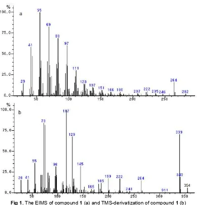
Fig 1.The EIMS of compound1(a) and TMS-derivatization of compound1(b)
RESULT AND DISCUSSION
Isolation and Characterization of Isolated Compounds
Bioassay-guided fractionation of the EtOAc extract of A. terreus LS07 by the general chromatographic techniques give four known isolated compounds. Compound1was colorless oil, at TLC analysis turned to yellow color when sprayed by Bromocresol green indicated organic acid group. From1H-NMR spectrum of compound 1, two olefinic protons at δH 5.33 (2H, m, H-9,10) and four allylic proton atδH 2.01 (4H, m, H8,11)
indicated a typical unsaturated fatty acid. Moreover, identified based on resemblance with fragmentation in EIMS by GC-MS showed characteristic pattern of unsaturated fatty acid (m/z 41, 55, 69, 83, 97, and 264 for [M-18] indicated lost of H2O) and molecular ion peak
at 282 was deduced to C18H34O2(Fig. 1a). In order to
verify this structure, compound 1 was treated with TMS
to derivatize a hydroxyl group (-OH) to GC analysis. Trimethylsilyl (TMS) ethers are a convenient way to derivatize a variety of functional groups prior to GC analysis. Pyridine was added as a basic catalyst to speed reaction with sterically hindered groups. The TMS derivatizations of compound 1 give molecular ion m/z 354 was indicated TMS instead a hydrogen of hydroxyl group (Fig. 1b). Based on those result, compound 1 was identified as oleic acid-9-octadecanoic acid (Z). Oleic acid is unsaturated fatty acid that occurs naturally in microorganism.
Compound2was obtained as colorless solid, give green-blue color when reacted with Liebermann-Burchard reagent indicated the compound is steroid or triterpene group. Structure of 2 was identified as
ergosterol (ergosta-5,7,22-triene-3β-ol) confirmed by
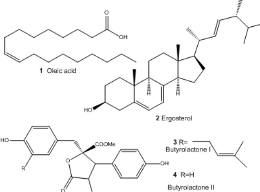
Fig 2.Chemical structures of the isolated active compounds fromA. terreusLS07
Table 1. Biological activity of isolated compounds of A. terreusLS07
IC50 (μM) a
Compound
Inhibitory of
α-glucosidase Antioxidant
1 8.54±0.61 n.d
2 n.d 378.79±3.42
3 52.17±5.68 51.39±3.68
4 96.01±3.70 17.64±6.41
Quercetin 10.92±3.72 39.63±5.21
n.d not detected ;aIC50value are shown as mean ± S.D. from three
independent experiment
absent or a minor component in higher plants plays an important role as inhibitor of lipid per-oxidation, active against HL-60 cells, MCF-7 cell line [16].
Compound 3 and 4 were the major active compounds in the EtOAc extract. The compound3 was obtained as colorless crystalline solid, which would eventually turn into a gummy material on storage for long time. The molecular formula 3 was determined to be C24H24O7 from its HRFABMS. It showed a
pseudomolecular ion peaks [M+H]+ at 425.1607 (calcd. for C24H25O7), indicating 13 degree of unsaturation. The 1
H-NMR spectrum revealed two methyl singlets at
δH 1.64 and 1.56, and one doublet at δH 3.43 and 3.10
representing a prenyl system. One triplet signal δH5.51, indicated an olefinic methine. Three aromatic proton
signal at δH 6.53, 6.52, and 6.49, being for
1,2,4-trisubtituted phenol along with two doublets at δH 7.61 and 6.95 representing 1,4-disubtituted phenolic moiety. The 13C-NMR showed the presence of ten aromatic signals for two aromatic rings, two ester carbonyls at
δC 171.0 and 168.7, olefenic carbon signals of
compound at δC 132.5 and 123.4. Two oxygenated
carbon at δC154.8 and 158.9 of phenolic system, along
with five quertenery carbon (δC 139.0-122.9). The
methine signals at δC 130.2 and 116.7 for 1,4-disubtituted phenol and three methine signals at
δC 132.4, 128.3, and 115.1 for 1,2,4-trisubtituted benzene ring. In the aliphatic region, signals for
oxygenated methine at δC 86.0, methoxy at δC 53.8,
two methylenes at δC 39.3 and 28.6, and two methyls 26.0 and 17.80 were assigned. Structure of compound 3 was further deduced on the basis of HMBC experimental data and comparison with literature, which confirms that compound 3, coincided with
butyrolactone I (α-oxo-β-(p-hydroxyphenyl)-γ-(p-hydroxy- m-3.3-dimethylallylbenzy l)-γ-methoxycarbonyl-γ-butyr
lactone I) as previous isolated from A. terreus MC751 [13]. This compound displays several interesting biological activities, such as antitumor effects (cytotoxicity), allergenic effects, inhibition of microbial and plant growth, and both convulsant and anticonvulsant activity [18]. Butyrolactone I also showed inhibitory activities against soybean lipoxygenese and had DPPH radical-scavenging activity [13,19].
Compound 4 was isolated as colorless gum. It gave spot on TLC more polar, but having color and appearance like compound 3, indicating it to have similar structure. The molecular formula C19H16O7 was
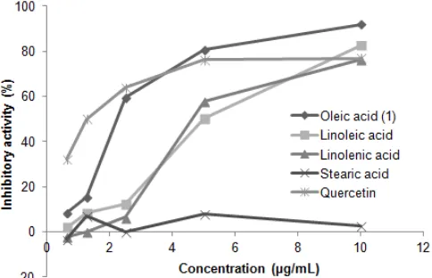
Fig 3. α-Glucosidase inhibitory activity of unsaturated and saturated fatty acid
Fig 4. Lineweaver-Burk plot for the inhibition of
α-glucosidase by compound 1
were coincided with butyrolactone II [methyl-4-hydroxy-2-(4-hydroxybenzyl)-3-(4-hydroxyphenyl)-5-oxo- 2,5-dihydro furan-2-carboxylate], which was isolated for the first time in 1982 from A. terreus IFO 4100 [20]. All the isolated compounds from EtOAc extract ofA. terreusLS07 were showed in Fig. 2.
Biological Activity of Isolated Compounds
The α-glucosidase inhibitory and antioxidant
activity of isolated compounds were investigated and compared with quercetin as referent standard. In thisstudy we used quercetin as standard due to several reports that quercetin have stronger inhibition of
α-glucosidase from yeast S. cerevisiae than acarbose [21]. The activities of isolated compounds against
α-glucosidase were shown in Table 1. In particular, oleic
acid (1) showed excellent inhibition on yeast
α-glucosidase with IC50value of 8.54 μM which is lower
than that of quercetin (IC50 value of 14.6 μM), however no activity on DPPH radicals.
To our best knowledge, this is the first report on the
inhibitory activity against α-glucosidase of oleic acid
which isolated from A. terreus. However, other unsaturated fatty acid were reported have potential
activity toward α-glucosidase such as 7(Z)-octadecanoic
acid and 7(Z),10(Z)-octadecanoic acid with IC50 were
1.81 and 2.86 μM, respectively. These compounds were
purified from the body wall of Stichopus japonicas [22] and 10-hydroxy-8(E)-octadecanoic acid, an intermediate of bioconversion of oleic acid [23].
In addition, we evaluated the inhibitory activities of saturated and unsaturated fatty acid to clarify whether inhibitory activity of compound 1 due to double bond in fatty acid or not. The result was presented in Fig. 3, the inhibitory activity of oleic acid (1), linoleic acid, and
linolenic acid exhibited high inhibition at 10 μg, with
91.92 ± 0.85%, 82.84 ± 1.51%, and 76.26 ± 2.41%,
respectively. On the other hand, stearic acid (C18:0) exhibited poor inhibitory activity (8 ± 2.01% at
10 μg/mL).
The inhibitory activity against α-glucosidase of the
unsaturated fatty acid were ranked as follows; oleic
acid > linoleic acid ≥ linolenic acid, while stearic acid,
saturated fatty acid, did not show significant activity. Therefore, we consider that a double bond in fatty acid is crucial for the activity; however increasing of the double bond number will decrease the inhibitory activity
on α-glucosidase. The investigation of α-glucosidase
activity of stearic acid compared to unsaturated fatty acid has not been reported.
This result accorded with previous studies that the presence of the double bond in the fatty acid affects the inhibitory potency [22-23]. The activity of unsaturated
fatty acid on α-glucosidase was assumed that the
binding of fatty acid may affect the secondary and tertiary structure of proteins because of their detergent effects alone, which was suggested for the effect of fatty alcohol sulfate and palmitic acid binding to bovine serum albumin [24]. The resultant alteration in the conformation of the protein molecule may effects its biological activity, which may be one of early effects involved in the inhibition of enzymes by fatty acids [22]. Moreover, Takahashi & Miyazawa also suggested that the olefin in serotonin derivatives is crucial for the
inhibition of α-glucosidase [25].
To determine the inhibition mode of compound 1
on α-glucosidase, series of experiments were carried
out in which the substrate concentration was varied, and several different concentration of the compound 1 were used. Compound 1 showed a mixed type
inhibition against α-glucosidase (Figure 4) with
In the contras, ergosterol (2) shows weak activities
both against of DPPH radicals and α-glucosidase. This
results similar with previous reported by Fatmawati et al.
[17] that ergosterol did not active against α-glucosidase.
On the other hand, butyrolactone I (3) and II (4) were
showed significant activities against α-glucosidase and
DPPH radical. The presence of a phenolic hydroxyl group in those compounds was assumed to contribute to
the α-glucosidase inhibitory effect and scavenging DPPH
radicals [26, 27].
CONCLUSION
The results presented here showed that butyrolactone I (3) and butyrolactone II (4) are the main
active compounds as α-glucosidase inhibitor and
antioxidant of A. terreus LS07, while oleic acid (1) was
displayed the strongest activity against α-glucosidase
but not active against DPPH free radical. Double bond in
oleic acid (unsaturated fatty acid) is crucial for the
α-glucosidase inhibitory activity; however increasing of the
double bond number will decrease the α-glucosidase
inhibitory activity.
REFERENCES
1. Kimura, A., Lee, J-H., Lee, I-S., Lee, H-S., Park, K-H., Chiba, S., and Kim, D., 2004, Carbohydr. Res., 339 (6), 1035–1040.
2. Du, Z-Y., Liu, R-R., Shao, W-Y., Mao, X-P., Ma, L., Gu, L-Q., Huang, Z-S., and Chan, A.S.C., 2006,Eur. J. Med. Chem., 41 (2), 213–218.
3. Cheng, A.Y.Y., and Josse R.G., 2004, Drug Discovery Today, 1 (2), 201–206.
4. Kim, K.Y., Nam, K.A., Kurihara, H., and Kim, S.M., 2008,Phytochemistry, 69 (16), 2820–2825.
5. Zhu, Y-P., Yin, L-J., Cheng, Y-Q., Yamaki, K., Mori, Y., Su, Y-C., and Li, L-T., 2008, Food Chem., 109 (4), 737–742.
6. Schmidt, D.D., Frommer, W., Junge, B., Müller, L., Wingender, W., Truscheit, E., and Schäfer, D., 1977, Naturwissenschaften, 64 (10), 535–536.
7. Meng, P., and Zhou, X., 2012,Med. Chem. Res., 21 (12), 4422–4429.
8. Ingavat, N., Dobereiner, J., Wiyakrutta, S., Mahidol, C., Ruchirawat, S., and Kittakoop, P., 2009, J. Nat. Prod., 72 (11), 2049–2052.
9. Krijgsheld, P., Bleichrodt, R., Veluw, G.J., Wang, F., Müller, W.H., Dijksterhuis, J., and Wösten, H.A.B., 2012,Stud. Mycol., 74 (1), 1–29.
10. Balajee, S.A., 2009,Med. Mycol., 47, S42–S46. 11. Hajjaj, H., Niederberger, P., and Duboc, P., 2001,
Appl. Environ. Microbiol., 67 (6), 2596–2602. 12. Dewi, R.T., Iskandar, Y., Hanafi, M., Kardono,
L.B.S., Angelina, M., Dewijanti, D.I, Banjarnahor, S.D.S., 2007, PJBS, 10:3131-3135.
13. Dewi, R.T., Tachibana, S., Kazutaka, I., and Ilyas, M., 2012,J. Microb. Biochem. Technol., 4 (1), 10– 14.
14. Kim, Y.M., Wang, M.H., and Rhee, H.I., 2004, Carbohydr. Res., 339 (3), 715–717.
15. Yen, G-C., and Chen, H-Y., 1995, J. Agric. Food. Chem., 43 (1), 27–32.
16. Nagia, M.M.S., Metwally, M.M., Shaaban. M., El-Zalabani, S.M., and Hanna, A.G., 2012,Org. Med. Chem. Lett., 2, 1–9.
17. Fatmawati, S., Shimizu, K., and Kondo, R., 2011, Phytomedicine, 18 (2), 1053–1055.
18. Cazar, M.E., Hirschman, S.G., and Astudillo, L., 2005, World J. Microbiol. Biotechnol., 21 (6-7), 1067–1075.
19. Sugiyama, Y., Yoshida, K., Abe, N., and Hirota, A., 2010, Biosci. Biotechnol. Biochem., 74 (4), 881– 883.
20. Nitta, K., Fujita, N., Yoshimura, T., Arai, K., and Yamamoto, Y., 1983, Chem. Pharm. Bull., 31 (5), 1528–1533.
21. Tadera, K., Minami, Y., Takamatsu, K., and Matsuoka, T., 2006,J. Nutr. Sci. Vitaminol., 52 (2), 149–153.
22. Nguyen, T.H., Um, B.H., and Kim, S.M., 2011, J. Food Sci., 76 (9), H208–H214.
23. Paul, S., Hou, C.T., and Kang, S.C., 2010, New Biotechnol., 27 (4), 419–423.
24. Hsia, J.C., Wong, L.T., Tan, C.T., Er, S.S., Kharouba, S., Balaskas, E., Tingker, D.O., and Feldhoff, R.C., 1984,Biochemistry, 23, 5930–5932. 25. Takahashi, T., and Miyazawa, M., 2012,Phytother.
Res., 26 (5), 722–726.
26. Dewi, R.T., Tachibana, S., and Darmawan, A., 2012, World Acad. Sci. Eng. Technol., 70, 882– 887.