review article
The new england journal of medicine
mechanisms of disease
Novel Therapies Based on Mechanisms
of HIV-1 Cell Entry
J. Michael Kilby, M.D., and Joseph J. Eron, M.D.
From the Department of Medicine, Univer-sity of Alabama, Birmingham (J.M.K.); and the Department of Medicine, University of North Carolina, Chapel Hill (J.J.E.). Address reprint requests to Dr. Kilby at 908 20th St. S., UAB, Birmingham, AL 35294-2050, or at [email protected].
N Engl J Med 2003;348:2228-38. Copyright © 2003 Massachusetts Medical Society.
nfection with human immunodeficiency virus type 1 (hiv-1), the retrovirus that causes the acquired immunodeficiency syndrome (AIDS), is one of the leading causes of death worldwide. All currently available antiretroviral agents inhibit essential HIV-1 enzymes — either the reverse transcriptase or the pro-tease (Fig. 1). Recent advances have markedly improved the outcome for many pa-tients who receive these classes of antiretroviral drugs. However, the success of cur-rent therapy is limited by the emergence of drug-resistant viruses, the necessity of sustained adherence to complex regimens, and the potential for toxic effects. Novel classes of safe and effective agents with a low risk of cross-resistance with other an-tiretroviral drugs are needed.
Targeting viral entry may have advantages over the inhibition of steps in the viral life cycle after the cell has been infected. A better understanding of how HIV-1 binds to and enters cells has prompted a reappraisal of previous attempts to block viral entry and an evaluation of new approaches (Table 1). In this article, we outline the steps involved in viral attachment and entry, provide an update on agents under development that have been designed to inhibit each of these steps, and consider the prospects of these com-pounds in the treatment of human immunodeficiency virus (HIV) infection.
early characterization of the viral envelope and cd4+ t-cell tropism
The initial characterization of HIV-1 centered on its tropism for mature human helper T lymphocytes, which express the CD4 (or T4) surface protein (also expressed on mono-cytes, dendritic cells, and brain microglia).1-4 Molecular studies demonstrated that, like
other retroviruses, the HIV-1 particle is surrounded by a lipid bilayer, derived from the host cell and studded with viral glycoproteins (Fig. 2). The infectivity of HIV requires the surface glycoprotein subunit (gp120) and the transmembrane glycoprotein subunit (gp41) of gp160, a viral precursor protein. The two subunits are cleaved from gp160 by host-cell proteases and then reassembled as oligomeric structures (trimers) on the viral
membrane (Fig. 3A).5,6
The amino acid sequence of gp120 contains five variable regions (V1 through V5), alternating with more conserved regions; the variable regions tend to be exposed on the
viral surface.7,8 Noncontiguous regions of the gp120 molecule come together to form
the CD4 binding site, and small deletions or substitutions in either CD4 or conserved regions of gp120 disrupt the binding of the virus.9-11
The design of inhibitors of viral entry must take into account the three-dimensional structure and the variability in the sequence of the intact wild-type HIV-1 envelope, rather than the linear sequences of denatured proteins or the envelopes of laboratory-adapted strains of HIV-1, which do not reliably predict the in vivo activity of investigational agents that block viral entry.
i
mechanisms of disease
the search for entry cofactors
Because CD4 alone is insufficient to permit the entry of HIV-1, it has long been suspected that additional receptors or other factors are required. Although most HIV-1 isolates successfully infect primary helper T cells, individual viral isolates have a range of tropisms (for example, in vitro, some preferen-tially infect macrophages over T-cell lines) and cause varying degrees of cell fusion (formation of syncytia or multinucleated giant cells) in T-cell lines. Amino acid residues within conserved V3-loop sites of
gp120 were known to affect membrane fusion,12,13
but the basis of the differences in tropism was until recently poorly understood. Important clues to the “coreceptor” mystery arose from the discovery that
b-chemokines (macrophage inflammatory
pro-teins 1a and 1b [MIP-1a and MIP-1b], as well as RANTES [regulated upon activation normal T-cell expressed and secreted]), which are chemotactic cy-tokines produced by macrophages, activated T cells, and natural killer cells, suppress the replication of some strains of HIV-1.14
chemokine coreceptors (cc chemokine receptor 5 and cxc chemokine receptor 4)
After the seminal discovery by Feng and colleagues that a G-protein–coupled chemokine receptor, CXC chemokine receptor 4 (CXCR4), was the key to cellu-lar entry for viruses that grow well in cultured T-cell lines (X4 viruses),15 several groups of researchers
rapidly confirmed that chemokine receptors were the missing link in our understanding of HIV-1 en-try (Fig. 2). The expression of CXCR4 made other-wise impenetrable CD4+ cell lines susceptible to productive HIV-1 infection. CC chemokine recep-tor 5 (CCR5), a b-chemokine receptor with a seven-transmembrane-protein structure similar to that of CXCR4, was found to serve as a coreceptor for non– syncytium-inducing or macrophage-tropic HIV-1 (R5 viruses).16,17 Chemokine receptors are the
pri-mary binding sites for many related retroviruses, and HIV-1 can be genetically modified to allow
CD4-independent cell entry,18 suggesting that CXCR4
and CCR5 are the primordial receptors, rather than just cofactors.19
HIV-1 isolates of the R5 type have been implicat-ed in most cases of sexually transmittimplicat-ed HIV infec-tion, whereas X4 viruses, which replicate best in T-cell lines, often predominate in the later stages of HIV disease and may be associated with rapid pro-gression to AIDS and death.20,21 Clinical isolates
may contain mixtures of R5 and X4 viruses, and
some individual viral strains (R5X4, or dual-trop-ic viruses) can use either the CXCR4 (X4-virus) or
CCR5 (R5-virus) receptor.22 As previous
experi-ments have suggested, V3-loop amino acid sequenc-es in gp120 are major determinants of
chemokine-receptor affinity.23,24 Although other chemokine
receptors (CC chemokine receptors 2, 3, and 8, BOB, and others) can facilitate the entry of specific HIV-1 variants in vitro, all clinical isolates of HIV-1
use CCR5, CXCR4, or both for entry.25
Naturally occurring host defects in CCR5 expres-sion have demonstrated the clinical significance of these receptors. A homozygous deletion that prevents CCR5 expression occurs disproportion-ately among persons who are frequently exposed to
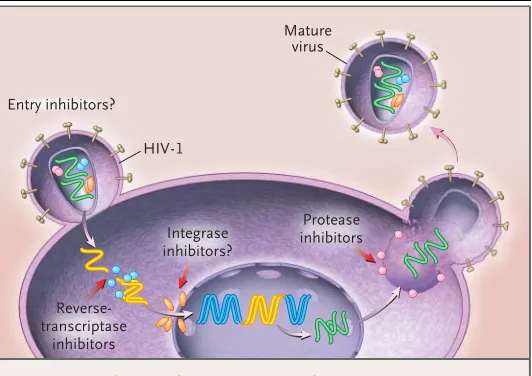
Figure 1. The Life Cycle of Human Immunodeficiency Virus Type 1 (HIV-1), Showing Potential Targets for Antiretroviral Therapy.
HIV-1 binds to receptors on the cell surface, undergoes membrane fusion, and then releases copies of the RNA genome into the cytoplasm. After suc-cessful invasion of the cell, the viral reverse-transcriptase enzyme transcribes single-stranded viral RNA into double-stranded DNA that can be integrated into the genetic material of the human host. Reverse-transcriptase inhibitors were the first agents approved for the treatment of HIV-1; currently available inhibitors of this enzyme are nucleoside antagonists (zidovudine, didano-sine, zalcitabine, lamivudine, stavudine, abacavir, and combined formula-tions), nonnucleoside competitive inhibitors (nevirapine, delavirdine, and efavirenz), and one nucleotide analogue (tenofovir). The viral integrase en-zyme is required for the integration of proviral DNA into the host genome before replication. Investigational integrase inhibitors are currently in early clinical trials. When the infected cell synthesizes new protein, integrated proviral DNA is also translated into the protein building blocks of new viral progeny. The viral components then assemble on the cell surface and bud out as immature viral particles. The final maturation of newly formed viruses requires the HIV-1 protease to digest larger components into the intricate pieces that make up an infectious virion. Several protease inhibitors (ritona-vir, indina(ritona-vir, nelfina(ritona-vir, amprena(ritona-vir, lopinavir–ritona(ritona-vir, and two formulations of saquinavir) are currently in clinical use.
Entry inhibitors?
HIV-1
Mature virus
Reverse-transcriptase
inhibitors
Integrase inhibitors?
The new england journal of medicine
HIV-1 but who nonetheless remain uninfected.26
Heterozygous or partial mutation of a gene respon-sible for CCR5 expression on the cell surface does not block infection but does provide some protec-tion from disease progression.27 Rare cases of
in-fection in persons with a homozygous deletion of the gene responsible for CCR5 expression appear to be caused by X4 viruses.28
membrane fusion
The final step in viral entry, the fusion of the viral envelope with the cell membrane, is mediated by gp41. The molecular sequence of gp41 includes
“heptad-repeat” regions (HR1 and HR2), reflecting the presence of periodic hydrophobic regions found in a-helical “coiled-coil” structures.29,30 Mutations
in the HR regions interfere with the fusion property of gp41.31
A model of gp41-mediated membrane fusion analogous to the “spring-loaded” mechanism of in-fluenzavirus has been proposed. After influenzavi-rus attaches to a target cell and enters the acid-rich endosome, the conformation of the hemagglutinin protein changes, shifting a “fusion peptide” into fa-vorable position for the mediation of fusion.32 The
model predicts that the gp120–gp41 trimer holds
* RANTES denotes regulated upon activation normal T-cell expressed and secreted, CCR5 CC chemokine receptor 5, gp glycoprotein, and CXCR4 CXC chemokine receptor 4, and FDA Food and Drug Administration.
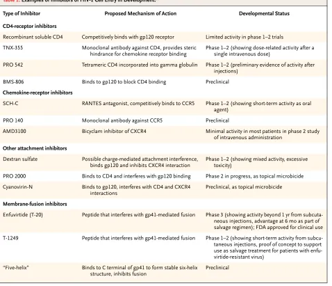
Table 1. Examples of Inhibitors of HIV-1 Cell Entry in Development.*
Type of Inhibitor Proposed Mechanism of Action Developmental Status
CD4-receptor inhibitors
Recombinant soluble CD4 Competitively binds with gp120 receptor Limited activity in phase 1–2 trials
TNX-355 Monoclonal antibody against CD4, provides steric hindrance for chemokine receptor binding
Phase 1–2 (showing dose-related activity after a single intravenous dose)
PRO 542 Tetrameric CD4 incorporated into gamma globulin Phase 1–2 (preliminary evidence of activity after injections)
BMS-806 Binds to gp120 to block CD4 binding Preclinical
Chemokine-receptor inhibitors
SCH-C RANTES antagonist, competitively binds to CCR5 Phase 1–2 (showing short-term activity as oral agent)
PRO 140 Monoclonal antibody against CCR5 Preclinical
AMD3100 Bicyclam inhibitor of CXCR4 Minimal activity in most patients in phase 2 study of intravenous administration
Other attachment inhibitors
Dextran sulfate Possible charge-mediated attachment interference, binds gp120 and inhibits CXCR4 interaction
Phase 1–2 (showing mixed activity, excessive toxicity)
PRO 2000 Binds to CD4 and interferes with gp120 binding Phase 2 in progress, as topical microbicide
Cyanovirin-N Binds to gp120, interferes with CD4 and CXCR4 interactions
Preclinical, as topical microbicide
Membrane-fusion inhibitors
Enfuvirtide (T-20) Peptide that interferes with gp41-mediated fusion Phase 3 (showing activity beyond 1 yr from subcuta-neous injections, advantage at 6 mo as part of salvage regimen); FDA approved for clinical use
T-1249 Peptide that interferes with gp41-mediated fusion Phase 1–2 (showing short-term activity from subcu-taneous injections, proof of concept to support use as salvage treatment for patients with enfu-virtide-resistant virus)
“Five-helix” Binds to C terminal of gp41 to form stable six-helix structure, inhibits fusion
mechanisms of disease
each gp41 in a high-energy configuration, with the fusion peptide pointed inward, toward the viral sur-face (Fig. 3A). The binding of gp120 to CD4 and chemokine receptors is thought to release gp41 from this configuration, causing the fusion peptide to spring outward toward the cell membrane (Fig. 3B). Still in their trimeric association, the HR1 re-gions then fold over into the hydrophobic groove formed by the three corresponding HR2 regions, forming a stable six-helix bundle, thus bringing vi-ral and cell membranes into proximity for fusion and entry (Fig. 3C).33
crystallizing the dynamic roles of the viral envelope proteins
Partial x-ray crystallization of the HIV-1 glycopro-teins, in complex with receptors, sheds light on the
three-dimensional interactions between viral and
host components during binding and entry.19 This
imaging of the complexes has confirmed the se-quential conformational changes that follow the binding of the virus to CD4 and chemokine recep-tors and has suggested the occurrence of several in-terdependent steps in the process of viral entry.
inhibiting the interaction of gp120 with the cd4 molecule
Initial attempts to block HIV entry focused on the interaction between gp120 and CD4. Recombinant soluble CD4 (rsCD4), which was developed as a vi-ral-attachment decoy, demonstrated potent inhibi-tion of HIV-1 infecinhibi-tion in vitro.34,35 In the clinical
i n h i b i t i o n o f v i r a l e n t r y
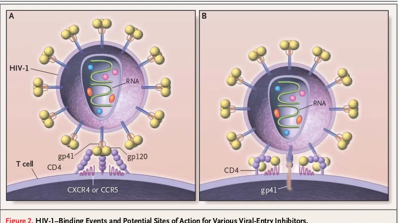
Figure 2. HIV-1–Binding Events and Potential Sites of Action for Various Viral-Entry Inhibitors.
HIV-1 is covered by a lipid bilayer derived from host-cell membranes. Incorporated into this bilayer are viral glycopro-teins as well as host adhesion molecules that may play a part in attachment to target cells. The viral-entry process con-sists of a series of coordinated interactions — binding to two different receptors (Panel A) and membrane fusion (Panel B). The viral envelope glycoproteins are synthesized as a single polyprotein that assembles into a trimer and then is broken down by host protease into surface glycoprotein subunits (gp120) and transmembrane glycoprotein subunits (gp41). Each gp120 monomer is a complex, folded structure, consisting of a series of variable loops formed by disulfide bonds, with noncontiguous segments brought together to form three-dimensional binding sites for the CD4 receptor and a che-mokine receptor (either CCR5 or CXCR4). Initial binding of gp120 to CD4 (Panel A) might be blocked by soluble CD4 de-coys, monoclonal antibodies against sequences on gp120 or CD4, or other small-molecular inhibitors. After CD4 binding, each gp120 undergoes a conformational change exposing the region that will bind to a seven-transmembrane chemo-kine receptor. Viral isolates have varying affinities for CCR5 or CXCR4 receptors. Binding of the chemochemo-kine coreceptors might be inhibited by natural ligands of these receptors or their derivatives, small-molecule inhibitors, monoclonal anti-bodies directed at the interacting sites, or down-regulation of receptor expression. It is hypothesized that binding of both the CD4 and chemokine receptors shifts away the steric hindrance of the heavily glycosylated gp120, allowing the gp41 segment to mediate membrane fusion and entry (Panel B).
gp41
HIV-1
T cell gp41 gp120
A B
CD4 CD4
RNA
gp41
CXCR4 or CCR5
CXCR4 or CCR5
The new england journal of medicine
setting, however, rsCD4 had negligible activity ex-cept at very high doses.36-39 A chimeric molecule
consisting of recombinant CD4 and gamma globu-lin had an extended half-life yet had little or no ac-tivity when administered with zidovudine.40 Despite
these failures, new inhibitors of interactions be-tween gp120 and CD4 continue to be pursued.
PRO 542, a hybrid tetramer, contains CD4 recep-tor domains within an IgG2 backbone and acts as a decoy for gp120 binding. It is active in vitro against diverse strains of HIV-1, including clinical
iso-lates.41 PRO 542 must be administered parenterally
but has a half-life in plasma of more than three days. In pilot studies, the compound was well tolerated and there was evidence of antiviral activity in adults42
and children.43
Monoclonal anti-CD4 antibodies can also block the interaction between gp120 and CD4; some of these monoclonal antibodies can inhibit the repli-cation of multiple subtypes of HIV-1 in vitro.44
Al-though anti-CD4 antibodies may have immunosup-pressive effects,45,46 a recent report on a humanized
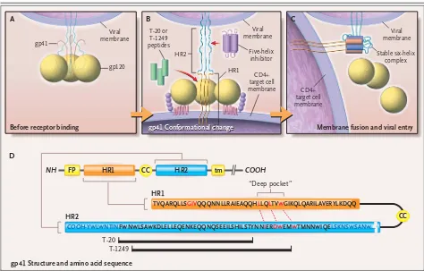
Figure 3. Proposed Model of the gp41-Mediated Membrane-Fusion Step, Showing Sites of Action for Fusion Inhibitors in Development.
Panel A shows the gp120–gp41 trimer before it binds to a cell. It is thought that after gp120 binds to CD4 and chemokine receptors, gp120 changes conformation (Panel B), probably breaking away entirely, which allows gp41 to spring out of its high-energy configuration. This re-lease of potential energy propels the fusion-peptide portion of gp41, which was previously pulled in close to the viral surface, outward to snag the target cell membrane. Within this extended configuration of gp41, intertwined heptad-repeat (HR2) regions form hydrophobic grooves that fold over and bind to corresponding HR1 coiled coils, collapsing gp41 into a stable six-helix or “trimer of hairpins” configuration (Panel C). This hinge action of gp41 brings the viral and cell lipid membranes into close proximity for viral entry.
Panel D shows the gp41 amino acid sequence (shown as a linear sequence, above, and with the relation between the HR1 and HR2 regions when gp41 is folded over into a hairpin configuration at a point where two cysteine residues [CC] form a disulfide-bonded loop, below). As shown at the bottom of the figure, T-20 and T-1249 are peptides derived from HR2 segments. These peptides may act as competitive decoys for the process through which the extended coiled-coil structure of HR1 folds back to bind to corresponding regions of HR2 (with the T-1249 binding region extending farther along into the “deep-pocket” sequence of HR1), thus disrupting the formation of the six-helix configuration required for membrane fusion. Similar approaches have been proposed, involving smaller peptides or a “five-helix” agent (essentially the converse of the enfuvirtide strategy) that bind HR2 segments, preventing viral entry through related mechanisms. FP denotes fusion peptide, and tm membrane-spanning region.
gp41 Conformational change
Viral membrane
Before receptor binding
gp41 Structure and amino acid sequence
gp41 Conformational change A
D
B C
Membrane fusion and viral entry
gp41
Viral membrane
Viral membrane
gp120
HR2
HR1 CD4+ target cell
membrane CD4+ target cell membrane Five-helix
inhibitor Stable six-helixcomplex T-20 or
T-1249 peptides
FP CC
CC tm
HR1
TVQARQLLSGIVQQQNNLLRAIEAQQHLLQLTVWGIKQLQARILAVERYLKDQQ
HR1
HR2
T-20 T-1249
“Deep pocket”
HR2 COOH
NH
mechanisms of disease
anti-CD4 antibody called TNX-355 demonstrated no serious adverse events in the short term and no CD4-cell depletion. This dose-escalation trial showed clinically significant reductions in the viral load (of >1.0 log10 copies of HIV-1 RNA per
millili-ter) two to three weeks after a single intravenous dose of 10 to 25 mg per kilogram of body weight.47
Certain small molecules may competitively and reversibly inhibit viral entry by binding gp120 and blocking the gp120–CD4 interaction.48 A lead
com-pound of this type (BMS-806) demonstrated potent HIV-1 inhibition in vitro, but activity varied among different subtypes (clades) of HIV-1 and even with-in the same clade. Mutations with-in the region of gp120 that binds to CD4 cause resistance to BMS-806,
sup-porting the proposed mechanism of action.49
nonspecific interference with attachment
Dextran sulfate and other polyanions can inhibit the replication of HIV-1 in vitro.50 Originally attributed
to nonspecific, charge-mediated interference with gp120–CD4 interactions, the effect of dextran sul-fate has been linked more recently to its ability to attach to the V3 loop of X4 or R5X4 viruses, thus blocking the binding of gp120 to coreceptors.51,52
Dextran sulfate inhibits the replication of X4 isolates but may enhance R5 infections in vitro.53 Clinically,
intravenous dextran sulfate was toxic and in some cases appeared to increase viral replication.54
PRO 2000 is a naphthalene polyanion that binds to CD4 but not to gp12055; it is currently in clinical
trials as a topical microbicide.56 Cyanovirin-N,
an-other topical compound with an active ingredient derived from blue-green algae, may interfere with several receptor-binding functions of gp120 simul-taneously.57
blocking chemokine-receptor binding
Several agents that bind the CCR5 and CXCR4 re-ceptors and block HIV-1 replication in vitro are in development.14,58-61 CCR5 may be a desirable
tar-get, because persons without CCR5 expression on the surface of cells are relatively resistant to HIV-1 infection and have no obvious immunologic defi-ciencies. However, the role of CCR5 in inflamma-tory and immune responses is not fully understood and may vary in different clinical settings. Because X4 variants are linked with a rapid decline in the number of CD4 cells,20,21 it is also potentially a
con-cern that CCR5 inhibitors may exert a selection pres-sure favoring CXCR4-tropic viruses.
The natural ligands for CCR5 — the b
-chemo-kines MIP 1-a, MIP 1-b, and RANTES — inhibit
HIV-1 replication in vitro. A series of small-molecule
b-chemokine antagonists and monoclonal
antibod-ies have potent in vitro activity (a 50 percent inhibi-tory concentration [IC50] of less than 10 nM) against
R5 variants, have no activity against X4 viruses, and act synergistically with approved antiretroviral drugs.62-65 Several compounds are now in clinical
development. In a short-term pilot study, one such compound, SCH-C, reduced the mean plasma vi-ral load by a factor of about three, although the ef-fect varied from subject to subject.66 Selection of
SCH-C–resistant HIV-1 in vitro has been observed without a switch from CCR5 to CXCR4 coreceptor usage.67
Because CXCR4 occurs on a wider range of types of cells than CCR5, there is concern about potential adverse effects of blocking this receptor. Indeed, CXCR4-knockout mice have fatal congenital defects (including abnormal B-cell development and
mul-tiple malformations).68 Several CXCR4 antagonists
(cationic bicyclams) with in vitro activity have been developed,69-71 but it has been difficult to
adminis-ter them, and their in vivo activity has been limited. One pilot study of a CXCR4 antagonist involving 40 HIV-infected subjects demonstrated selective pres-sure against X4 variants, but a response in the
plas-ma viral load was observed in only one subject.72
Amino acid changes in gp120 (some in and around the V3 loop) that confer resistance to CXCR4 an-tagonists have been described.73 The a-defensins,
recently described endogenous HIV-1–inhibitory factors, are small cationic proteins that act predom-inantly against X4 variants74; at least part of this
an-tiviral activity may be related to blocking the entry of the virus.
blocking the fusion of virus with the cell membrane
Synthetic peptides that mimic HR2 segments of gp41 and probably block fusion by binding compet-itively to the hydrophobic groove formed by inter-twined HR1 regions when gp41 is in its extended conformation (Fig. 3) have significant antiretroviral effects in vitro.29-31 Two peptides, T-20 and T-1249,
are currently being studied in clinical trials. Several groups of investigators have demonstrated that other compounds in preclinical development — smaller peptides as well as a “five-helix” protein75-77
The new england journal of medicine
t-20 (enfuvirtide)
Enfuvirtide (T-20), a 36-amino-acid peptide derived from the HR2 sequence of a laboratory strain of HIV-1, has broad activity against X4, R5, and dual-tropic variants of HIV-178 and is furthest along in
clinical development. Oral treatment with this large peptide is not feasible. The initial clinical study of intravenous enfuvirtide monotherapy demonstrated potent antiretroviral effects without clinically sig-nificant short-term toxic effects.79 Further studies
were undertaken to evaluate the use of enfuvirtide by subcutaneous injection. In a 28-day phase 2 study involving 78 HIV-infected adults in whom conven-tional regimens had failed, enfuvirtide was delivered by continuous subcutaneous infusion with the use of an insulin pump or by twice-daily subcutaneous injection.80 Dose-related decreases in the viral load
were observed with both methods of administra-tion, but continuous subcutaneous infusion was hampered by technical difficulties. The largest
re-ductions in viral load (mean reduction, 1.6 log10
copies of HIV-1 RNA per milliliter) were observed in the group that received twice-daily injections of 100 mg of enfuvirtide. Viral-load rebound during therapy was noted in some subjects, and drug re-sistance was demonstrated in viruses from some of these subjects.81
Longer-term activity and tolerability appeared favorable in 70 subjects who had previously been in-volved in short-term clinical trials, who were then offered further open-label therapy with enfuvirtide (50 mg twice daily by subcutaneous injection).82 A
randomized, open-label trial involving patients who had received protease inhibitors but had not had a clinical response to them suggested that switching to a salvage regimen containing a nonnucleoside reverse-transcriptase inhibitor plus enfuvirtide was more effective than the same salvage regimen with-out enfuvirtide.83 These results were similar to, or
better than, those of other trials involving salvage regimens for subjects who had been treated with multiple antiretroviral regimens, but the limited size of these studies and their designs preclude definitive conclusions.
The results of two parallel phase 3 studies of enfuvirtide in patients with extensive previous treatment, one (T-20 vs. Optimized Regimen Only Study 1 [TORO 1])84 involving 491 subjects in North
America and South America and the other (T-20 vs.
Optimized Regimen Only Study 2 [TORO 2])85
in-volving 504 subjects in Europe and Australia, are published in this issue of the Journal. All participants
underwent genotypic and phenotypic resistance testing to assist with the individualized selection of the best available antiretroviral regimen (the opti-mized background regimen), which consisted of three to five drugs, and then were randomly as-signed in a 2:1 ratio to receive the optimized back-ground regimen plus subcutaneous enfuvirtide or the optimized background regimen alone. At week 24, the mean reductions in viral load in these pa-tients with relatively advanced and treatment-resist-ant disease were significtreatment-resist-antly greater among enfu-virtide recipients than among controls.
The most common adverse events in all studies of subcutaneous enfuvirtide have been injection-site reactions, which are typically mild but occur in the majority of patients. These reactions usually result in pruritic subcutaneous nodules, although larger painful inflammatory masses are occasionally ob-served. In the phase 3 trials,84,85 self-administration
of enfuvirtide was generally successful; approxi-mately 3 percent of the patients discontinued treat-ment because of local reactions. A complex process of synthesis is required to produce enfuvirtide, but methods are being streamlined, and the drug has recently received approval from the Food and Drug Administration.
t-1249
The second peptide inhibitor of fusion that is now in development, T-1249, binds to a region partially overlapping with the region to which enfuvirtide binds but extending into a “deep-pocket” region of HR1 that is important for the formation of the six-helix structure required for fusion.86 T-1249 has
been studied in a 14-day phase 1–2 trial involving 115 HIV-1–infected subjects. Subjects received T-1249 alone at a total daily dose ranging from 6.25 to 200 mg, with some groups receiving once-daily injections and others twice-once-daily injections.87
The largest median declines in plasma HIV-1 RNA levels (2.0 log10 copies per milliliter) were observed
in subjects receiving 150 to 200 mg once daily. Three serious adverse events thought to be related to T-1249 were observed.
resistance to enfuvirtide and t-1249
Analyses of HIV-1 from patients in the middle-dose groups of the original phase 1 trial of enfuvirtide demonstrated a rapid evolution of changes in the HR1 coding region, which correlated with resist-ance to the drug.88 The mutations cluster around a
mechanisms of disease
mechanism of action of enfuvirtide. Two amino acid changes in this region may lead to decreases by a factor of 100 in susceptibility. Most variants that are resistant to enfuvirtide maintain susceptibility to T-1249 in vitro.81 Viruses containing mutations that
confer resistance to enfuvirtide may have disadvan-tages of replication capacity (decreased “viral fit-ness”) in the absence of selection pressure from the drug, as compared with wild-type virus.91
Recent-ly, antiviral activity of T-1249 was demonstrated in subjects with prolonged previous exposure to en-fuvirtide and documented enen-fuvirtide-resistant virus.92
A new, diverse class of compounds designed or se-lected to inhibit the entry of HIV-1 into host cells is approaching clinical application. Early clinical ex-perience with some of these compounds has been favorable, and toxic effects or drug-resistance pat-terns that overlap with those of currently available therapies have not been observed. Randomized clinical trials have recently demonstrated a benefit when a fusion inhibitor, enfuvirtide, is given as part of a salvage regimen for patients with drug-resist-ant HIV-1 who currently have limited therapeutic options.
As with all available antiretroviral agents, the clinical activity of viral-entry inhibitors will be limit-ed by selection for drug-resistant viral variants un-less the compounds can be used together with other effective drugs. Because dual-tropic virus or mixed populations of viruses may be present within the
same host, CCR5 and CXCR4 inhibitors may have a greater likelihood of clinical effectiveness if they can be safely administered in combination. The avail-ability of chemokine receptors modulates the sus-ceptibility to membrane-fusion inhibitors in some in vitro assays90,93,94; however, there is no evidence
thus far that this observation will have clinically sig-nificant implications.95 Several groups have
demon-strated potent synergism between viral-entry inhib-itors when various combinations of agents directed at the gp120–CD4 interaction (PRO 542), chemo-kine-receptor antagonists, and peptide-fusion in-hibitors are evaluated in vitro.96-98
Observations regarding the range of suscepti-bility of different viral strains under different assay conditions and regarding the different degrees of synergism between agents that block viral entry may have similar pathogenetic explanations: factors that impede the initial steps toward entry, especially in-teractions with CCR5 or CXCR4, may increase the window of opportunity for peptide inhibitors such as enfuvirtide to interfere with gp41-mediated
fu-sion.94 These findings suggest that there may be
good prospects for potent combinations of entry inhibitors, just as HIV-1 protease inhibitors or re-verse-transcriptase inhibitors are administered to-gether today.
Supported by grants (AI50410 and AI27767) to the Centers for AIDS Research at the University of North Carolina and the Universi-ty of Alabama, Birmingham; by a grant (5MO1 RR00032-38) to the Pittman General Clinical Research Center at the University of Ala-bama, Birmingham; and by grants (AI41530-05 and AI32775-07) from the National Institutes of Health.
We are indebted to Michael S. Saag, M.D., for constant support and guidance over the years; and to Eric Hunter, Ph.D., for expert as-sistance with figures and mechanistic explanations.
f u t u r e c o n s i d e r a t i o n s
r e f e r e n c e s
1. Barre-Sinoussi F, Chermann JC, Rey F, et al. Isolation of a T-lymphotropic retrovirus from a patient at risk for acquired immune deficiency syndrome (AIDS). Science 1983; 220:868-71.
2. Gallo RC, Sarin PS, Gelmann EP, et al.
Isolation of human T-cell leukemia virus in acquired immune deficiency syndrome (AIDS). Science 1983;220:865-7.
3. Klatzmann D, Champagne E, Chamaret
S, et al. T-lymphocyte T4 molecule behaves as the receptor for human retrovirus LAV. Na-ture 1984;312:767-8.
4. Dalgleish AG, Beverley PCL, Clapham
PR, et al. The CD4 (T4) antigen is an essen-tial component of the receptor for the AIDS retrovirus. Nature 1984;312:763-7.
5. Earl PL, Doms RW, Moss B. Oligomeric
structure of the human immunodeficiency virus type 1 envelope glycoprotein. Proc Natl Acad Sci U S A 1990;87:648-52.
6. Weiss CD, Levy JA, White JM.
Oligomer-ic organization of gp120 on infectious hu-man immunodeficiency virus type 1 parti-cles. J Virol 1990;64:5674-7.
7. Wyatt R, Sullivan N, Thali M, et al. Func-tional and immunologic characterization of human immunodeficiency virus type 1 enve-lope glycoproteins containing deletions of the major variable regions. J Virol 1993;67: 4557-65.
8. Moore JP, Sattentau QJ, Wyatt R, Sodros-ki J. Probing the structure of the human im-munodeficiency virus surface glycoprotein gp120 with a panel of monoclonal antibod-ies. J Virol 1994;68:469-84.
9. Peterson A, Seed B. Genetic analysis of monoclonal antibody and HIV binding sites on the human lymphocyte antigen CD4. Cell 1988;54:65-72.
10.Lasky LA, Nakamura G, Smith DH, et al. Delineation of a region of the human
immu-nodeficiency virus type 1 gp120 glycopro-tein critical for interaction with the CD4 re-ceptor. Cell 1987;50:975-85.
11.Kowalski M, Potz J, Basiripour L, et al. Functional regions of the envelope glyco-protein of human immunodeficiency virus type 1. Science 1987;237:1351-5.
12.Skinner MA, Langlois AJ, McDanal CB,
McDougal JS, Bolognesi DP, Matthews TJ. Neutralizing antibodies to an immunodom-inant envelope sequence do not prevent gp120 binding to CD4. J Virol 1988;62: 4195-200.
13.Freed EO, Myers DJ, Risser R. Identifi-cation of the principal neutralizing deter-minant of human immunodeficiency virus type 1 as a fusion domain. J Virol 1991;65: 190-4.
14.Cocchi F, DeVico AL, Garzino-Demo A,
The new england journal of medicine
the major HIV-suppressive factors produced by CD8+ T cells. Science 1995;270:1811-5.
15.Feng Y, Broder CC, Kennedy PE, Berger
EA. HIV-1 entry cofactor: functional cDNA cloning of a seven-transmembrane, G pro-tein-coupled receptor. Science 1996;272: 872-7.
16.Dragic T, Litwin V, Allaway GP, et al. HIV-1 entry into CD4+ cells is mediated by the chemokine receptor CC-CKR-5. Nature 1996;381:667-73.
17.Deng H, Liu R, Ellmeier W, et al. Identi-fication of a major co-receptor for primary isolates of HIV-1. Nature 1996;381:661-6.
18.Hoxie JA, LaBranche CC, Endres MJ, et
al. CD4-independent utilization of the CXCR4 chemokine receptor by HIV-1 and HIV-2. J Reprod Immunol 1998;41:197-211. 19.Wyatt R, Sodroski J. The HIV-1 envelope glycoproteins: fusogens, antigens, and im-munogens. Science 1998;280:1884-8. 20.Koot M, Keet IP, Vos AH, et al. Prognostic value of HIV-1 syncytium-inducing pheno-type for rate of CD4+ cell depletion and pro-gression to AIDS. Ann Intern Med 1993; 118:681-8.
21.Richman DD, Bozzette SA. The impact
of the syncytium-inducing phenotype of hu-man immunodeficiency virus on disease pro-gression. J Infect Dis 1994;169:968-74.
22.Singh A, Collman RG. Heterogeneous
spectrum of coreceptor usage among vari-ants within a dualtropic human immunode-ficiency virus type 1 primary-isolate quasispe-cies. J Virol 2000;74:10229-35.
23.Choe H, Farzan M, Sun Y, et al. The
beta-chemokine receptors CCR3 and CCR5 facilitate infection by primary HIV-1 isolates. Cell 1996;85:1135-48.
24.Cocchi F, DeVico AL, Garzino-Demo A,
Cara A, Gallo RC, Lusso P. The V3 domain of the HIV-1 gp120 envelope glycoprotein is critical for chemokine-mediated blockade of infection. Nat Med 1996;2:1244-7.
25.Clapham PR, McNight A. Cell surface
receptors, virus entry and tropism of primate lentiviruses. J Gen Virol 2002;83:1809-29. 26.Liu R, Paxton WA, Choe S, et al. Homozy-gous defect in HIV-1 coreceptor accounts for resistance of some multiply-exposed individ-uals to HIV-1 infection. Cell 1996;86:367-77. 27.Dean M, Carrington M, Winkler C, et al. Genetic restriction of HIV-1 infection and progression to AIDS by a deletion allele of the CKR5 structural gene. Science 1996;273: 1856-62. [Erratum, Science 1996;274:1069.]
28.Michael NL, Nelson JA, KewalRamani
VN, et al. Exclusive and persistent use of the entry coreceptor CXCR4 by human immu-nodeficiency virus type 1 from a subject ho-mozygous for CCR5 delta32. J Virol 1998; 72:6040-7.
29.Gallaher WR, Ball JM, Garry RF, Griffin MC, Montelaro RC. A general model of the transmembrane proteins of HIV and other retroviruses. AIDS Res Hum Retroviruses 1989;5:431-40.
30.Delwart EL, Mosialos G, Gilmore T. Ret-roviral envelope glycoproteins contain a
“leu-cine zipper”-like repeat. AIDS Res Hum Ret-roviruses 1990;6:703-6.
31.Dubay JW, Roberts SJ, Brody B, Hunter
E. Mutations in the leucine zipper of the hu-man immunodeficiency virus type 1 trans-membrane glycoprotein affect fusion and infectivity. J Virol 1992;66:4748-56. 32.Carr C, Kim PS. A spring-loaded mecha-nism for the conformational change of in-fluenza hemagglutinin. Cell 1993;73:823-32.
33.Chen CH, Matthews TJ, McDanal CB,
Bolognesi DP, Greenberg ML. A molecular clasp in the human immunodeficiency virus (HIV) type 1 TM protein determines the anti-HIV activity of gp41 derivatives: implication for viral fusion. J Virol 1995;69:3771-7. 34.Fisher RA, Bertonis JM, Meier W, et al. HIV infection is blocked in vitro by recombi-nant soluble CD4. Nature 1988;331:76-8.
35.Hussey RE, Richardson NE, Kowalski M,
et al. A soluble CD4 protein selectively in-hibits HIV replication and syncytium forma-tion. Nature 1988;331:78-81.
36.Schooley RT, Merigan TC, Gaut P, et al. Recombinant soluble CD4 therapy in pa-tients with the acquired immunodeficiency syndrome (AIDS) and AIDS-related com-plex: a phase I-II escalating dosage trial. Ann Intern Med 1990;112:247-53.
37.Schacker T, Collier AC, Coombs R, et al. Phase I study of high-dose, intravenous rsCD4 in subjects with advanced HIV-1 in-fection. J Acquir Immune Defic Syndr Hum Retrovirol 1995;9:145-52.
38.O’Brien WA, Chen IS, Ho DD, Daar ES.
Mapping genetic determinants for human immunodeficiency virus type 1 resistance to soluble CD4. J Virol 1992;66:3125-30.
39.Moore JP, McKeating JA, Huang YX,
Ashkenazi A, Ho DD. Virions of primary hu-man immunodeficiency virus type 1 isolates resistant to soluble CD4 (sCD4) neutraliza-tion differ in sCD4 binding and glycoprotein gp120 retention from sCD4-sensitive iso-lates. J Virol 1992;66:235-43.
40.Meng T-C, Fischl MA, Cheeseman SH,
et al. Combination therapy with recombi-nant human soluble CD4-immunoglobulin G and zidovudine in patients with HIV infec-tion: a phase I study. J Acquir Immune Defic Syndr Hum Retrovirol 1995;8:152-60.
41.Trkola A, Pomales AB, Yuan H, et al.
Cross-clade neutralization of primary iso-lates of human immunodeficiency virus type 1 by human monoclonal antibodies and tet-rameric CD4-IgG. J Virol 1995;69:6609-17. 42.Jacobson JM, Lowy I, Fletcher CV, et al. Single-dose safety, pharmacology, and anti-viral activity of the human immunodeficien-cy virus (HIV) type 1 entry inhibitor PRO 542 in HIV-infected adults. J Infect Dis 2000; 182:326-9.
43.Shearer WT, Israel RJ, Starr S, et al. Re-combinant CD4-IgG2 in human immunode-ficiency virus type 1-infected children: phase 1/2 study. J Infect Dis 2000;182:1774-9.
44.Shearer MH, Timanus DK, Benton PA,
Lee DR, Kennedy RC. Cross-clade inhibition of human immunodeficiency virus type 1
primary isolates by monoclonal anti-CD4. J Infect Dis 1998;177:1727-9.
45.Moreland LW, Bucy RP, Koopman WJ.
Regeneration of T cells after chemotherapy. N Engl J Med 1995;332:1651-2.
46.Wofsy D. Treatment of murine lupus
with anti-CD4 monoclonal antibodies. Im-munol Ser 1993;59:221-36.
47.Kuritzkes DR, Jacobson JM, Powderly
WG, et al. Safety and preliminary anti-HIV activity of an anti-CD4 mAb (TNX-355; for-merly Hu5A8) in HIV-infected patients. In: Programs and abstracts of the 10th Confer-ence on Retroviruses and Opportunistic In-fections, Boston, February 10–14, 2003. Al-exandria, Va.: Foundation for Retrovirology and Human Health, 2003:62. abstract. 48.Lin P-F, Guo K, Fridell R, Ho H-T, Ya-manaka G, Colonno R. Identification and characterization of a novel inhibitor of HIV-1 entry. II. Mechanism of action. In: Programs and abstracts of the Ninth Conference on Retroviruses and Opportunistic Infections, Seattle, February 24–28, 2002. Alexandria, Va.: Foundation for Retrovirology and Hu-man Health, 2001:56. abstract.
49.Lin P-F, Gong YF, Rose B, et al. Genera-tion and characterizaGenera-tion of HIV-1 variants resistant to BMS 806, a novel HIV-1 entry in-hibitor. Antiviral Ther 2002;7:Suppl 1:S8. abstract.
50.Mitsuya H, Looney DJ, Kuno S, Ueno R,
Wong-Staal F, Broder S. Dextran sulfate sup-pression of viruses in the HIV family: inhibi-tion of virion binding to CD4+ cells. Science 1988;240:646-9.
51.Moulard M, Lortat-Jacob H, Mondor I,
et al. Selective interactions of polyanions with basic surfaces on human immunodeficiency virus type 1 gp120. J Virol 2000;74:1948-60. 52.Este JA, Schols D, De Vreese K, et al. De-velopment of resistance of human immuno-deficiency virus type 1 to dextran sulfate as-sociated with the emergence of specific mutations in the envelope gp120 glycopro-tein. Mol Pharmacol 1997;52:98-104. 53.Meylan P, Kornbluth RS, Zbinden I, Rich-man DD. Influence of host cell type and V3 loop of the surface glycoprotein on suscep-tibility of human immunodeficiency virus type 1 to polyanion compounds. Antimi-crob Agents Chemother 1994;38:2910-6.
54.Flexner C, Barditch-Crovo PA,
Korn-hauser DM, et al. Pharmacokinetics, toxicity, and activity of intravenous dextran sulfate in human immunodeficiency infection. Antimi-crob Agents Chemother 1991;35:2544-50. 55.Rusconi S, Moonis M, Merrill DP, et al. Naphthalene sulfonate polymers with CD4-blocking and anti-human immunodeficien-cy virus type 1 activities. Antimicrob Agents Chemother 1996;40:234-6.
56.Van Damme L, Wright A, Depraetere K,
et al. A phase I study of a novel potential in-travaginal microbicide, PRO 2000, in healthy sexually inactive women. Sex Transm Infect 2000;76:126-30.
mechanisms of disease
protein, blocks both CD4-dependent and CD4-independent binding of soluble gp120 (sgp120) to target cells, inhibits sCD4-induced binding of sgp120 to cell-associat-ed CXCR4, and dissociates bound sgp120 from target cells. Antimicrob Agents Chemother 2001;45:664-72.
58.Dragic T, Trkola A, Thompson DA, et al. A binding pocket for a small molecule in-hibitor of HIV-1 entry within the transmem-brane helices of CCR5. Proc Natl Acad Sci U S A 2000;97:5639-44.
59.Rusconi S, La Seta Catamancio S, Citte-rio P, et al. Combination of CCR5 and CXCR4 inhibitors in therapy of human immunode-ficiency virus type 1 infection: in vitro stud-ies of mixed virus infections. J Virol 2000; 74:9328-32.
60.Simmons G, Clapham PR, Picard L.
Po-tent inhibition of HIV-1 infectivity in macro-phages and lymphocytes by a novel CCR5 antagonist. Science 1997;276:276-9.
61.Cairns JS, D’Souza MP. Chemokines
and HIV-1 second receptors: the therapeutic connection. Nat Med 1998;4:563-8. 62.Mosier DE, Picchio GR, Gulizia RJ, et al. Highly potent RANTES analogues either prevent CCR5-using human immunodefi-ciency virus type 1 infection in vivo or rapidly select for CXCR4-using variants. J Virol 1999;73:3544-50.
63.Strizki JM, Xu S, Wagner NE, et al.
SCH-C (SCH 351125), an orally bioavail-able, small molecule antagonist of the che-mokine receptor CCR5, is a potent inhibitor of HIV-1 infection in vitro and in vivo. Proc Natl Acad Sci U S A 2001;98:12718-23.
64.Reyes G. Development of CCR5
antago-nists as a new class of anti-HIV therapeutic. In: Programs and abstracts of the Eighth Conference on Retroviruses and Opportun-istic Infections, Chicago, February 4–8, 2001. Alexandria, Va.: Foundation for Retrovirolo-gy and Human Health, 2001:285. abstract. 65.Trkola A, Ketas TJ, Nagashima KA, et al. Potent broad-spectrum inhibition of human immunodeficiency virus type 1 by the CCR5 monoclonal antibody PRO 140. J Virol 2001; 75:579-88.
66.Reynes J, Rouzier R, Kanouni T, et al. Safety and antiviral effects of a CCR5 recep-tor antagonist in HIV-1 infected subjects. In: Programs and abstracts of the Ninth Con-ference on Retroviruses and Opportunistic Infections, Seattle, February 24–28, 2002. Alexandria, Va.: Foundation for Retrovirolo-gy and Human Health, 2002:53. abstract. 67.Trkola A, Kuhmann SE, Strizki JM, et al. HIV-1 escape from a small molecule, CCR5-specific entry inhibitor does not involve CXCR4 use. Proc Natl Acad Sci U S A 2002; 99:395-400.
68.Nagasawa T, Hirota S, Tachibana K, et al. Defects of B-cell lymphopoiesis and bone-marrow myelopoiesis in mice lacking the CXC chemokine PBSF/SDF-1. Nature 1996; 382:635-8.
69.Doranz BJ, Filion LG, Diaz-Mitoma F, et al. Safe use of the CXCR4 inhibitor ALX40-4C
in humans. AIDS Res Hum Retroviruses 2001;17:475-86.
70.Donzella GA, Schols D, Lin SW.
AMD3100, a small molecule inhibitor of HIV-1 entry via the CXCR4 co-receptor. Nat Med 1998;4:72-7.
71.Hendrix CW, Flexner C, MacFarland RT,
et al. Pharmacokinetics and safety of AMD-3100, a novel antagonist of the CXCR4 che-mokine receptor, in human volunteers. Antimicrob Agents Chemother 2000;44: 1667-73.
72.Schols D, Claes S, De Clercq E, et al. AMD-3100, a CXCR4 antagonist, reduced HIV viral load and X4 levels in humans. In: Programs and abstracts of the Ninth Con-ference on Retroviruses and Opportunistic Infections, Seattle, February 24–28, 2002. Alexandria, Va.: Foundation for Retrovirolo-gy and Human Health, 2002:53. abstract. 73.de Vreese K, Kofler-Mongold V, Leutgeb C, et al. The molecular target of bicyclams, potent inhibitors of human immunodefi-ciency virus replication. J Virol 1996;70:689-96.
74.Zhang L, Yu W, He T, et al. Contribution of human alpha-defensin 1, 2, and 3 to the anti-HIV-1 activity of CD8 antiviral factor. Science 2002;298:995-1000.
75.Eckert DM, Malashkevich VN, Hong
LH, Carr PA, Kim PS. Inhibiting HIV-1 entry: discovery of D-peptide inhibitors that target the gp41 coiled-coil pocket. Cell 1999;99: 103-15.
76.Ferrer M, Kapoor TM, Strassmaier T, et al. Selection of gp41-mediated HIV-1 cell entry inhibitors from biased combinatorial libraries of non-natural binding elements. Nat Struct Biol 1999;6:953-60.
77.Judice JK, Tom JY, Huang W, et al. Inhi-bition of HIV type 1 infectivity by constrained alpha-helical peptides: implications for the viral fusion mechanism. Proc Natl Acad Sci U S A 1997;94:13426-30.
78.Wild C, Greenwell T, Matthews T. A syn-thetic peptide from HIV-1 gp41 is a potent inhibitor of virus-mediated cell-cell fusion. AIDS Res Hum Retroviruses 1993;9:1051-3. 79.Kilby JM, Hopkins S, Venetta TM, et al. Potent suppression of HIV-1 replication in humans by T-20, a peptide inhibitor of gp41-mediated virus entry. Nat Med 1998;4:1302-7.
80.Kilby JM, Lalezari JP, Eron JJ, et al. The safety, plasma pharmacokinetics, and anti-viral activity of subcutaneous enfuvirtide (T-20), a peptide inhibitor of gp41-mediat-ed virus fusion, in HIV-infectgp41-mediat-ed adults. AIDS Res Hum Retroviruses 2002;18:685-93. [Erratum, AIDS Res Hum Retroviruses 2003;19:83.]
81.Greenberg M, Sista P, Miralles G, et al. Enfuvirtide (T-20) and T-1249 resistance: observations from phase II clinical trials of enfuvirtide in combination with oral antiret-rovirals and a phase I/II dose-ranging mono-therapy trial of T-1249. Antiviral Ther 2002; 7:Suppl:S140. abstract.
82.Lalezari JP, Eron JJ, Carlson M, et al.
A phase II clinical study of the long-term safety and antiviral activity of enfuvirtide-based antiretroviral therapy. AIDS 2003;17: 691-8.
83.Lalezari J, Drucker J, Demasi R,
Hop-kins S, Salgo M. A controlled phase II trial assessing three doses of T-20 in combina-tion with abacavir, amprenavir, low dose ri-tonavir, and efavirenz in non-nucleoside naive, protease inhibitor-experienced, HIV-1-infected adults: In: Programs and abstracts of the Eighth Conference on Retroviruses and Opportunistic Infections, Chicago, Feb-ruary 4–8, 2001. Alexandria, Va.: Founda-tion for Retrovirology and Human Health, 2001:277. abstract.
84.Lalezari JP, Henry K, O’Hearn M, et al. Enfuvirtide, an HIV-1 fusion inhibitor, for drug-resistant HIV infection in North and South America. N Engl J Med 2003;348: 2175-85.
85.Lazzarin A, Clotet B, Cooper D, et al. Ef-ficacy of enfuvirtide in patients infected with drug-resistant HIV-1 in Europe and Austral-ia. N Engl J Med 2003;348:2186-95. 86.Greenberg M, Davison D, Jin L, et al. In vitro antiviral activity of T-1249, a second generation fusion inhibitor. Antiviral Ther 2002;7:Suppl:S14. abstract.
87.Eron J, Merigan T, Kilby M, et al. A 14-day assessment of the safety, pharmacokinetics, and antiviral activity of T-1249, a peptide in-hibitor of membrane fusion. In: Programs and abstracts of the Eighth Conference on Retroviruses and Opportunistic Infections, Chicago, February 4–8, 2001. Alexandria, Va.: Foundation for Retrovirology and Hu-man Health, 2001:47. abstract.
88.Wei X, Decker JM, Liu H, et al.
Emer-gence of resistant human immunodeficien-cy virus type 1 in patients receiving fusion inhibitor (T-20) monotherapy. Antimicrob Agents Chemother 2002;46:1896-905.
89.Rimsky LT, Shugars DC, Matthews TJ.
Determinants of human immunodeficiency virus type 1 resistance to gp41-derived in-hibitory peptides. J Virol 1998;72:986-93.
90.Derdeyn CA, Decker JM, Sfakianos JN,
et al. Sensitivity of human immunodeficien-cy virus type 1 to the fusion inhibitor T-20 is modulated by coreceptor specificity defined by the V3 loop of gp120. J Virol 2000;74: 8358-67.
91.Lu J, Sista P, Cammack N, Kurtizkes D. Fitness of HIV-1 clinical isolates resistant to T-20 (enfuvirtide). Antiviral Ther 2002;7: Suppl:S74. abstract.
92.Miralles G, Lalezari J, Bellos N, et al. T-1249 demonstrates potent antiviral activity over 10-day dosing in most patients who have failed a regimen containing enfuvirtide: planned interim analysis of T1249-102, a phase I/II study. In: Programs and abstracts of the 10th Conference on Retroviruses and Opportunistic Infections, Boston, February 10–14, 2003. Alexandria, Va.: Foundation for Retrovirology and Human Health, 2003:63. abstract.
mechanisms of disease
et al. Sensitivity of human immunodeficien-cy virus type 1 to fusion inhibitors targeted to the gp41 first heptad repeat involves dis-tinct regions of gp41 and is consistently modulated by gp120 interactions with the coreceptor. J Virol 2001;75:8605-14.
94.Reeves JD, Gallo SA, Ahmad N, et al.
Sensitivity of HIV-1 to entry inhibitors corlates with envelope/coreceptor affinity, re-ceptor density, and fusion kinetics. Proc Natl Acad Sci U S A 2002;99:16249-54.
95.Greenberg ML, McDanal CB,
Stanfield-Oakley SA, et al. Virus sensitivity to T-20 and T-1249 is independent of coreceptor usage. In: Programs and abstracts of the Eighth
Conference on Retroviruses and Opportun-istic Infections, Chicago, February 4–8, 2001. Alexandria, Va.: Foundation for Retrovirolo-gy and Human Health, 2001:184. abstract.
96.Tremblay C, Kollman C, Giguel F, Chou
TC, Hirsch MS. Strong in vitro synergy ob-served between the fusion inhibitor T-20 and a CXCR4 blocker, AMD3100. In: Pro-grams and abstracts of the Seventh Confer-ence on Retroviruses and Opportunistic In-fections, San Francisco, January 30–February 4, 2000. Alexandria, Va.: Foundation for Ret-rovirology and Human Health, 2001:170. abstract.
97.Nagashima KA, Thompson DA,
Rosen-field SI, Maddon PJ, Dragic T, Olson WC. Human immunodeficiency virus type 1 entry inhibitors PRO 542 and T-20 are potently synergistic in blocking virus-cell and cell-cell fusion. J Infect Dis 2001;183:1121-5.
98.Tremblay CL, Giguel F, Kollmann C, et
al. Anti-human immunodeficiency virus in-teractions of SCH-C (SCH 351125), a CCR5 antagonist, with other antiretroviral agents in vitro. Antimicrob Agents Chemother 2002;46:1336-9.
Copyright © 2003 Massachusetts Medical Society.
full text of all journal articles on the world wide web