www.elsevier.com/locate/jinsphys
An antidiuretic factor in the forest ant: purification and
physiological effects on the Malpighian tubules
Bart Laenen
a, Nadine De Decker
a, Paul Steels
a, Emmy Van Kerkhove
a,*,
Sue Nicolson
baLimburgs Universitair Centrum, Department MBW-Fysiologie, B-3590 Diepenbeek, Belgium bZoology Department, University of Cape Town, Rondebosch 7701, South Africa
Received 26 November 1999; accepted 8 June 2000
Abstract
Formica polyctena antidiuretic factor (FopADF) was purified from a 15% trifluoroacetic acid (TFA) extract of the abdomens of
150,000 worker ants. After solid phase extraction of the crude extract and reversed-phase HPLC on two C18columns, an antidiuretic
factor was isolated. Tested at a concentration of 1.0 ant-equivalents/µl (ant-eq/µl), the factor reversibly inhibited fluid secretion of isolated Malpighian tubules to 29±5% (mean±SE, n=24) of the control value. The same concentration of FopADF reversibly depolar-ized both the basolateral membrane potential (Vbl), from221±2 mV to23±1 mV (n=5), and the apical membrane potential (Vap),
from265±5 mV to220±5 mV (n=5). Similar effects on fluid secretion and Vapwere caused by a TFA extract of the haemolymph
of ants with non-secreting tubules. Unfortunately, further purification of FopADF on a C4column led to a loss of activity in the
fluid secretion assay. This is the first time an endogenous antidiuretic factor acting directly on Malpighian tubules has been partially purified and shown to depolarize the tubule cell membranes. 2000 Elsevier Science Ltd. All rights reserved.
Keywords: Formica polyctena; High performance liquid chromatography; Fluid secretion; Electrophysiology
1. Introduction
The osmotic and ionic composition of an insect’s hae-molymph is regulated by diuretic and antidiuretic factors acting on the Malpighian tubules and hindgut. Some ambiguity exists regarding the definition of the terms ‘diuretic’ and ‘antidiuretic’ in insects (Spring, 1990; Nic-olson, 1991). For the sake of simplicity, ‘diuretic’ should refer to an increase in tubule secretion, decrease in hindgut reabsorption, or increase in water loss from the whole animal (Ga¨de et al., 1997). Conversely, ‘antidi-uretic’ should refer to a decrease in tubule secretion, an increase in hindgut reabsorption, or a decrease in water loss. The greater complexity of the hindgut, compared to Malpighian tubules, has resulted in the antidiuretic factors receiving far less research attention (Ga¨de et al., 1997).
* Corresponding author. Tel.:+32-11-268533; fax:+32-11-268599.
E-mail address: [email protected] (E. Van Kerkhove).
0022-1910/00/$ - see front matter2000 Elsevier Science Ltd. All rights reserved. PII: S 0 0 2 2 - 1 9 1 0 ( 0 0 ) 0 0 1 0 4 - 9
Declining rates of fluid secretion by Malpighian tubules are normally assumed to be the result of excretion or inactivation of diuretic hormones. However, there are a few reports of the existence of antidiuretic factors which act directly on isolated Malpighian tubules. Such effects have been demonstrated in haemo-lymph and corpora cardiaca extracts of the cricket
Ach-eta domesticus (Spring et al., 1988), whole body extracts
of the mosquito Aedes aegypti (Petzel and Conlon, 1991) and head extracts of forest ants, F. polyctena (De Decker et al., 1994); but the identity of the factors involved was unknown. Quinlan et al. (1997) recently showed that fluid secretion by Rhodnius prolixus Malpighian tubules is inhibited by the cardioacceleratory peptide CAP2b, originally isolated from Manduca sexta (Huesmann et al., 1995).
The presence of an antidiuretic factor in the forest ant
F. polyctena is suggested by the variability in fluid
white and opaque appearance is not due to concretions in the lumen, because at higher magnification the cells are granular and the lumen is closed. The following day tubules of the same batch of animals resume fluid secretion and regain the translucent appearance of secreting tubules. This observation led to the hypothesis that stress may induce the release of antidiuretic factors into the ant’s hemolymph.
Two types of stress could trigger this release. When predators disturb the nest, the ants spray formic acid and may empty their poison glands (Lo¨fqvist, 1976). The mass of a full gland is approx. 10% of body mass in formicine ants (Graedel and Eisner, 1988). To counteract this water loss, excretory water loss must be suppressed. Secondly, during winter the worker ants hibernate at the base of the nest, before reactivating the colony in spring (Ho¨lldobler and Wilson, 1990). Water preservation is of prime importance during hibernation and antidiuretic factors may help to reduce the loss of body fluid.
In the present study we have purified an antidiuretic factor from the ant Formica polyctena, using extracts of haemolymph and whole abdomens, and demonstrated its effects on the fluid secretion rates and membrane poten-tials of the ant’s Malpighian tubules.
2. Materials and methods
2.1. Insects
Ants were collected from nests in the forest of Sled-erlo (Genk, Belgium). They were maintained at 25°C and 65% r.h. under a 16 h:8 h light:dark cycle, and fed a diet of sugar cubes and water ad libitum.
2.2. Saline
The composition of the insect saline was based on the composition of the ant’s haemolymph (Van Kerkhove et al., 1989), and contained (in mmol/l): 27.0 KCl, 8.0 K3citrate, 2.0 CaCl2, 13.0 MgCl2, 16.8 Na2fumarate, 14.4 Na2succinate, 2.8 l-alanine, 10.5 trehalose, 11.7 maltose, 138.8 glucose and 12.1 4-(2-hydroxyethyl)-1-piperazine-ethanesulphonic acid (HEPES). The saline had an osmolality of 350 mOsm/kg and was adjusted to pH 7.2 with NaOH. The K+and Cl2concentrations were somewhat higher than the mean concentrations measured in the haemolymph of F. polyctena (Van Kerkhove et al., 1989), which permitted us to start a secretion experi-ment at an easily measured rate, providing a reliable con-trol value for the tubule under study.
2.3. Preparation of a 15% trifluoroacetic acid (TFA) haemolymph extract
Ants were collected from their nests in batches of approx. 500. Within 2 h of collection, the Malpighian
tubules of 100 ants in each batch were examined under a binocular microscope. Non-secreting tubules could easily be recognized since they were white and opaque compared to brownish-yellow and translucent secreting tubules. If more than 65% of the ants had non-secreting tubules, we used the batch to prepare a haemolymph extract.
To collect the hemolymph ants were decapitated and transferred to a small Petri dish filled with 15 ml of 15% TFA, kept on ice. The abdomens were opened by means of two pairs of forceps and the hemolymph collected by gently stirring the abdomen in the solution. The cells may have lysed in the solvent. In order to remove debris and remaining hemocytes the mixture was centrifuged in a Beckmann JA14 rotor at 14,000 rpm for 15 min. The supernatant was collected and filtered in sequence over Whatman No. 42 and Whatman No. 1 filter paper, prior to prepurification with SepPak Vac 35cc C18 Car-tridges (Waters) as described below. The prepurified fractions were pooled and tested for effects on fluid secretion and membrane potentials (see below).
2.4. Preparation of a 15% TFA abdomen extract
Over a period of 3 weeks 150,000 ants were collected. They were frozen instantly and kept in liquid nitrogen until further use. The frozen body parts could be separ-ated by sieving: the abdomens were retained with a 1.7 mm sieve, the heads and thoraces with a 1.4 mm sieve. Batches of 5000 abdomens (50 g) were transferred to 250 ml centrifugation bottles and crushed by means of a pestle. An ice-cold 15% TFA solution (120 ml) was added, and the mixture shaken vigorously and left on ice for 10 min, prior to centrifugation for 15 min in a Beckmann JA14 rotor at 14,000 rpm. The supernatant was collected and the pellet washed twice with 120 ml of ice-cold 15% TFA.
The collected supernatant was filtered in sequence over Whatman No. 42 and Whatman No. 1 filter paper, prior to prepurification with SepPak Vac 35cc C18 Car-tridges (Waters). The carCar-tridges had been wetted pre-viously with acetonitrile (CH3CN) and were equilibrated in milliQ (Millipore) water. The cartridges were eluted stepwise with 90 ml of 20%, 40%, 60% and 80% CH3CN in 0.1% TFA. The fractions were lyophilized in a Heto-Vac system and stored in the freezer at 270°C for further use. The dried fractions were redissolved in insect saline and tested for their effect on fluid secretion by isolated Malpighian tubules (see below).
2.5. Chromatographic purification of the abdomen extract
pumps and a 486 tunable absorbance detector set at 214 nm. Data acquisition and analysis was performed using the MillenniumTM 2.15 chromatography manager software. Samples, containing 10,000–15,000 ant-eq of the lyophilized 60% CH3CN fraction, were resuspended in 40 ml of 5% CH3CN, 0.1% TFA. This solution was loaded on a preparative Waters Delta-Pak C18 column (15 µm, 300 A˚ , 25×100 mm) with one of the HLPC solvent delivery pumps. A linear gradient of 5–80% CH3CN in constant 0.1% aqueous TFA over 160 min was used for the chromatographic separation (flow rate 6 ml/min). Fractions were collected every 2 min with a Waters 5.302 Fraction Collector. Samples of these frac-tions, containing 2000 ant-eq, were dried in poly-propylene Eppendorf tubes by centrifugal evaporation. The dried fractions were redissolved in insect saline and used in the fluid secretion bioassay (see below).
The fraction with the most pronounced antidiuretic effect in the bioassay was concentrated by means of lyophilization. The lyophilized material was redissolved in 5% CH3CN in 0.1% aqueous TFA and loaded on an analytical Waters Delta-Pak HPI C18column (5µm, 300 A˚ , 3.9×150 mm) as described above. The column was eluted with a linear gradient of 5–80% CH3CN in con-stant 0.1% aqueous TFA over 50 min, with a flow rate of 1 ml/min. Peak fractions were collected manually and prepared for assay as described above.
Only one fraction from the analytical C18 column showed significant activity and this was finally run on a Pharmacia Sephasil Protein C4 column (5 µm, 300 A˚ , 4.6×100 mm). A linear gradient of 20–80% CH3CN in constant 0.05% aqueous TFA over 60 min, at a flow rate of 0.5 ml/min, was used. Peak fractions were collected manually and prepared for assay as described above. When the peak fraction from the C4 column which con-tained the antidiuretic activity was run once again on the C4 column, only one peak was obtained, suggesting the factor was relatively pure.
2.6. Fluid secretion assay
The technique for measuring fluid secretion rates in single isolated Malpighian tubules of F. polyctena has been described previously (De Decker et al., 1994). The experiment consisted of three periods of 3×10 min. Dur-ing the initial control period the third 10 min collection was taken as a reference. Secretion rate was expressed as a percentage of this reference value. Subsequently the perfusion was switched off and the bathing droplet replaced with 50 µl of test solution. Stopping the bath perfusion was necessary because of the limited quantity of test solution available, and did not affect the fluid secretion rate in a set of 35 control experiments (results not shown). Bath perfusion was switched on again dur-ing the washout period. Only one fraction was tested on each tubule.
2.7. Measurement of membrane potentials
A freshly dissected Malpighian tubule was transferred to a 50 µl bathing fluid under paraffin oil. This drop was continuously perfused with insect saline, and two holding pipettes were used to immobilize the tubule. Intracellular (Vbl) and transepithelial (Vte) potentials were measured simultaneously in the same tubule using two conventional microelectrodes (borosilicate filament glass, Hilgenberg, FRG; OD 1.5 mm, ID 1 mm; tip diameter ,0.5 µm) filled with 3 M KCl. These high resistance (20–40 MV) microelectrodes were connected to a dual probe electrometer (WPI Model M750) via a Ag/AgCl wire. A low resistance (1 MV) reference microelectrode filled with 3 M KCl and connected to the ground via a Ag/AgCl wire closed the electrical circuit. The first microelectrode was inserted through the cell layer into the lumen and measured Vte. Subsequently a cell was impaled with the second microelectrode to mea-sure Vbl. The impalement was accepted if a sudden nega-tive deflection occurred and remained stable for at least a few minutes.
The results were recorded on a dual-pen recorder (Philips Model PMB 8252). They were accepted only if the potential differed not more than ±4 mV from zero after withdrawal of the microelectrode. The apical mem-brane potential (Vap) was calculated as the difference between Vbl and Vte(Vap=Vbl2Vte).
Each experiment consisted of three periods each last-ing 10–15 min: a control, a test and a washout period. As with the fluid secretion experiments, bath perfusion was stopped during the test period. This did not affect the membrane potentials as seen in a set of ten control experiments (results not shown).
2.8. Cross-reactivity with Tenebrio molitor
A 10% TFA abdomen extract was made from a batch of 5000 ants. Half of the 65% CH3CN fraction was pur-ified on an analytical Waters Delta-Pak HPI C18column (5 µm, 300 A˚ , 3.9×150 mm) using the conditions described above. The peak fraction containing FopADF was collected manually and 2µl of a 0.5% bovine serum albumin solution was added before lyophilization. One sample was tested in the fluid secretion assay on ant Malpighian tubules to verify its activity. The rest of the material was tested at a concentration of 4 ant-eq/µl on isolated Malpighian tubules of the mealworm T. molitor, using the methods of Nicolson (1992). At the end of the test period, FopADF was washed out and replaced by 1 mmol/l cyclic AMP.
2.9. Weight loss due to formic acid release
10 worker ants with nest material in a beaker (Sartorius balance, precision 0.1 mg), then reweighing after each of the ants had been disturbed briefly and the formic acid had volatilized. The nest material and beaker were then weighed after removal of the ants. This procedure was repeated with four further batches of 10 ants.
2.10. Statistics
Results are given as mean values±standard error (SE), with the number of tubules in parentheses. Statistical sig-nificance was calculated using the paired Student’s t-test, unless indicated otherwise. A difference was considered significant if p,0.05.
3. Results
3.1. Physiological effects of the haemolymph extract
In the fluid secretion assay, isolated active Malpighian tubules were challenged with a test solution containing 1.0 ant-eq/µl of the crude haemolymph extract. The extract reduced the fluid secretion of active tubules by 76±4% (n=8, p,0.001). Upon washout the fluid secretion recovered slowly and only partially to a value of 47±8% of the control rate (Fig. 1). This slow recovery may be due to the presence of toxic components in the crude extract.
At the same concentration the extract depolarized Vte by 14.8±2.6 mV (n=8) (Fig. 2). The effect was not reversible. The extract had no significant effect on Vbl, so the drop in Vte was due to a depolarization of Vap.
3.2. Prepurification of the abdominal extract
Collection of hemolymph is time consuming. To obtain a large amount of antidiuretic material, a 15% TFA extract was made of whole abdomens of 150,000 ants that had been frozen and sieved. Prepurification of the crude extract over the C18cartridges resulted in four
Fig. 1. Effect of 1.0 ant-eq/µl of crude haemolymph extract (CHE) on the fluid secretion rate of isolated Malpighian tubules (n=8). The extract was present during collection periods 4–6. Mean values±SE.
Fig. 2. Running mean for the effect of crude haemolymph extract (CHE), tested at a concentration of 1 ant-eq/µl, on the transepithelial (Vte) and the basolateral (Vbl) membrane potentials. Presence of the
extract is indicated by the bar. Arrows indicate impalement (ON) or withdrawal (OFF) of the microelectrodes. Mean values±SE, n=number of tubules.
separate CH3CN fractions (20, 40, 60, and 80%) charac-terized by a stepwise increase in hydrophobicity of the eluted material. The different prepurified fractions were tested in the fluid secretion assay (Table 1).
The 60% SepPak fraction had an irreversible effect on fluid secretion which became evident only during wash-out, when the rate of fluid secretion was significantly reduced. Possibly some of the diuretic material, extracted mostly in the 40% fraction (see below), over-lapped with the 60% fraction and may have obscured the antidiuretic effect of FopADF. The 20% fraction had a stimulatory effect which persisted throughout the wash-out period. The 40% fraction provoked a dual response: the stimulatory response seen during the test period was followed by inhibition of fluid secretion during the wash-out period. The 80% fraction had no significant effect
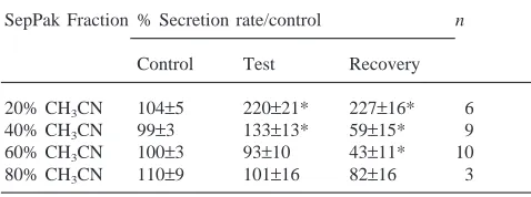
Table 1
Effect of 0.1 ant-eq/µl of the different SepPak fractions on the fluid secretion rate of single isolated Malpighian tubules of the anta
SepPak Fraction % Secretion rate/control n
Control Test Recovery
20% CH3CN 104±5 220±21* 227±16* 6
40% CH3CN 99±3 133±13* 59±15* 9
60% CH3CN 100±3 93±10 43±11* 10
80% CH3CN 110±9 101±16 82±16 3
a The third collection of secreted fluid during the control period
on fluid secretion. These results are similar to those obtained with a 10% TFA head/thorax extract of the ant (Laenen et al., 1999) and suggest that the abdominal extract contained both diuretic and antidiuretic factors. The 60% fraction seemed to have the highest content of antidiuretic factor(s) and was consequently used for further purification.
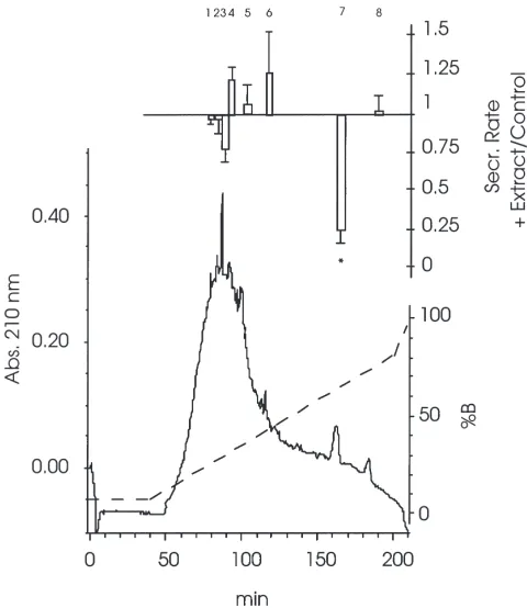
3.3. Chromatographic fractionation of the 60% CH3CN SepPak fraction
Running the 60% prepurified fraction on the prepara-tive Delta-Pak C18column yielded different fractions that were tested for their effect on fluid secretion in isolated Malpighian tubules (Fig. 3). Only one of them, fraction 7, significantly inhibited fluid secretion. This fraction had a retention time of 156 min. It eluted at 62% CH3CN and, when tested at a concentration of 1.0 ant-eq/µl, it irreversibly reduced the fluid secretion rate to a value of 25±5% of the control (n=4, p,0.01).
To process the total crude extract nine preparative runs had to be performed. Fraction 7 was pooled, con-centrated by centrifugal evaporation and temporarily stored in 50% CH3CN at220°C, prior to the
lyophiliz-Fig. 3. UV absorbance profile of 10,000 ant-eq of the 60% SepPak fraction separated on the Waters PrePak DP C18 column (300 A˚ ,
25×100 mm). Conditions are described in Section 2. The dashed line represents the concentration of CH3CN. The upper part shows the
effect of the fractions, tested at a concentration of 1.0 ant-eq/µl, on fluid secretion by isolated Malpighian tubules (means±SE, n=4). The effect is expressed as the test secretion rate relative to the control secretion rate. *Significantly different from 1 (p,0.01).
Fig. 4. Effect of FopADF, tested at a concentration of 1 ant-eq/µl, on fluid secretion by isolated Malpighian tubules of F. polyctena. The extract was present during collection periods 4–6. Values are means±SE, n=24.
ation needed to prepare the material for the next purifi-cation step. Chromatographic separation of the fraction 7 pool on the analytical Waters Delta-Pak HPI C18column revealed only one major UV-absorbing peak. This peak fraction had an inhibitory effect on the primary urine production, but in contrast to fraction 7 the effect was reversible (Fig. 4). Applied at a concentration of 1.0 ant-eq/µl it reduced the fluid secretion rate to 29±5% (n=24,
p,0.01)of the control, with recovery to 83±5% after washout. Based on this biological effect we named this factor F. polyctena antidiuretic factor (FopADF).
FopADF was finally fractionated on a Sephasil Protein C4 column (Fig. 5). However, after this last step we needed to apply it at a much higher concentration to pick up the antidiuretic effect in the bioassay. A concentration of 20 ant-eq/µl was needed to reduce the fluid secretion
Fig. 5. UV absorbance profile of 40,000 ant-eq of FopADF separated on the Sephasil Protein C4(5µm, 300 A˚ , 4.6×100 mm) column.
rate to a value of 45±15% of the control (n=4, p,0.05), with recovery to 93±5% after washout.
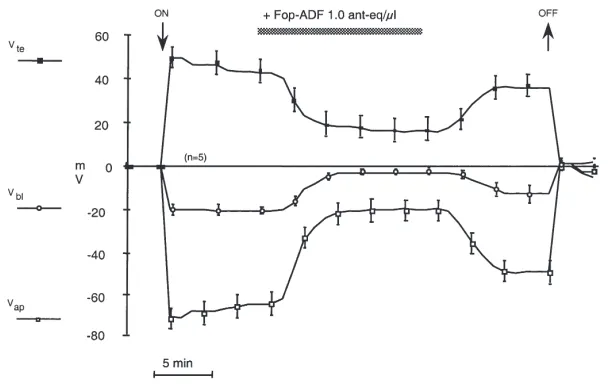
3.4. Effect of FopADF on membrane potentials
Freshly extracted FopADF, purified on the analytical C18column, was applied at a concentration of 1 ant-eq/µl to active tubules (Fig. 6). A reversible depolarization of both Vbl (from 221±2 mV to 23±1 mV, n=5) and of Vap (from 265±5 mV to 220±5 mV, n=5), measured simultaneously in the same tubule, was observed. After treatment with FopADF the potential profile was compa-rable to that measured by De Decker (1993) in non-secreting tubules: in these tubules Vbl was 25±5 mV (n=20) and Vte was +8±2 mV (n=5), resulting in Vap equal to 213 mV.
3.5. Cross-reactivity with T. molitor
When tested at a concentration of 1 ant-eq/µl the pur-ified extract reduced the fluid secretion rate of ant tubules to 43% ±8 (n=5, p,0.01) of the control. The same extract, tested at a concentration of 4 ant-eq/µl on mealworm tubules, reduced the fluid secretion rate to 37±16% (n=4) of the control (Fig. 7). At the end of the test period, FopADF was washed out and replaced by 1 mmol/l cyclic AMP. As expected, the fluid secretion rate was stimulated to 179±26% of the control (n=4). So FopADF inhibited the fluid secretion to the same extent as in the ant and the effect was not toxic: fluid secretion resumed after washout and could be stimulated with cyc-lic AMP.
Fig. 6. Running mean for the effect of FopADF, tested at a concentration of 1 ant-eq/µl, on the transepithelial (Vte) and the basolateral (Vbl)
membrane potentials measured simultaneously with two microelectrodes in the same tubule. The apical membrane potential (Vap) was calculated
as Vap=Vbl2Vte. Presence of the factor is indicated by the bar. Arrows indicate impalement (ON) or withdrawal (OFF) of the microelectrodes.
Means±SE, n=5 tubules.
Fig. 7. Effect of FopADF, tested at a concentration of 4 ant-eq/µl, on fluid secretion by isolated Malpighian tubules of the mealworm T.
molitor. FopADF was present during the period indicated by the bar,
then replaced by 1 mmol/l cyclic AMP. Values represent means±SE (n=4).
3.6. Weight loss due to formic acid release
Batches of 10 worker ants weighed 163±6 mg (n=5). The mean weight loss due to formic acid release when the ants were disturbed was 15.7±3.6 mg, or 9.4±1.8% of body mass (n=5).
4. Discussion
4.1. Purification of FopADF
Decker at al, 1994). Since antidiuretic effects were obtained with head extracts, the antidiuretic factor(s) may be released by the corpora cardiaca into the haemo-lymph. In the present study antidiuretic activity was demonstrated in extracts of both haemolymph and whole abdomens (containing haemolymph). Although activity was lost in the final HPLC step during purification of the abdomen extract, an antidiuretic factor was clearly present, causing a completely reversible inhibition (to about 50%) of the control secretion rate. Loss of activity always occurs during purification. Peptides can and do stick to plastic surfaces. This is especially true for small quantities of highly purified peptides in solution contain-ing small amounts of acetonitrile (Holman and Hayes, 1997).
An antidiuretic factor, demonstrated in the haemo-lymph of ants with inactive tubules, was purified from whole abdomens with a three step HPLC protocol. FopADF had a reversible, inhibitory effect on the fluid secretion of ant (and mealworm) Malpighian tubules. So far no endogenous factor with a direct inhibitory effect on fluid secretion has been purified, although antidiuretic effects have been previously described for crude extracts of A. domesticus (Spring et al., 1988), A. aegypti (Petzel and Conlon, 1991) and F. polyctena (De Decker et al., 1994). Inhibitory effects of exogenous peptides have also been demonstrated: destruxins reduce fluid secretion of
Schistocerca gregaria tubules (James et al., 1993) and
destruxin B has been shown to inhibit the activity of V-type H+-ATPases in a reversible manner (Muroi et al., 1994). The cardioacceleratory peptide CAP2b inhibits fluid secretion by tubules of R. prolixus (Quinlan et al., 1997) and T. molitor (R.A. Eigenheer, D.A. Schooley and S.W. Nicolson, unpublished data), although it has a stimulatory effect on those of Drosophila (Davies et al., 1995).
CAP2b, originally isolated from the hawkmoth
Mand-uca sexta (Davies et al., 1995), belongs to a group of
peptides that are believed to be involved in the regulation of cardiac function after adult emergence and in thermor-egulation during flight. So far only two CAPs have been sequenced: CAP2a (Cheung et al., 1992), a cyclic non-apeptide and CAP2b (Huesmann et al., 1995), an octa-peptide blocked at both the amino and carboxyl ends. CAP2aand CAP2bhave no sequence homology. Destrux-ins are cyclic peptide toxDestrux-ins isolated from the Destrux-insect pathogenic fungus Metarhizium anisopliae (James et al., 1993). Both CAP2b and destruxins are well protected from peptidase activity: CAP2b, since both the amino and carboxyl terminus are blocked, and destruxins, since they are cyclic peptides. Although they are small mol-ecules, they elute in the 60% CH3CN fraction (Cheung et al., 1992; Huesmann et al., 1995; James et al., 1993) indicating that these substances are quite hydrophobic. The importance of this characteristic in relation to their physiological function still has to be elucidated.
The elution behaviour of FopADF is comparable to that of the peptides described above. Consequently it is possible that FopADF might be a small amidated peptide blocked at the amino terminus with pyroglutamate or blocked by cyclization. Treatment of the purified material with pyroglutamate aminopeptidase would show whether it is N-terminally blocked. Shielding of the N-terminus would explain why we could not obtain sequence information using standard Edman degradation techniques (results not shown). Other methods may be needed (e.g. mass spectrometry) in order to obtain an amino acid sequence for FopADF.
4.2. Is FopADF a toxic substance or a metabolic inhibitor?
Since our antidiuretic factor was purified from whole abdomens, it could be argued that toxins from the poison gland may be responsible for the antidiuretic effects on the tubules. However, this can be discounted for two reasons. Firstly, formic acid comprises 99% of the poi-son gland secretion of formicine ants (Lo¨fqvist, 1976), the rest being minor hydrocarbons in negligible quan-tities. Secondly, formic acid is highly volatile and would probably disappear from the extract before an HPLC run. If not, it would pass through the column very rapidly.
Dufour’s gland is also located in the abdomen of for-micine ants, and its secretion, which is dominated by saturated hydrocarbons, is probably emitted together with formic acid as an alarm/defence signal (Lo¨fqvist, 1976). We do not consider the components of the Dufour’s gland secretion likely to be responsible for antidiuretic effects on ant Malpighian tubules, however, because the 15% TFA used in our solid-phase extraction procedure extracts water-soluble, low molecular weight substances, not membrane lipids or hydrocarbons.
Furthermore, in a study of second messengers (Laenen, in preparation) applying a Ca2+ ionophore A23187 in the presence of FopADF still had a small, but significant effect on fluid secretion on Formica tubules. Ca2+ seems to be the second messenger for F.
polyctena diuretic peptide, FopDP (Laenen et al., 1999).
Cyclic AMP had no stimulatory effect in the same con-ditions, but on washout of both factors, a typical over-shoot after cyclic AMP (Laenen, in preparation) was seen during the recovery. Possibly FopADF acts on cell properties beyond the effect of cyclic AMP, which can still be activated by Ca2+. These findings seem to indi-cate that FopADF is neither toxic or a metabolic inhibi-tor.
4.3. Mode of action of FopADF
second messengers (O’Donnell and Spring, 2000). Sero-tonin (in Rhodnius) and the CRF-like diuretic peptide Tem-DH1 (in Tenebrio) are stimulatory and act via cyc-lic AMP. The cardioacceleratory peptide CAP2b (in
Rhodnius) and an antidiuretic peptide (in Tenebrio) are
inhibitory and act via cyclic GMP (Quinlan et al., 1997; Furuya et al., 1995; Eigenheer et al., 1999). On the other hand, both nucleotides stimulate secretion in tubules of
Drosophila (Davies et al., 1995) and F. polyctena (De
Decker, 1993; Laenen, 1999), so neither of them can be second messengers for FopADF.
In Formica tubules at least part of the K+ uptake by the cell seems to be via a conductive channel: a favor-able electrochemical gradient for K+ has been demon-strated and Ba2+, blocking the K+ channels, severely inhibits fluid secretion (Weltens et al., 1992; Leyssens et al., 1993a). Although Ba2+reduces fluid secretion, it hyperpolarizes both apical and basolateral membrane potentials (Weltens et al., 1992). In the presence of Ba2+ the electrogenic pump at the level of the apical mem-brane continues to function and draws a current across the increased resistance of the basolateral membrane, hyperpolarizing it. Previously, it was also found that use of a metabolic inhibitor such as DNP inhibits the pump and completely abolishes fluid secretion, but does not affect Vbl(Leyssens et al., 1993b). Only when Ba2+and DNP are applied together does secretion stop and do all membranes reversibly depolarize.
FopADF mimicks this situation and would therefore be an efficient inhibitor of secretion by blocking both one of the entry pathways of K+ across the basolateral membrane and by inhibiting the pump across the apical membrane. Further electrophysiological experiments are needed to corroborate the decrease of K+ conductance in the basolateral membrane.
4.4. Physiological roles of antidiuretic factors
The physiological role of antidiuretic factors with a direct inhibitory effect on the Malpighian tubules of insects remains to be elucidated. In crickets the release of an antidiuretic hormone from corpora cardiaca was triggered by dehydration (Spring et al., 1988). The release could be triggered indirectly by the decline in hemolymph volume and reduced distention of the abdo-men, sensed by abdominal stretch receptors. Release of regulatory factors after a change in abdominal volume is well known for blood feeders which need to reduce their hemolymph volume after a blood meal (Maddrell, 1964).
In ants FopADF may be released after emptying the contents of the poison gland, of which formic acid makes up about 20% by volume (Graedel and Eisner, 1988). Assuming the rest of the gland contents to be water, and the water content of adult F. polyctena to be 77% (Coenen-Stass, 1986), the 10% mass loss of disturbed
ants represents an 10.4% reduction in body water. It is noteworthy that the ants drank a lot of water during the first 12 h after collection from their nests (unpublished observations). So a change in abdominal volume could also explain the release of FopADF in F. polyctena. The antidiuretic factor(s) may be important for inactivating the tubules and preventing a further reduction in body fluid. This idea is supported by the antidiuretic activity of the haemolymph extract from stressed ants. Further research is necessary to confirm this hypothesis, and antidiuretic effects at the level of the hindgut and the whole insect should not be overlooked.
Acknowledgements
The authors thank Mrs A. Roosen for preparing sol-utions, Mr P. Pirotte for making holding pipettes, Mr R. Van Werde for help with electronics and Mr W. Leys-sens for administrative tasks. We also thank Professor G.M. Coast for useful discussions. This work was sup-ported by a B.O.F.-grant (Bijzonder Onderzoeks Fonds) (1994–1998) from the Limburgs Universitair Centrum, and by a bilateral award (to EVK and SWN) under the Flemish–South African agreement on Science and Tech-nology Cooperation.
References
Cheung, C.C., Loi, P.K., Sylwester, A.W., Lee, T.D., Tublitz, N.J., 1992. Primary structure of a cardioactive neuropeptide from the tobacco hawkmoth, Manduca sexta. FEBS Letters 313, 165–168. Coenen-Stass, D., 1986. Investigations on the water balance in the red
wood ant, Formica polyctena (Hymenoptera, Formicidae): workers, their larvae and pupae. Comparative Biochemistry and Physiology 83A, 141–147.
Davies, S.A., Huesmann, G.R., Maddrell, S.H.P., O’Donnell, M.J., Skaer, N.J.V., Dow, J.A.T., Tublitz, N.J., 1995. CAP2b, a
cardioac-celeratory peptide, is present in Drosophila and stimulates tubule fluid secretion via cGMP. American Journal of Physiology 269, R1321–R1326.
De Decker, N., 1993. Regulation of fluid secretion in Malpighain tubules of Formica polyctena by exo- and endogenous factors. Ph.D. thesis, Limburgs Universitair Centrum, Belgium.
De Decker, N., Hayes, T.K., Van Kerkhove, E., Steels, P., 1994. Stimulatory and inhibitory effects of endogenous factors in head extracts of Formica polyctena (Hymenoptera) on the fluid secretion of Malpighian tubules. Journal of Insect Physiology 40, 1025– 1036.
Eigenheer, R.A., Nicolson, S.W., Schooley, D.A., 1999. Isolation and characterization of a cyclic GMP-elevating neuropeptide from
Tenebrio molitor. Abstract, paper presented at the 20th Winter
Neu-ropeptide Conference, Breckenridge, CO.
Furuya, K., Schegg, K.M., Wang, H., King, D.S., Schooley, D.S., 1995. Isolation and identification of a diuretic hormone from the mealworm Tenebrio molitor. Proceedings of the National Academy of Sciences of the USA 92, 12323–12327.
Graedel, T.E., Eisner, T., 1988. Atmospheric formic acid from for-micine ants: a preliminary assessment. Tellus 40B, 335–339. Ho¨lldobler, B., Wilson, E.O., 1990. The Ants. Springer, Berlin. Holman, G.M., Hayes, T.K., 1997. HPLC methods to isolate peptide
neurotransmitters. Methods in Molecular Biology 72, 205–218. Huesmann, G.R., Cheung, C.C., Loi, P.K., Lee, T.D., Swiderek, K.M.,
Tublitz, N.J., 1995. Amino acid sequence of CAP2ban insect
car-dioacceleratory peptide from the tobacco hawkmoth Manduca
sexta. FEBS Letters 371, 331.
James, P.J., Kershaw, M.J., Reynolds, S.E., Charnley, A.K., 1993. Inhibition of desert locust (Schistocerca gregaria) Malpighian tubule fluid secretion by destruxins, cyclic peptide toxins from the insect pathogenic fungus Metarhizium anisopliae. Journal of Insect Physiology 39, 797–804.
Laenen, B., 1999. Purification, characterization and mode of action of endogenous neuroendocrine factors in the forest ant, Formica
polyctena. Ph.D. thesis, Limburgs Universitair Centrum, Belgium.
Laenen, B., Verhaert, P., Schoofs, L., Steels, P., Van Kerkhove, E., 1999. Partial identification of a peptide that stimulates the primary urine production of single isolated Malpighian tubules of the forest ant Formica polyctena. Journal of Insect Physiology 45, 743–753. Leyssens, A., Van Kerkhove, E., Zhang, S.-L., Weltens, R., Steels, P., 1993a. Measurement of Intracellular and luminal K+concentrations in a Malpighian tubule (Formica). Estimate of basal and luminal electrochemical K+ gradients. Journal of Insect Physiology 39, 945–958.
Leyssens, A., Zhang, S.-L., Van Kerkhove, E., Steels, P., 1993b. Both dinitrophenol and Ba2+reduce KCl and fluid secretion in
Malpigh-ian tubules of Formica polyctena: the role of the apical H+and K+ concentration gradient. Journal of Insect Physiology 39, 1061– 1073.
Lo¨fqvist, J., 1976. Formic acid and saturated hydrocarbons as alarm pheromones for the ant Formica rufa. Journal of Insect Physiology 22, 1331–1346.
Maddrell, S.H.P., 1964. Excretion in the blood-sucking bug, Rhodnius
prolixus Sta˚l. III. The control of the release of the diuretic hormone.
Journal of Experimental Biology 41, 459–472.
Muroi, M., Shiragami, N., Takatsuki, A., 1994. Destruxin B, a specific and readily reversible inhibitor of vacuolar-type H(+)-translocating ATPase. Biochemical and Biophysical Research Communications 205, 1358–1365.
Nicolson, S.W., 1991. Diuresis or clearance: is there a physiological role for the ‘diuretic hormone’ of the desert beetle Onymacris? Journal of Insect Physiology 37, 447–452.
Nicolson, S., 1992. Excretory function in Tenebrio molitor: fast tubular secretion in a vapour-absorbing insect. Journal of Insect Physiology 38, 139–146.
O’Donnell, M.J., Spring, J.H., 2000. Modes of control of insect Mal-pighian tubules: synergism, antagonism, cooperation and auton-omous regulation. Journal of Insect Physiology. 46, 107–117. Petzel, D.H., Conlon, J.M., 1991. Evidence for an antidiuretic factor
affecting fluid secretion in mosquito Malpighian tubules. FASEB Journal 5, Abstract 1059.
Quinlan, M.C., Tublitz, N.J., O’Donnell, M.J., 1997. Anti-diuresis in the blood-feeding insect Rhodnius proxilus Sta˚l: the peptide CAP2b
and cyclic GMP inhibit Malpighian tubule fluid secretion. Journal of Experimental Biology 200, 2363–2367.
Spring, J.H., 1990. Endocrine regulation of diuresis in insects. Journal of Insect Physiology 36, 13–22.
Spring, J.H., Morgan, A.M., Hazelton, S.R., 1988. A novel target for antidiuretic hormone in insects. Science 241, 1096–1098. Van Kerkhove, E., Weltens, R., Roinel, N., De Decker, N., 1989.
Hae-molymph composition in Formica (Hymenoptera) and urine forma-tion by the short isolated Malpighian tubules: electrochemical gradients for ion transport. Journal of Insect Physiology 35, 991– 1003.