ANTIMICROBIAL SUBSTANCES FROM ENDOPHYTIC FUNGI IN
TAMARIND (
Tamarindus indica
, Linn), MALAY APPLE (
Eugenia malaccensis
,
Linn), RAMBUTAN (
Nephelium lappaceum
), AND INDIAN MULBERRY
(
Morindacitrifolia
, Linn)
Abdullah Dolah Dalee*, Saranyu Mukhurah, Khosiya Sali, Nurainee Hayeeyusoh, Zubaidah Hajiwangoh and Phurqanni Salaeh
Microbiology Program, Department of Science, Faculty of Science, Technology and Agriculture, Yala Province, Southern Thailand
Abstract
Endophytic fungi are known to produce useful substances including antibiotics and other active compounds. Endophytic fungi from local plants; Tamarind (Tamarindus indica, Linn), Malay apple (Eugenia malaccensis, Linn), Rambutan (Nephelium lappaceum), and Indian mulberry (Morinda citrifolia, Linn) were investigated for their ability to produce antimicrobial substances. Plant parts were sterilized cut, inoculated on Potato Dextrose Agar (PDA) plates, and incubated at 27OC for weeks until the appearance of endophytic growth. It was then found that the highest total endophytic fungal count was observed from Tamarind (39.47%). Upon screening of antimicrobial activity, not all but MA1, MA5, RB2, BU1 and YB1 isolates showed growth inhibition activity. Antimicrobial production in liquid (PDB) and solid (PDA) condition were then compared, and results showed that in liquid condition of PDB, the fungi gave higher production. Extraction of antimicrobial substances by culturing the isolates and distilling the cell-free filtrate with chloroform (1:3) yielded partially-purified extract (PPE) with different degree of antimicrobial activity on Escherichia coli, Salmonella typhi, Staphylococcus aureus, Bacillus cereus, and Candida albicans. Minimum Inhibitory Concentration (MIC) of these extracts in comparison with antibiotics as evaluated by broth microdilution technique showed that MA5 and YB1 gave the lowest value i.e. 1.709µg/mL, whereas BU1 gave the highest i.e. 437.5 µg/ml. RB2 gave similar MIC value with that of Chloramphenicol. As regard activity spectrum, all PPE were of broad type as it showed inhibition activity to all tested bacteria including C. albicans. Macroscopically, colonies of those isolates were white in color except for YB1, which was slightly reddish purple. Microscopically, all isolates showed aseptated hypha and non-sporulation in PDA. This primary study on local endophytic fungi and their plant sources is believed to be very useful for initiating further and advanced investigation with pharmaceutical application.
Key words: Antimicrobial, Endophytic fungi, Minimal Inhibitory Concentration (MIC), local plants
B-2
INTRODUCTION
Endophytic fungi residing in plant tissues are known by nature to produce active substance(s) with in vitroability to act as cancer, antiviral, antibacterial, antifungal, anti-oxidant, plant growth hormones, insecticides and other biochemical agents (Worapong et al., 2001a-b; Strobel, 2003 & 2004; Agut & Calvo, 2004; Liu et al., 2007; Pandi et al., 2010; Ramos et al., 2010; Ahmad et al., 2011; Bhimba et al., 2012; McCutcheon & Moran, 2012; Raffaele & Kamoun, 2012; Teiten et al., 2013; Zilla et al., 2013; Yadap et al., 2014; Agusta et al., 2014; Wei et al., 2014; Hussain et al., 2007, 2009, 2011, 2014 & 2015; Gao et al., 2015; Syamsia et al., 2015) with possible applications as starting materials for pharmaceutical, industrial and agrochemical products (Sturz et al., 2000; Strobel, 2006; Hardoim et al., 2008; Kaul et al., 2012; Brader et al., 2014). As a world has faced the wide spread resistant bacterial strains due to wide and extensive use of antibiotics in the treatment of infectious illness (Kumarasamy et al., 2010; D’Costa et al., 2011; Kempf & Rolain, 2012; Nordmann et al., 2012), endophytic microoragisms in particular the fungi have become the alternative resources for new antimicrobials. For decades the exploration of these fungi has brought about promising achievement (Tan & Zou, 2001; Strobel, 2003, 2006; Zhang et al., 2006; Guo et al., 2008; Priti et. al., 2009; Aly et. al., 2011; Kharwar et. al., 2011; Radic &Strukelj, 2012; Zhang et. al., 2012) These antimicrobial-producing endophytes are as diverse as wide world forests, and so, they become unlimited sources of these microbial endophytes (Strobel, 2003, 2006; Debbab et al., 2012; Radic & Strukelj, 2012; Mousa &Raizada, 2013).
vivo effect on cardiovascular disease and cancer (Wang & Su, 2001; Wang et al., 2002; Furusawa et al., 2003; Jasril et al., 2003; Wang et al., 2009, 2011; Nualsanit et al., 2012), fertility and inflammation (Hirazumi & Furusawa, 1999; Nualsanit et al., 2011), and infectious diseases (Saludes et al., 2002; Pawlus et al., 2010; Baque, 2011; Baque et al., 2011; Lv et al., 2011).
In the present study, tamarind, Malay apple, rambutan and Indian mulberry had been subjected to isolate endophytic fungi with antimicrobial activity against Esherichia coli, Salmonella typhi, Staphylococcus aureus and Bacillus cereus as well as Candida albicans. Efforts made included also preliminary characterization, MIC determination with reference to several antimicrobial agent. Subsequently, several endophytic fungi and partially putified extracts (PPEs) with efficacy in growth inhibition of test organisms, their characteristics, and MIC were expected to achieve.
RESEARCH METHOD
Plant materials. Plant samples were collected from sides of highway around Yala Capital District, Yala Province, Southern Thailand. Only disease-free leaf and branch stem of tamarind (Tamarindus indica, Linn.), Malay apple (Eugenia malaccensis, Linn.), rambutan (Nephelium lappaceum), and Indian mulberry (Morinda citrifolia, Linn.) were selected.
Test organisms.E. coli, S. typhi, St. aureus and B. cereus, and C. albicans, was supplied as a courtesy from Department of Microbiology, Faculty of Science, Prince of Songkla University, Hadyai, Songkhla Province, Southern Thailand. Bacteria were maintained on Nutrient Agar (NA, Merck, Germany) slant and yeast on PDA slants at 4OC in refrigerator after confirming their purity.
Reference antimicrobial agents. Chloramphenicol (Cloman, Thailand), Streptomycin (M & H, Thailand), Rifampicin (Sigma, USA), Penicillin V (Sigma, USA), Tetracycline (Sigma, USA), and Nystatin (Cloman, Thailand) used as reference in minimal inhibition concentration (MIC) determination were purchased from suppliers in the locality. Their preparation was done as guided by manufacturers’ description, and used as per requirement of experimental procedures.
Isolation of endophytic fungi. Leaf and stem samples were washed, surface-sterilized, blot dried, excised (~ 0.2 cm3), and inoculated (40 pc/plate) using both aqueous agar (AA) and potato dextrose agar (PDA, Merck, Germany), supplemented by 0.3% (w/v) each of Chloramphenicol andAmpicillin sodium salt (Merck, Germany) (Nalini et al., 2014; Arnold et. al., 2000). Fungal colony from each segment developed following several days of incubation at 27OC was subsequently transferred to antibiotic-free PDA for culture purifyingand identifying purposes. Morphological and reproductive structure and spore characteristics was determined(Barnett& Hunter, 1998; Domsch et. al., 2003; Leslie&Summerell, 2006; Mulloch, 2014).Culture stocks on PDA slants were maintained at 4OC until further uses.
Preliminary Screening for antimicrobial-producing endophytes. Modified agar plate-based assay was employed in screening of endophyte isolates(Arasu et. al., 2009). Aseptically, hyphal plugs from maximal growth on PDAwasplaced on Mueller Hinton Agar (MHA, Merck, Germany)lawn of each test bacteria (1x10⁸ cfu/mL)spread or PDA lawn of C. albicans (1x10⁴ cfu/mL) spread. Clear zone surrounding the endophyte plugs developed after 24 hours at 27OC incubation, was measured for inhibition activity evaluation.
B-4
Hayeewangoh, 2002). Both solid medium and fungal mycelium were harvested, and distillingextracted by soaking in chloroform(Sigma, USA) for 72 hours at 27OC with 150-rpm agitation. Finally, resulting biomass as partially-purified extract (PPE) was filtered, vapour-dried using Rotary Evaporator (Buchi, Switzerland), kept in sterile capped bottles in refegerator until further use. Fungal biomass from 200-mL PDB was treated, filterred and distilled in the same manner as previously described.
Evaluation of Antimicrobial activity. , Antimicrobial activity was evaluated against test bacteria on MHA, and yeast on PDAby employing disc diffusion technique(Schwalbe et. al., 2007). Aseptically, sterile paper discs (6 mm-diameter) mounted with 10 µL of each endophyte extract (10 mG/mL), was firmly placed on the prepared test cultures, and incubated at 35OC(28OC for yeast) for 24-48 hours. Diameter of inhibition zone was measured.
Minimal Inhibition Concentration (MIC) of fungal extracts. MICs of each PPE and reference antimicrobial agent were determinedusing broth microdilution method (Pawthong et. al., 2012).Each PPE and reference agentin 96-well of polystyrene microtitre plates (Thermoscientific, USA)was serially 2-fold dilutedto make concentration ranged from 875
μG/mL to 0.43 μG/mL. Into each wells, 10 μL suspension of each test organism(cfu/mL: 1x10⁸ or 1x10⁴) was added to diluted mixtures of PPE-reference agents (350 μL) and 2-strength
growth media (350 μL ). After incubation at 28O
C for 24-48 hours, growth inhibition was evaluated based on developed turbidity.
Morphological characteristics of endophytes. Each of MA1, MA5, RU2, BU1 and YB1endophyticisolates were characterized macroscopically and microscopically using conventional techniques including slide culture technique.
RESULT AND DISCUSSION
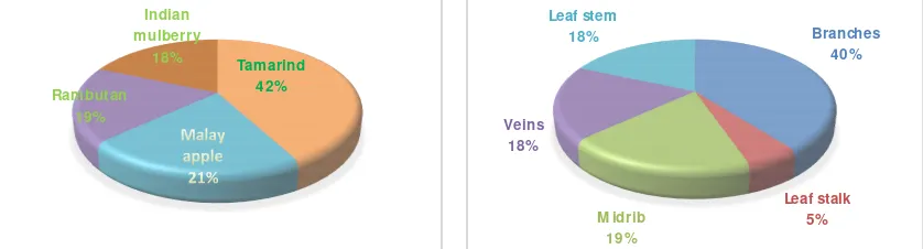
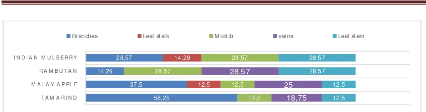
EndophyticDistribution.Counts of endophytic fungi recoveredfrom organ parts of tamarind, Malay apple, rambutan, and Indian mulberry were varied with the highest were from that of tamarind, 42.10%. Lowest count was recovered from rambutan and Indian mulberry, 18.32%each (Figure 1). Endophyte count from branches washigh, and from leaf stalk was low. Moderate count was observed from midrib, vein and stem sections of leaf (Figure 1). There was no uniformity in occurrence of fungal endophyte for every host plant (Figure 2). Higher number of fungal endophytes were recovered from tamarind branches (56.25%), and lower numbers were from midrib, veins and leaf stem of rambutan and Indian mulberry (28.5% each). Detailed result was shown in Figure 2.
Figure 1. Percentage of endophytic fungi recovered from (Left) disease-free parts of tamarind, Malay apple, rambutan and Indian mulberry, and from (Right) different disease-free parts of host plants.
Tamarind 42%
Rambutan 19%
Indian mulberry
18%
Branches 40%
Leaf stalk 5%
M idrib 19%
Veins 18%
Figure 2. Distribution percentage of fungal endophytes in various organal parts of host plants, i.e. tamarind, Malay apple, rambutan, and Indian mulberry.
Varying distribution of endophytic fungi in different parts of plants was common, and determined by factors like ecology, nutrient availability, plant physiology and experimental parameters (Madigan et. al., 2015). Despite not many reports described this variation, Waenawae (2009) reported similar result of endophyte distribution in organ parts of cashew nut tree (Anacardium occidenfale) and common jujube (Zizyphus mauritiana, Lamk), cussud tree (Cassia siamea, Lamk), Khraimanpoo (Glochidion sphaerogynum), and Longkong (Lanseum domesticum correa).Dolah (2005) reported varying percentage distribution in branches, and leaf stalk, midrib, vein and stem of endophytic fungi recovered from pomelo (Citrus maxima, Merr) and bullet wood (Mimusops elengi, Linn.). Sama (3013) and Bangosatoo (2013) comparatively described endophytic fungal distribution in parts of pomelo (Citrus maxima, Merr), bullet wood (Mimusops elengi, Linn.), guava (Psidium guajava), marian plum (Bouea macrophylla), Santol (Sandoricum koetjape), tamarind (Tamarindus indica, Linn), Malay apple (Eugenia malaccensis, Linn), Indian mulberry and (Morinda citrifolia, Linn).
In Angelica sinensis root, stem and leaf, 3 separate collectionsof endophytic fungi recorded a total recovery of 206 isolates representing 22 species with 24.27%, 26.21% and 49.51% distribution (Shu et. al., 2013). Khan and colleagues (2010) reported the distribution of endophytic flora in medicinal Withania somnifera plant employing 643 segments (202 leaf, 391 stem, and 50 root) from 20 different plants, and claimed 20species within 12 genera of fungi, including 9 fungi from leaves, 20 from stems and 4 from roots.Leaf and stem segments of N. arbor-tristis were reported to give recovery of 19 endophytic fungal species in 15 taxa and 10 species in 9 taxa, with Alternaria alternata showed highest colonization in leaf tissues (15.0%), and Cladosporium cladosporioides mostly (12%)colonized stem tissues (Gond et. al., 2010). Occurrence of different groups of endophytes in halophytes from an estuarine mangrove forest was reported as their percentage occurrence and species richness (Suryanarayanan & Kumaresan, 2000).
Antimicrobial-producing endophytes.Results of preliminary screening of endophytic isolates for the antimicrobial production were shown in Table 1. There was neither all isolates exhibitinginhibition zone on growth lawn of any tested bacteria and yeast, norsingle isolate showed inhibition activity on all growth lawns of organisms.MA1, MA5, MC3, MC5, MD2, and ME4 tamarind isolatesdemonstrated varying degree of inhibition zones. Malay apple isolates showing inhibition zones were RA1, RA2, RB2, and RB5. Rambutan-related BU1 and BU4, and Indian mulberry-related YB1, YB2, and YE1 were also antimicrobial producers. PPEs with wider activity spectrum were from MC5, RA1, BU1, and YE1.
56,25
B-6
Total Antimicrobial producers Antimicrobial non-producers Antimicrobial Spectrum
Percentage of antimicrobial producers in endophyte fungal population colonizing plant tissues was not only varied but also time-dependent. Waenawae (2009) reported the screening of endophytic fungi from Anacardium occidenfale and Lanseum domesticum correa, and isolated GU2M, GU2B, LA2P, LA1B, and RM1M with narrow spectrum of activity against only single test organism, i.e. E. coli or St. aureus. Dolah (2005) reported endophytic fungi isolation from pomelo (Citrus maxima, Merr) and bullet wood (Mimusops elengi, Linn) that only 8.33% of isolates wereantimicrobial producers, and of these only 1 was active against St. aureus and E. coli, but not S. typhi, B. cereus nor C. albicans. Casella and colleagues (2013) screened tropical leaf endophytes, and described 4 of 138 extracts (2.9%) possessing significant antibacterial activity against S. aureus, and 22 extracts (15.9%) were active against C. albicans, S. flexnii, S. boydii, S. enteritidis, S. paratyphi, P. aeruginosa, C. freundii, M. morganii, and P. vulgaris. This difference in distribution finding partly due to the nature of endophyte-host plant association, which in turn determined their natural trait (Madigan et. al., 2015). Producers fromTamarind (37.5%), Malay apple (50%), rambutan (28.57%), and Indian mulberry (42.86%) were all ethnomedicinal plants, contrasting with A. occidenfale and L. domesticum correa as well aspomelo and bullet wood, which were not. Such nature of association effected their distribution.
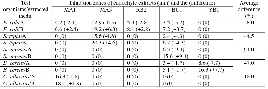
varying degree. S. typhi were however found sensitive to MA5 and BU1, while St. aureus to BU1, and B. cereus to BU1 and YB1. As for C. albicans, it was only MA1 was effective (Table 2). As the actvity spectrum was concerned, BU1 had broader spectrum than those of MA1 and MA5. MA1 had wider range of actvity encompassing inhibition activity against fungi, C. albicans.
Table 2.Inhibition zone of extracts from endophyte grown in solid (A) and liquid (B) media of PDA and PDB evaluated by disc diffusion method against test organisms.
Test organisms/extracted
media
Inhibition zones of endophyte extracts (mm) and the (difference) Average difference
(%)
MA1 MA5 RB2 BU1 YB1
E. coli/A 4.2 (-2.4) 12.9 (-6.3) 5.3 (-2.8) 3.5 (-3.7) 0 (0) 38.0
E. coli/B 6.6 (+2.4) 19.2 (+6.3) 8.1 (+2.8) 7.2 (+3.7) 0 (0)
S. typhi/A 0 (0) 15.6 (-4.6) 0 (0) 2.4 (-4.3) 0 (0) 44.5
S. typhi/B 0 (0) 20.3 (+4.6) 0 (0) 6.7 (+4.3) 0 (0)
St. aureus/A 0 (0) 0 (0) 0 (0) 6.3 (-9.4) 0 (0) 94.0
St. aureus/B 0 (0) 0 (0) 0 (0) 15.6 (+9.4) 0 (0)
B. cereus/A 0 (0) 0 (0) 0 (0) 3.4 (-1.7) 8.6 (-7.7) 47.0
B. cereus/B 0 (0) 0 (0) 0 (0) 5.1 (+1.7) 16.3 (+7.7)
C. albicans/A 16.3 (-1.8) 0 (0) 0 (0) 0 (0) 0 (0) 18.0
C. albicans/B 18.1 (+1.8) 0 (0) 0 (0) 0 (0) 0 (0)
Many works on antimicrobial activity of endophytic fungi from plants were brought out, and claimed more or less similar degree of activity and spectrum. Bhimba& colleaques (2012) described the extract having activity against E.coli, Pseudomonas, Enterococcus, Staphylococcus, and Bacillus with zone inhibition ranged from 12 to 30 mm. Filtrate of endophytic fungal culture broth of Talaromyces flavus, Mortierella hyaline, Paecilomyces variabilis, Penicillium sp., which were isolated from ethnomedicinal host plants, Potentilla fulgens, Osbeckia stellate, Osbeckia chinensis, Camellia caduca, and Schima khasiana was reported to show antimicrobial activity against B. cereus (MTCC 430), St. aureus (MTCC 740), E. coli (MTCC 116), S. typhi (MTCC 733), and C. albicans (MTCC 227) with inhibition zone ranging from 8.60 mm to 32.67 mm (Bhagobaty & Joshi, 2012). Interestingly, those extracts were active against both bacteria and fungi, a case of which was in consistent with this current finding. Antibacterial activity of extracts from Kigelia Africana-associated endophytic fungi Aspergillus flavus, Aspergillus sp., Curvularia lunata, Cladosporium sp. and 3 unidentified were found active against B. subtilis, St. aureus and E. coli with 14-37 mm inhibition zone diameter, and broader spectrum(Idris et. al., 2013). Plectophomellasp., Physalosporasp., and Crataegus monogyna extracts showed fungistatic and bacteriostatic activities with 1.0-30 mm inhibition zones (Hussain et. al., 2014). Moreover, , Yang and co-workers (2014) demonstrated by using agar diffusion assay that Camellia sinensisbranch-associated endophytic fungus, Alternaria alternata displayed bacteriostatic effect against St. aureus and B. subtilis with 10–25 mm inhibition zones but insensitive to E. coli. Ficus pumila Linn-associated endophytic Phomopsis sp. was active against a panel of human and phytopathogenic microbes with 16-30.66 mm and 14-30 mm inhibition zones for test fungi and for test bacteria, respectively (Rakshith et. al., 2013).
B-8
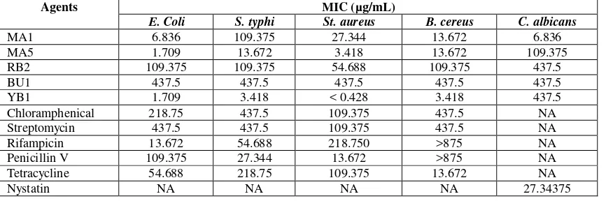
organisms, including C. albicans. Malay apple-associated endophyte RB1 PPE exhibited moderate inhibition activity (µg/mL MIC ranged 54.688-437.5). Most effective bacteriostatic activity was demonstrated by Indian mulberry-associated endophyte PPE, YB1, whose MICs ranged from < 0.427 µg/mL to 3.418 µg/mL. Whereas, rambutan-associated BU1 showed highest MICs at 437.5 µg/mL. In addition, all PPEs had both bactericidal and fungical activity as seen by C. albicanssensitivity (µg/mL MICs, 6.836 to 437.5). Such MIC range was comparable with reference fungistatic Nystatin. Also, bacteriostatis of PPEs was comparable with reference agents, whose MICs ranged from 13.672 µg/mL to >875 µg/mL. As far as the efficacy of these PPEs were concerned, MA1 was found to have lower MIC than those of reference antimicrobial agents, whose MIC ranged 13.672 µg/mL to >875 µg/mL. For E. coli, MA5 and YB1 PPEs were the most effective (MIC, 1.709 µg/mL). Furthermore, YB1 was seen to be the most effective to all test bacteria (MIC range, < 0.428 µg/mL to 3.418 µg/mL).
MIC evaluation of endophyte biological substance(s) in comparison with the reference agents was very useful in determining their efficacy. That was why almost all reports on antimicrobial activity included MIC determination. Casella and colleaques (2013) reported MIC for C. albicans and St. aureus of crude extract from tropical leaf endophytes were ≤ 128 µg/mL. MIC ranging from 0.49 µg/mL to 15.625 µg/mL was reported by Powthong and co-workers (2012), who determined antimicrobial activities of endophytic fungi crude extract recovered from Sesbania grandiflora (L.) Pers. Against St. aureus ATCC 25923 B. subtilis ATCC6633, E. coli ATCC 25922, Pseudomonas aeruginosa ATCC 27853, C. albicans, Cryptococcus neoformans using broth microdilution method.
Table 3. MICs of the endophyte PPEs with reference to antimicrobial agents. NA standed for Not Applicable, i.e. tests were not performed.
Agents MIC (µg/mL)
E. Coli S. typhi St. aureus B. cereus C. albicans
MA1 6.836 109.375 27.344 13.672 6.836
MA5 1.709 13.672 3.418 13.672 109.375
RB2 109.375 109.375 54.688 109.375 437.5
BU1 437.5 437.5 437.5 437.5 437.5
YB1 1.709 3.418 < 0.428 3.418 437.5
Chloramphenical 218.75 437.5 109.375 437.5 NA
Streptomycin 437.5 437.5 109.375 437.5 NA
Rifampicin 13.672 54.688 218.750 >875 NA
Penicillin V 109.375 27.344 13.672 >875 NA
Tetracycline 54.688 218.75 109.375 13.672 NA
Nystatin NA NA NA NA 27.34375
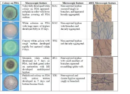
Macroscopic and microscopic characteristics of endophytes. Endophyte isolates MA1, MA5, RB2, BU1, and YB1were macroscopically and microscopically studies using conventional technique including slide culture, and it was found that these fungi on PDA medium were non-septate and did not produce any reproductive spores (Table 3). Consequently, it was unable and with doubt to identify the fungal identity based on the available characteristics. However, their characteristics were summarized in Table 3.
CONCLUSION AND SUGGESTION
contrasting with leaf stalks that were low count. There was neither uniformity in their
distribution in every host plant, nor equality in their ability to produce antimicrobial
substances. Hence, highest total endophytic fungal count was recovered from
Tamarind (39.47%), and its associated MA1, MA5, MC3, MC5, MD2, and ME4
manifested varying inhibition zone sizes. Malay apple-associated RA1, RA2, RB2, and
RB5, rambutan-associated BU1 and BU4, and Indian mulberry-associated YB1, YB2,
and YE1 displayed unequal antimicrobial activity too. Extracts with wider spectrum
included MC5, RA1, BU1, and YE1.Antimicrobial production in liquid (PDB) media
gave higher yield than that of solid (PDA) condition. Partially-purified extract (PPE)
that resulted from extracting and distilling using 1/3 chloroform showed different
degree of antimicrobial activity against
E. coli
,
S. typhi
,
St. aureus
,
B. cereus
, and
C.
albicans
with MICas lowest as 1.71 µg/ml. This signified the higher efficacy, and
happened to be lower MICs than those of reference antimicrobial agents, whose MIC
rangedfrom 13.671875 µg/mL to >875 µg/mL. All PPE showed broad type of
spectrum. It was unfortunately unable to identify the isolates because their
macroscopical and microscopical characteristics were not in line with those described in
identification key of simple fungi. Therefore, we designated as unidentified isolates.
This preliminary study on local endophytic fungi and their plant sources is believed to
be very useful for initiating further and advanced investigation with pharmaceutical
application
B-10
Acknowledgement. This work was funded by Microbiology Program, Department of Science, Faculty of Science, Technology and Agriculture, Yala Rajabhat University, Yala Province, South Thailand. All concerns are appreciated.
REFERENCES
A. Ramos, A. Visozo, J. Piloto, A. Garc´ıa, C. A. Rodr´ıguez, R. Rivero. (2003). Screening of
antimutagenicity via antioxidant activity in Cuban medicinal plants. J. Ethnopharmacol. 87, 241– 246.
Agusta, A., D. Wulansari, Praptiwi, A. Nurkanto, A. Fathoni. (2014). Biotransformation of Protoberberine Alkaloids by the Endophytic Fungus Coelomycetes AFKR-3 Isolated from Yellow Moonsheed Plant (Archangelisia flava (L.) Merr.). Procedia Chem. 13, 38 – 43.
Agut, M & M. A. Calvo. (2004). In vitro conidial germination inArthrinium aureum and Arthrinium phaeospermum.Mycopathologia. 157, 363-367.
Ahmed, I., H. Hussain, B. Schulz, S. Draeger, D. Padula, G. Pescitelli,et al. Three new antimicrobial metabolites from the endophyticfungus Phomopsis sp. European J Org Chem 2011; 15: 2867-2873. Al-Fatimi, M., M. Wurster, G. Schroder, U. Lindequist. (2007). Antioxidant,antimicrobial and cytotoxic
activities of selected medicinal plants fromYemen. J. Ethnopharmacol. 111, 657–666.
Aly, A. H., A. Debbab, P. Proksch. (2011). Fungal endophytes: unique plant inhabitants with great promises. Appl. Microbiol. Biotechnol. 90, 1829–1845.
Arasu, M. V., V. Duraipandiyan, P. Agastian, S. Ignacimuthu. (2009). In vitro antimicrobial activity of Streptomyces spp. ERI-3 isolated from Western Ghats rock soil (India). J. de Mycologie Medicale. (19)1, 22–28.
Arnold, A. E., Z. Maynard, G. S. Gilbert, P. D. Coley, T. A. Kursar. (2000). Are tropical fungal endophytes hyperdiverse? Ecol. Lett. (3)4, 267–274.
Bangosatoo, K. (2013). Endophytic fungi and antimicrobial products from tamarind (Tamarindus indica, Linn), Malay apple (Eugenia malaccensis, Linn), rambutan (Nephelium lappaceum), Indian mulberry (Morinda citrifolia, Linn), and marian plum (Bouae burmanica, Griff). B. Sc thesis. Yala Rajabhat University. Yala. Thailand.
Baque, M. A. (2011). Production of biomass and secondary metabolites through adventitious root cultures of Morinda citrifolia using bioreactors. Ph. D thesis, Chungbuk National Univeristy, Republic of Korea.
Baque, M. A., A. Elgirban, E. J. Lee, K. Y. Paek. (2011). Sucrose regulated enhanced induction of anthraquinone, phenolics, flavonoids biosynthesis and activities of antioxidant enzymes in adventitious root suspension cultures of Morinda citrifolia (L.). Acta Physiol. Plant. 34(2), 405-415.
Barbosa-Filho, J. M., T. H. C. Vasconcelos, A. A. Alencar, L. M. Batista; R. A.G. Oliveira; D. N. Guedes; H. S. Falcão; M. D. Moura; M. F.F.M. Diniz; J. Modesto-Filho. (2005). Plants and their active constituents from South, Central, and North America with hypoglycemic activity. Revista Brasileira de Farmacognosia. 15, 392–413.
Barnett, H., B. Hunter. (1998). Illustrated Genera of Imperfect Fungi. Minneapolis: Burgess Publishing. USA.
Bhagobaty, R. K., S.R. Joshi. (2012). Antimicrobial and antioxidant activity of endophytic fungi isolated from ethnomedicinal plants of the “Sacred forests” of Meghalaya, India. Mikologia Lekarska. 19(1), 5-11.
Bhat. R. S., S. Al-daihan. (2014). Antimicrobial activity of Litchi chinensis and Nephelium lappaceum aqueous seed extracts against some pathogenic bacterial strains. J. King Saud University– Sci. 26, 79–82.
Bhimba, B. V, D. A. A. D. Franco, J. M. Mathew, G. M. Jose, E. L. Joel, M. Thangaraj. (2012).Anticancer and antimicrobial activity of mangrove derived fungi Hypocrea lixii VB1. Chin. J. Natur. Med. 10, 0077-0080
Brader, G., S. Compant, B. Mitter, F. Trognitz, A. Sessitsch. (2014). Metabolic potential of endophytic bacteria. Curr. Opin. Biotechnol. Sci. 27, 30–37.
endophytes: Isolation of antibacterial agent pyrrocidine C from Lewia infectoria SNB-GTC2402. Phytochem. 96, 370–377.
Consolini, A. E., O. A. N. Baldini, A. G. Amat. (1999). Pharmacological basis for the empirical use of Eugenia uniflora, L. (Myrtaceae) as antihypertensive. J. Ethnopharmacol. 66(1), 33–39.
D’Costa, V. M., C. E. King, L. Kalan, M. Morar, W. W. L. Sung, C. Schwarz, D. Froese, G. Zazula, F. Calmels, R. Debruyne, G. B. Golding, H. N. Poinar, G. D. Wright. (2011). Antibiotic resistance is ancient. Nature 477, 457–461.
Dalimartha, S. (2003) Atlas of Indonesian medicinal plants (In Bahasa Indonesia), 2nd Ed., Jakarta: Puspa Swara.
Davidson, J. L., A. Davidson, H. Saberi, T. Jaine. (2006). The Oxford Companion to Food. Oxford [Oxfordshire]: Oxford University Press.
Debbab, A., A. H. Aly, P. Proksch. (2012). Endophytes and associated marine derived fungi ecological and chemical perspectives. Fungal Diversity. 57, 45–83.
Dolah, Nurizan. (2005). Antimicrobial substances from endophytic fungi in pomelo (Citrus maxima, Merr) and bullet wood (Mimusops elengi, Linn). B. Sc thesis. Yala Rajabhat University. Yala, Thailand.
Domsch, K. H., W. Gams, T. Anderson. (2003). Compendium of Soil Fungi. New York: Academic Press, USA.
Falcão, H. S., I. O. Lima, V. L. Santos, H. F. Dantas; M. F.F.M. Diniz; J. M. Barbosa-Filho; L. M. Batista. (2005). Review of the plants with anti-inflammatory activity studied in Brazil. Revista Brasileira de Farmacognosia. 15, 381–391.
Furusawa, E., A. Hirazumi, S. Story, J. Jensen. (2003). Antitumour potential of a polysaccharide-rich substance from the fruit juice of Morinda citrifolia (Noni) on sarcoma 180 ascites tumour in mice. Phytother Res. 17(10), 1158–1164.
Gao, Y., Q. Liu, P. Zang, X. Li, Q. Ji, Z. He, Y. Zhao, H. Yang, X. Zhao, L. Zhang. (2015). An endophytic bacterium isolated from Panax ginseng C.A. Meyer enhances growth, reduces morbidity, and stimulates ginsenoside biosynthesis. Phytochem. Lett. 11, 132–138.
Gond, S. K., A. Mishra, V.K. Sharma, S. K. Verma, J. Kumar, R. N. Kharwar, A. Kumar. (2010). Diversity and antimicrobial activity of endophytic fungi isolatedfrom Nyctanthes arbor-tristis, a well-known medicinal plant of India. Mycoscience. 53, 113–121.
Guo, B., Y. Wang, X. Sun, K. Tang. (2008). Bioactive natural products from endophytes: a review. Appl. Biochem. Microbiol. 44, 136–142.
Hardoim, P.R., L. S. V. Overbeek, J. D. Elsas. (2008). Properties of bacterial endophytes and their proposed role in plant growth. Trends Microbiol. Sci. 16, 463–471.
Haruenkeit, R., S. Poovarodom, S. Vearasilp, J. Namiesnik, M. Sliwka-Kaszynska, Y-S. Park, B.-G. Heo, J-Y. Cho, H. G. Jang, S. Gorinstein. (2010). Comparison of bioactive compounds, antioxidant and antiproliferative activities of Mon Thong Durian during ripening, Food Chem. 118(3), 540-547. Havinga, R. M., A. Hartl, J. Putscher, S. Prehsler, C. Buchmann, C. R. Vogl. (2010). Tamarindus
indicaL. (Fabaceae): Patterns of use in traditional African medicine. J. Ethnopharmacol. 127, 573– 588.
Hayeewangoh, Z. (2002). Screening of antimicrobial metabolites from filamentous fungi. M. Sc thesis, Prince of Songkhla University. Songkhla, Thailand.
Hemshekhar, M., K. Kemparaju, K. S. Girish. (2011). Tamarind (Tamarindus indica) Seeds: An Overview on Remedial Qualities. In Nuts and Seeds in Health and Disease Prevention, pp.1107-1114. Hirazumi, A., E. Furusawa. (1999). An immunomodulatory polysaccharide-rich substance from the fruit
juice of Morinda citrifolia (Noni) with antitumor activity. Phytother. Res. 13(5), 380–387.
Hussain H, Tchimene MK, Ahmed I, Meier K, Steinert M, DraegerS, et al. Antimicrobial chemical constituents from the endophyticfungus from Phomopsis sp. from Notobasis syriaca. Nat ProdCommun 2011; 6: 1905-1906.
B-12
Hussain, H., K. Krohn, S. Draeger, K. Meier, B. Schulz. (2009). Bioactive chemical constituents of a sterile endophytic fungus from Meliotus dentatus. Rec Nat Prod. 3, 114-117.
Hussain, H., K. Krohn, Z. Ullah, S. Draeger, B. Schulz. (2007). Bioactive chemical constituents of two endophytic fungi. Biochem Syst Ecol. 35, 898-900.
Hussain, H., M. John, A. Al-Harrasi, A. Shah, Z. Hassan, G. Abbas, U. A. Rana, I. R. Green, B. Schulz, K. Krohn. (2015). Phytochemical investigation and antimicrobial activity of an endophytic fungus Phoma sp. J. King Saud Univ. – Sci. 27, 92–95.
Idris, A., I. Al-tahir, E. Idris. (2013). Antibacterial activity of endophytic fungi extracts from the medicinal plant Kigelia Africana. Egypt. Acad. J. Biol. Sci. 5(1), 1-9.
Jalikop, S. H. (2013). Rambutan. <http://www.fruitipedia.com/>.
Jasril, L. N. H., L. Y. Mooi, M. A. Abdullah, M. A. Sukari, A. M. Ali. (2003). Antitumor promoting and antioxidant activities of anthraquinones isolated from the cell suspension cultures of Morinda elliptica. Asian Pac. J. Mol. Biol. Biotechnol. 11, 3–7.
Kaul, S., S. Gupta, M. Ahmed, M. K. Dhar. (2012). Endophytic fungi frommedicinal plants: a treasure hunt for bioactive metabolites.Phytochem Rev.11, 487-505.
Kaushik, G., S. Satya, R. K. Khandelwal, S. N. Naik. (2010). Commonlyconsumed Indian plant food materials in the management of diabetes mellitus. Diabetes and metabolic syndrome. Clin. Res. Rev. 4(1), 21–40.
Kempf, M. & J. M. Rolain. (2012). Emergence of resistance to carbapenems in Acinetobacter baumannii in Europe: clinical impact and therapeutic options. Int. J. Antimicrob. Agents 39, 105–114. Khan, R., S. Shahzad, M. I. Choudhary, S. A. Khan, A. Ahmad. (2010). Communities of endophytic fungi
in medicinal plant Withania somnifera. Pak. J. Bot., 42(2), 1281-1287.
Kharwar, R. N., A. Mishra, S. K. Gond, A. Stierle, D. Stierle. (2011). Anti-cancer compounds derived from fungal endophytes: their importance and future challenges. Nat. Prod. Rep. 28, 1208–1228 Khonkarn, R., S. Okonogi, C. Ampasavate, S. Anuchapreeda. (2010). Investigation of fruit peel extracts
as sources for compounds with antioxidant and antiproliferative activities against human cell lines. Food Chem. Toxicol. 48, 2122–2129.
Komutarin, T., L. Azadi, L. Butterworth, D. Keil, B. Chitsomboon, M.Suttajit, B. J. Meade. (2004). Extract of the seed coat of Tamarindus indica inhibits nitricoxide production by murine macrophages in vitro and in vivo. Food Chem.Toxicol. 42, 649–658.
Kumarasamy, K. K., M. A. Toleman, T. R. Walsh, J. Bagaria, F. Butt, R. Balakrishnan, U. Chaudhary,. M. Doumith, C. G. Giske, S. Irfan, P. Krishnan, A. V. Kumar, S. Maharjan, S. Mushtaq, T. Noorie, D. L. Paterson, A. Pearson, C. Perry, R. Pike, B. Rao, U. Ray, J. B. Sarma, M. Sharma, E. Sheridan, M. A. Thirunarayan, J. Turton, S. Upadhyay, M. Warner, W. Welfare, D. M. Livermore, N. Woodford. (2010). Emergence of a new antibiotic resistance mechanism in India, Pakistan, and the UK: a molecular, biological, and epidemiological study. Lancet Infect. Dis. 10, 597–602.
Leslie, J. F., B. A. Summerell. (2006). The Fusarium Laboratory Manual. London: Blackwell Publishing, UK.
Lestari, S. R., M. S. Djati, A. Rudijanto, F. Fatchiyah. (2014). The physiological response of obese rat model with rambutan peel extract treatment. Asian Pac J Trop Dis. 4(S2), S780-S785
Lima, I. O., R. A. G. Oliveira, E. O. Lima, N. M. P. Farias, E. L. Souza. (2006). Atividade antifúngica de óleos essenciais sobre espécies de Candida. Revista Brasileira de Farmacognosia. 16, 197–201. Liu, X., M. Dong, X. Chen, M. Jiang, X. Lv, G. Yan. (2007). Antioxidant activity and phenolics of an
endophytic Xylaria sp. from Ginkgo biloba. Food Chem. 105, 548–554.
Lunardi, I., J. L. B. Peixoto, C. C. da Silva, I. T. A. Shuquel, E. A. Basso, G. J. Vidotti. (2001). Triterpenic acids from Eugenia moraviana. J. Brazilian Chem. Soc. 12(2), 180–183.
Lv, L., H. Chen, C-T. Ho, S. Sang. (2011). Chemical components of the roots of Noni (Morinda citrifolia) and their cytotoxic effects. Fitoterapia. 82(4), 704–708.
Madigan,M. T., J. M. Martinko, K. S. Bender, D. H. Buckley, D. A. Stahl. (2015). Brock biology of microorganisms. 14th Ed. New York: Pearson Education, Inc.
Mahmoud, I. I., M. S. A. Marzouk, F. A. Moharram, M. R. El-Gindi, A. M. K. Hassan. (2001). Acylated flavonol glycosides from Eugenia jambolana leaves. Phytochem. 58(8), 1239–1244.
Martinello, F., S. M. Soares, J. J. Franco, A. C. Santos, A. Sugohara, Garcia, S. B., C. Curti, S. A. Uyemura. (2006). Hypolipemic and antioxidant activities from Tamarindus indica L. pulpfruit extract in hypercholesterolemic hamsters. Food Chem. Toxicol.44, 810–818.
McCutcheon, J. P. & N. A. Moran (2012). Extreme genome reduction in symbiotic bacteria. Nat. Rev. Microbiol. 10, 13–26.
Mousa, W. K.& M. N. Raizada. (2013). The diversity of anti-microbial secondary metabolites produced by fungal endophytes: an interdisciplinary perspective. Front. Microbiol. 4, 65.
Muhtadi, A. U. Primarianti, T. A. Sujono. (2015). Antidiabetic activity of Durian (Durio zibethinus Murr.) and Rambutan (Nephelium lappaceum L.) fruit peels in alloxan diabetic rats. Procedia Food Sci. 3, 255–261.
Muhtadi, Haryoto, T. A. Sujono, A. Suhendi, K. H. Yen. (2014). Antioxidant and chemical constituents of some Indonesian fruit peels, Med. Plants–Inter. J. Phytomed. Related Industries. 6(1), 43-46. Mulloch, D. (2014). Moulds; Isolation, cultivation and identification.
http://website.nbm-mnb.ca/mycologywebpages/Moulds/Moulds.html.
Muthu, S. E., S. Nandakumar, U. A. Rao. (2005). The effect of methanolic extract ofTamarindus indica Linn. on the growth of clinical isolates of Burkholderiapseudomallei. Indian J. Med. Res. 122, 525– 528.
Nalini, M. S., N. Sunayana, H. S. Prakash. (2014). Endophytic Fungal Diversity in Medicinal Plants of Western Ghats, India. International Journal of Biodiversity. 2014, http://dx.doi.org/10.1155/2014/494213
Nawawi, A., N. Nakamura, M. Hattori, M. Kurokawa, K. Shiraki. (1999). Inhibitory effects of Indonesian medicinal plants on the infection of herpes simplex virus type 1. Phytother. Res. 13, 37–41. Nordmann, P., L. Dortet, L. Poirel. ( 2012). Carbapenem resistance in Enterobacteriaceae: here is the
storm. Trends Mol. Med. 18, 263–272.
Nualsanit, T., P. Rojanapanthu, W. Gritsanapan, S. H. Lee, D. Lawson, S. J. Baek. (2012). Damnacanthal, a noni component, exhibits antitumorigenic activity in human colorectal cancer cells. J. Nutr. Biochem. 23(8):915–923.
Nualsanit, T., P. Rojanapanthu, W. Gritsanapan, T. Kwankitpraniti, K. W. Min, S. J. Baek. (2011). Damnacanthal-induced anti-inflammation is associated with inhibition of NF-kappa B activity. Inflamm. Allergy Drug Targets. 10(6), 455–463.
Okonogi, S., C. Duangrat, S. Anuchpreeda, S. Tachakittirungrod, S. Chowwanapoonpohn. (2007). Comparison of antioxidant capacities and cytotoxicities of certain fruit peels. Food Chem. 103, 839–846.
Oliveira, R. N., I. J. M. Dias, C. A. G. Câmara. (2005). Estudo comparativo do óleo essencial de Eugenia punicifolia (HBK) DC. de diferentes localidades de Pernambuco. Revista Brasileira de Farmacognosia. 15, 39–43.
Palanisamy, U. D., L. T. Ling, T. Manaharan, D. Appleton. (2011a). Rapid isolation of Geraniin from Nephelium lappaceum rind waste and its anti-hyperglycemic activity. Food Chem. 127, 21–27. Palanisamy, U., H. M. Cheng, T. Masilamani, T. Subramaniam, L. T. Ling, A. K. Radhakrishnan. (2008).
Rind of the rambutan, Nephelium lappaceum, a potential source of natural antioxidants. Food Chem. 109(1), 54–63.
Palanisamy, U., T. Manaharan, L. L. Teng, A. K. C. Radhakrishnan. T. Subramaniam, T. Masilamani. (2011b). Rambutan rind in the management of hyperglycemia. Food Res. Inter. 44, 2278–2282. Pandi, M., R. Manikandan, J. Muthumary. (2010). Anticancer activity of fungal taxol derived from
Botryodiplodia theobromae Pat.; an endophytic fungus, against 7, 12 dimethyl benz(a)anthracene (DMBA)-induced mammary gland carcinogenesis in Sprague dawley rats. Biomed. & Pharmacother. 64, 48–53.
Pawlus, A. D., A. D. Kinghorn. (2007). Review of the ethnobotany, chemistry, biological activity and safety of the botanical dietary supplement Morinda citrifolia (noni). J. Pharm. Pharmacol. 59, 1587–1609.
B-14
Potterat, O., M. Hamburger. (2007). Morinda citrifolia (noni) fruit: phytochemistry, pharmacology, safety. Planta Med.73, 191–199.
Powthong, P., B. Jantrapanukorn, A. Thongmee, P. Suntornthiticharoen. (2012). Evaluation of endophytic fungi extract for their antimicrobial activity from Sesbania grandiflora (L.) Pers. Int. J. Pharm. Biomed. Res. 3(2), 132-136.
Priti, V., B. T. Ramesha, S. Singh, G. Ravikanth, K. N. Ganeshaiah, T. S. Suryanarayanan, R. Umashaanker. (2009). How promising are endophytic fungi as alternative sources of plant secondary metabolites? Curr. Sci. 97, 477–478.
Radic, N. & B. Strukelj. (2012). Endophytic fungi—The treasure chest of antibacterial substances. Phytomed. 19, 1270–1284.
Raffaele, S. & S. Kamoun. (2012). Genome evolution in filamentous plant pathogens: why bigger can be better. Nat. Rev. Microbiol. 10, 417–430.
Rakshith, D., P. Santosh, S. Satish. (2013). Isolation and characterization of antimicrobial metabolite producing endophytic Phomopsis sp. from Ficus pumila Linn. (Moraceae). Internation. J. Chem. Anal. Sc. 4, 156-160.
Ramos, H. P., G. H. Braun, M. T. Pupo, S. Said. (2010). Antimicrobial activity from endophytic fungi Arthrinium state of Apiospora montagnei Sacc. and Papula sporaimmersa. Brazilian Arch. Biol. Technol. 53, 629-632.
Ravi, K., S. Rajasekaran, S. Subramanian. (2005). Antihyperlipidemic effect of Eugenia jambolana seed kernel on streptozotocin-induced diabetes in rats. Food Chem. Toxicol. 43(9), 1433–1439.
Roosita, K., C. M. Kusharto, M. Sekiyama, Y. Fachrurozi, R. Ohtsuka. (2008). Medicinal plants used by the villagers of a Sundanese community in West Java, Indonesia. J. Ethnopharmacol. 115(1), 72–81.
Rosa, L. H., N. Tabanca, N. Techen, Z. Pan, D. E. Wedge, R. M. Moraes. (2012). Antifungal activity of extracts from endophytic fungi associated with Smallanthus maintained in vitro as autotrophic cultures and as pot plants in the greenhouse. Can. J. Microbiol. 58, 1202–1211.
Saad, S., M. Taher, D. Susanti, H. Qaralleh, N. A. Abdul Rahim. (2011). Antimicrobial activity of mangrove plant (Lumnitzera littorea). Asian Pac. J. Trop. Med. 523-525.
Saludes, J. P., M. J. Garson, S. G. Franzblau, A. M. Aguinaldo. (2002). Antitubercular constituents from the hexane fraction of Morinda citrifolia Linn. (Rubiaceae). Phytother. Res. 16, 683–685.
Sama, F. (2013). Antimicrobial substances from endophytic fungi in pomelo (Citrus maxima, Merr), bullet wood (Mimusops elengi, Linn), guava (Psidium guajava), marian plum (Bouae burmanica, Griff), and santol (Sandoricum koetjape). B. Sc Thesis. Yala Rajabhat University. Yala, Thailand. Schwalbe, R., L. S. Moore, A. C. Goodwin. (2007). Antimicrobial Susceptibility Testing Protocols.
Boca Raton, London, New York: CRC Press, Taylor and Francis Group.
Shu, J., Q. Da-wei, Y. Nian-yun, T. Jin-hua, D. Jin-ao. (2013). Biodiversity and Antimicrobial Activity of Endophytic Fungi in Angelica sinensis. Chinese Herbal Med. 5(4), 264-271.
Strobel, G. A. (2003). Endophytes as sources of bioactive products. Microb. & Inf. 5, 535–544 Strobel, G. A. (2004). Natural products from endophytic microorganism. J. Natur. Prod. 67, 257-268. Strobel, G., (2006). Harnessing endophytes for industrial microbiology. Curr. Opin. Microbiol. 9, 240–
244.
Sturz, A.V., B. R. Christie, J. Nowak. (2000). Bacterial endophytes: potential role in developing sustainable systems of crop production. Crit. Rev. Plant Sci. 19, 1–30.
Sudjaroen, Y., R. Haubner, G. Würtele, W. E. Hull, G. Erben, B. Spiegelhalder, S. Changbumrung, H. Bartsch, R. W. Owen. (2005). Isolation and structureelucidation of phenolic antioxidants from tamarind (Tamarindus indica L.) seedsand pericarp. Food Chem. Toxicol. 43, 1673–1682.
Suryanarayanan, T. S., V. Kumaresan. (2000). Endophytic fungi of some halophytes from an estuarine mangrove forest. Mycol. Res. 104(12), 1465-1467.
Syamsia, T. Kuswinanti, E. Syam’un & A. Masniawati. (2015). The Potency of Endophytic Fungal Isolates Collected from Local Aromatic Rice as Indole Acetic Acid (IAA) Producer. Procedia Food Sci. 3, 96–103
Tachakittirungrod, S., S. Okonogi, S. Chowwanapoonpohn. (2007). Study on antioxidant activity of certain plants in Thailand: Mechanism of antioxidant action of guava leaf extract. Food Chem. 103, 381–388.
Tamimy. (2006). Antioxidant activity of ethanol extract of Rambutan (Nephellium lappaceum, L.) fruit peel against DPPH free radical suppression by spectrophotometer (In Bahasa Indonesia), Yogyakarta.
Tan, R.X. & W. X. Zou (2001). Endophytes: a rich source of functional metabolites. Nat. Prod. Rep. 18, 448–459.
Teiten, M-H., F. Mack, A. Debbab, A. H. Aly, M. Dicato, P. Proksch, M. Diederich. (2013). Anticancer effect of altersolanol A, a metabolite produced by the endophytic fungus Stemphylium globuliferum, mediated by its pro-apoptotic and anti-invasive potential via the inhibition of NF-ƙB activity. Bioorg. & Med. Chem. 21, 3850–3858.
Thitilertdecha, N., A. Teerawutgulrag, J. D. Kilburn, N. Rakariyatham. (2010). Identification of major phenolic compounds from Nephelium lappaceum L. and their antioxidant activities. Molecules. 15(3), 1453–1465.
Thitilertdecha, N., A. Teerawutgulrag, N. Rakariyatham. (2008). Antioxidant and antibacterial activities of Nephelium lappaceum L. extracts. Food Sci. Technol. 41, 2029–2035.
Ushanandini, S., S. Nagaraju, K. H. Kumar, M. Vedavathi, D. K. Machiah, K. Kemparaju,B. S. Vishwanath, T. V. Gowda, K. S. Girish. (2006). The anti-snake venomproperties of Tamarindus indica (Leguminosae) seed extract. Phytother. Res. 20,851–858.
Velázquez, E., H. A. Tournier, P. M. de Buschiazzo, G. Saavedra, G. R. Schinella. (2003). Antioxidant activity of Paraguayan plant extracts. Fitoterapia. 74(1-2), 91–97.
Waenawae, N. (2009). Antimicrobial substances from endophytic fungi in Anacardium occidenfale and Lanseum domesticum correa. B. Sc thesis. Yala Rajabhat University. Yala, Thailand.
Waltner-Law M, E., X, L. Wang, B, K. Law, R, K. Hall, M. Nawano. (2002). Epigallocatechin gallate, a constituent of green tea represses hepatic glucose production, J. Bio. Chem. 277(38), 34933–34940. Wang, M. Y., B. J. West, C. J. Jensen, D. Nowicki, C. Su, A. K. Palu, G. Anderson. (2002). Morinda citrifolia (Noni): a literature review and recent advances in Noni research. Acta Pharmacol. Sin. 23(12), 1127–1141.
Wang, M. Y., C. Su. (2001). Cancer preventive effect of Morinda citrifolia (Noni). Ann. New York Acad. Sci. 952, 161–168.
Wang, W., E. R. Rayburn, Y. Zhao, H. Wang, R. Zhang. (2011). Novel ginsenosides OH-PPD and 25-OCH3-PPD as experimental therapy for pancreatic cancer: anticancer activity and mechanisms of action. Cancer Lett. 278, 241–248.
Wang, Y., M-H. Yang, X-B. Wang, T-X. Li, L-Y. Kong. (2014). Bioactive metabolites from the endophytic fungus Alternaria alternata. Fitoterapia, 99, 153–158.
Wei, W., N. Jiang, Y. N. Mei, Y. L. Chu, H. M. Ge, Y. C. Song, S. W. Ng, R. X. Tan. (2014). An antibacterial metabolite from Lasiodiplodia pseudotheobromae F2. Phytochem. 100, 103–109. Worapong, J., G. A. Strobel, B. Daisy, U. Castillo, G. Baird, W.M. Hess. (2001a). Muscodor roseus
anam.. nov. an endophyte from Grevillea pteridifolia. Mycotaxon. 81, 463-475.
Worapong, J., G.A. Strobel, E.J. Ford, J.Y. Li, G. Baird, W.M. Hess. (2001b) Muscodor albus gen. et sp. nov. an endophyte from Cinnamomum Zeylanicum. Mycotaxon. 79, 67-79.
Yadav, M., A. Yadav, J. P. Yadav. (2014). In vitro antioxidant activity and total phenolic content of endophytic fungi isolated from Eugenia jambolana Lam. Asian Pac J Trop Med. 7(S1), S256-S261. Zhang, H.W., Y. C. Song, R. X. Tan. (2006). Biology and chemistry of endophytes. Nat. Prod. Rep. 23,
753–771.
Zhang, Y., T. Han, Q. Ming, L. Wu, K. Rahman, L. Qin, 2012. Alkaloids produced by endophytic fungi: a review. Nat. Prod. Commun. 7, 963–968.
Zilla, M. K., M. Qadri, A. S. Pathania, G. A. Strobel, Y. Nalli, S. Kumar, S. K. Guru, S. Bhushan, S. K. Singh, R. A. Vishwakarma, S. Riyaz-Ul-Hassan, A. Ali. (2013). Bioactive metabolites from an endophytic Cryptosporiopsis sp. inhabiting Clidemia hirta. Phytochem. 95, 291–297.