INFLUENCE OF JAMU MADURA “EMPOT SUPER” ON THE VAGINAL EPITHELIUM THICKNESS OF WHITE MICE (Rattus norvegicus) – AN IN VIVO
STUDY
Anik Listiyana
Department of Biology Faculty of Science and Technology Universutas Islam Negeri Maulana Malik Ibrahim Malang
Corresponding author: [email protected]
ABSTRACT
The aim of this research is to determine the influence of jamu Madura “Empot Super” (JMES) on the vaginal epithelium thickness of Rattus norvegicus in vivo. This research is kind of “true experimental-post test only control group design”. The rats were given drinking JMES once daily PS (Per-Sonde) for a month, then the vagina was taken to be sample for HE colouring. The sample was observed by the binocular microscope (100 times magnification) to identify the changes in the thickness of their vaginal epithelium. Calculation of the vaginal epithelium thickness was counted on the 10 field of view chosen randomly by the blind method. The result show that the vaginal epithelium thickness increased with dose 0,17mg/BW, 0,34mg/BW, and 0,68mg/BW of JMES compared with negative control group. But, the vaginal epithelium thickness decrease at the dose 0,51mg/BW compared with negative control group.
Keywords: Jamu Madura “Empot Super” (JMES), vaginal epithelium thickness, white mice (Rattus norvegicus), In Vivo study
INTRODUCTION
Indonesia is known as an archipelago with megabiodiversity. These biodiversity are used to fulfill population’s needs, including for medication. Indonesian has utilized medicinal plants to make a jamu. Madura’s people have also used medicinal plant to make a jamu and healed so many diseases. Then these jamu were handed down to the next generation. Now, jamu used by Madura’s people is developed and disseminated from local environment to the global civilization. So, people around the world can take the benefits of Jamu Madura (Holil, 2015). One of them is jamu Madura which can heal and treat for men and women reproduction health (Handayani, dkk., 1998).
There are so many kind of Jamu Madura used for treating and healing of men and women
reproduction, one of them is Jamu Madura “Empot Super” (JMES) that has so many brand
names. JMES is also known as Jamu “Sari Rapat” (also called “rapet”, “pakak”, “sari rapet”, “empot ayam”, “empot-empot”) that is mostly consumed by women in Madura. JMES is used for reducing vaginal mucous physiologically to becomes drier, to overcome the itching and not good smell on women, preserving the marital relationship and always be youthful. It can be
Beside that, it can strengthen woman reproduction organ and look so youthful (Handayani, dkk., 1998).
JMES is marketed with various brand name and has same efficacy, but it is contained various medicinal plants. Based on Saleh (2009), JMES is formulated of some plants, which are
cortex of pomegranate stem (Punica granatum L), pronojiwo seed (Euchresta horsfieldii (Lesch.)
Benn.), majakani (Quercus lusitanica Lam.), kayu rapet (Parameria laevigata (Juss.) Moldenke),
and other materials ad 100%.
The plants used for JMES were ordinary medicinal plants that were known long ago for
women reproduction treatment. For example, cortex of pomegranate stem (Punica granatum L) is
used for medication of candidiasis; pronojiwo seed (Euchresta horsfieldii (Lesch.) Benn.) for
tonic and treating impotence (Dalimartha, 2003). There is of kayu rapet’s cortex (Parameria
laevigata (Juss.) Moldenke) that is used to relief a pain after giving birth (Sundari et al, 2005)
and also majakani (Quercus lusitanica Lam.) for treating candidiasis and irregular menstruation
cycle. Some of those plants have been tested phytochemically.
Based on phytochemical screening, majakani (Quercus lusitanica Lam.) contains active
compound, including galotannins, essentials oil, starch, and tannic acid (Roshni R and Ramesh G,
2013). Kayu rapet (P. laevigata) has some active compound; they are flavonoid, polyphenol,
saponin, and tannins. Phytochemical screening of (P. laevigata) shows that it contains 6 kind of
prothosianidin (Kamiya et al., 2001). Then, pomegranate (P. granatum) contains alkaloid, reason, this jamu allegedly related to reproduction hormone, including estrogen. Estrogen is reproduction hormone that has fucntion to maintain the thickness of vaginal epithelium and make it red (Sturdee D.W. et al., 2010). This research aims to determine the effect of jamu Madura “Empot Super” on the vaginal epithelium thickness of white mice (Rattus norvegicus) in vivo.
MATERIAL AND METHOD
This research was conducted in the Physiology Laboratory and Biosistematic & Experimental Animal Laboratory, Science and Technology Faculty, State Islamic University of
Maulana Malik Ibrahim Malang by true eksperimental-post test only control group design that
aimed to determine and compare some experimental groups. It used Rattus norvegicus as
experimental animal and was treated by jamu Madura Madura “Empot Super” per sonde once
daily for 30 days.
The samples used for this research are 30 female Rattus norvegicus (8-10 weeks old).
Those rats were not fed for 24 hours, but they were given water ad libitum and acclimatized for 7
days. Then they were screened by same criteria. Every rat was given an identity tag. They were placed in the cages; each cage contains 3-5 rats. The enviromental temperature was setted at the range 23-26°C with 12 hours dark-light cycle. Those rats were divided to be 5 groups and each
group consist of 5 rats. First group, the rats were treated by H2O 5ml as negative control group.
Group I, the rats were treated by H2O 5ml + 0,17mg/gBW jamu Madura “Empot Super”. Group
II, the rats were treated by H2O 5ml + 0,34mg/gBW jamu Madura “Empot Super”. Group III, the
were treated by H2O 5ml + 0,68mg/gBW jamu Madura “Empot Super”. Body weights of the rats minor surgical instrument, sterofoam, straight pin, waterbath, microtome, incubator, paraffin block cassett, oven paraffin, alat tulis, binocular microscope with camera, tube 15ml, cotton bud, and magnifying glass.
Materials
Buffer formaldehyde 10%, PBS, Ethanol absolute 50%, 70%, 80%, 90%, Xylol 100%, Hematoxilin-Eosin (HE) stain, sterile Aquadest, pellet BR-1, Bleomycin, sterile NaCl 0,9%, chaff, tissue, cotton, Alcohol 70%, chloroform.
The experiment is started by given an identity tag to the each rat. They were placed in the cages; each cage contains 3-5 rats. The enviromental temperature was setted at the range 23-26°C with
12 hours dark-light cycle. The rats were fed by pellet BR-1 and water ad libitum.
The treatment of Jamu Madura “Empot Super”
The tablet of jamu Madura “Empot Super” measured was based on the needs each group.
Then that was mixed with H2O, and given peroral by sonde. The treatment was conducted for 30
days. After 30 days, the rats were sacrificed with chloroform. Then they were dissected on the abdominal cavity and the vaginal was taken carefully to analyse the epithelial thickness. Those organs were washed by sterile PBS and fixated by buffer formaldehyde 10%. Then they were made as preparations of HE hystological staining.
Data Analysis
The results from HE hystological staining were analyzed by measuring the vaginal epithelium thickness in µm. The average measurement results will be compared between treatment groups. The data obtained is ratio data and presented as table. Hypothesis was proven
by different test; but before that, the data was analysed by normality test using
Kolmogorov-Smirnov to determine normality of data distribution. If the data had normal distribution, so it will be conducted a data analysis using parametric statistical test. But, non-parametric statistical test was applicated for non-normal data distribution. One Way ANOVA (Parametric statistical test) is applicated to determine the distinction between control group and treatment groups. The results
obtained were used to determine the influence of doses given of jamu Madura “Empot Super” on
the thickness of rat vaginal epithelium. This treatment was approved when p value was less than
0, 05 (p ≤ 0, 05). Then, PostHoc analysis was conducted with Turkey test. That test was applied
to determine the groups with significant distinction.
RESULT AND DISCUSSION
The thickness of rat’s vaginal epithelium from HE staining is observed by binocular light
microscope with 100 magnification. The results showed that there were significant differences
in the some treatment groups.
The vaginal epithelium thickness of Rattus norvegicus after treatment showed that the
Control (-)
Dosage1 Dosage 2
Dosage 3 Dosage 4
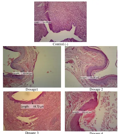
Figure 1. hystological and longitudinal cuts of the thickness of rat (Rattus norvegicus) vaginal epithelium after treatment (HE staining and 100 magnification) Information:
Control (-)
: Rat was treated with H2O 5ml (epithelium thickness 54,88 µm)
Dosage 1 : Rat was treated with H2O 5ml + 0,17mg/gBW jamu Madura “Empot Super” (epithelium
thickness itel 63,08 µm)
Dosage 2 : Rat was treated with H2O 5ml + 0,34mg/gBW jamu Madura “Empot Super” (epithelium
thickness 74,78 µm)
Dosage 3 : Rat was treated with H2O 5ml + 0,51mg/gBW jamu Madura “Empot Super” (epithelium
thickness 44,70 µm)
Dosage 4 : Rat was treated with H2O 5ml + 0,68mg/gBW jamu Madura “Empot Super” (epithelium
Figure 2. Graphic about the measurement average of rat vaginal epithelium after treatment (the data is presentated as mean SD)
Based on the statistical test, the data had normal distribution, and homogen. Generally, there were significant difference between each group, except in the negative control group and group I. In the group II, group III, and group IV exhibited that the thikcness of vaginal epithelium increased stastically.
In each group was given different treatment dosage of jamu Madura “Empot Super”. Jamu Madura “Empot Super” was known as tonic for organ reproduction, including vagina. It was known can thighten the vaginal muscles, but not proven scientifically. The information was just about the advantages of medicinal plant contained in that jamu.
Medicinal plants contained in the jamu Madura “Empot Super” have active compounds
including gallotannin, essential oil, starch, tannic acid, flavonoid, polyphenol, saponin, tannin,
pronthocyanidin, alkaloid, glycoside, and terpen (Kamiya, et al., (2001) Roshni R and Ramesh G,
(2013), Andriani, (2011)). Scientist suspected that this jamu was related to the activity of women
hormone, estrogen. Accordance to the study related to that by Satpathy S, et al., (2013), Punica
granatum L. had ability related to the estrogen activity. Punica granatum L. is medicinal plant
contained in the jamu Madura “Empot Super”. Estrogen has effect to increase collagen tissue on
the vagina, increasing the thickness of vaginal epithelium, and decreasing the atrophy of vaginal
epithelium (Sturdee D.W. et al., 2010).
Estrogen caused enhancement of mitosis and proliferation of epithelium cells and
hardening process of the epithelium surface (Kumar, et al, 2005). Estrogen is natural hormone
produced by ovary that can cause proliferation of vaginal epithelium. Estrogen worked by
sticking to the estrogen reseptor α (ER-α) on the target cell (cells in the vagina) to influence the
activity of cell proliferation. Conformational changes caused the bond between estrogen and its receptor going to be active, and then they were bonded with binding site of DNA. This interaction caused the increasing of gen expressions. Gen expression was catalyzed by RNA polymerase (enzym) that also can increase mRNA. Beside that, tRNA also increased that causes increasing of cel proliferation on the vaginal epithelium and escalating the thickness of vaginal
epithelium. Estrogen also can bond with estrigen receptor α stroma indirectly to activate
paracrine factor such as epidermal growth factor (EGF). Activated EGF such as tyroxine kinase
can be found in the epithelium. EGF and tyroxine kinase activated protein kinase in the cell
citoplasm. Protein kinase such as mitogen-activated protein kinase (MAPK) wich became the
main signal of trancription activator and active translation. Then, the synthesis of protein was conducted that needed for epithelium mitosis. Mitosis of epithelial cell caused sel proliferation (Cooke. et al., 1998). The development and ability of ovary to produce estrogen can be seen indirectly from the effect of estrogen produced on the changes of vaginal epitheliuum cell. Estrogen caused the increasing of mitosis, escalating cells proliferation and hardening process on the epithelial usurface (Astirin and Mutmainah, 2002).
Generally, it can be concluded that increasing of epithelium thickness was proportional with incresing of the dosage, except for third dosage (in the group III). This condition was fathomed because of the increasing of estrogen level. This experiment was similiar with study
conducted by Raden (2011), the thickness of vaginal epithelium of Rattus norvegicus Wistar
increased after be treated by pegagan extract. Pegagan (kind of medicinal plant) was known as phytoestrogen that can increase the collagen level and the vaginal epithelium thickness of ovariectomized rats. Proliferation and maturation of epithelial cell tend to increase (in micrometer scale) caused by the treatment of pegagan extract and that had significant disticntion from control group (without treatment of pegagan extract). Based on microscopical observation, incision of the cervix (vaginal wall) coloured by HE staining proved that there was the thickening of the rats cervix after treated by pegagan.
In this experiment, generally it can be concluded that increasing of epithelium thickness
was proportional with incresing of the dosage, except for third dosage (H2O 5ml + 0,51mg/gBW
jamu Madura “Empot Super”). In the third dosage, the thickness of epithelium decreased under the thickening in negative control group. It was often found in traditional medicine composed by some medicinal plants or multicomponents. Those components can produced sinergistic activity,
additive, or antagonistic activity. The third dosage was sugested that didn’t give positive effect.
Beside that, there was antioxidant compound in the jamu Madura “Empot Super” (Holil, 2015).
Antioxidant compound found in jamu can canged to be prooxidant (Suryani et al, 2013). In the
jamu Madura “Empot Super”, there were antioxidant compounds, including flavonoid, gallotannin, etc (Holil, 2015). Antioxidant level added took effect on oxidation rate. In high concentration, antioxidant activity of phenol function group was lost or canged to be prooxidant. Therefore, the third dosage was sugested that antioxidant compounds were changed to be
prooxidant. Prooxidant was toxic and damaging cells (Wijaya et al., 2014).
CONCLUSION
The treatment of jamu Madura “Empot” Super in the white mice (Rattus norvegicus) can
increase the thickness of its vaginal epithelium. This experiment shows that the increasing of vaginal epithelial thickness is in line with the increasing of dosage of jamu Madura “Empot Super” compared to the negative control group. But, that is not happen in the dosage 0,51mg/BW
jamu Madura “Empot Super”. In that dosage, the thickness of vaginal epithelium decreases
compared to the negative control group.
REFERENCES
[1] Andriani Ary. 2011. Skrining Fitokimia dan Uji Penghambatan Aktivitas α-Glukosidase
pada Ekstrak Etanol dari Beberapa Tanaman yang Digunakan Sebagai ObatAntidiabetes. Skripsi Diterbitkan. Fakultas Matematika dan Ilmu Pengetahuan Alam (FMIPA). Universitas Indonesia. Jakarta.
[2] Astirin O. P dan Mutmainah. 2002. Struktur Histologi Ovarium Tikus (Rattus
[3] Cooke, Paul S., Buchanan, David L., Lubahn, Dennis B., Cunha, Gerald R. 1998.
Mechanism of Estrogen Action: Lessons from the Estrogen Receptor-α Knockout Mouse.
Biology of Reproduction 59: 470-475. Urbana.
[4] Dalimartha S. 2003. Atlas Tumbuhan Obat Indonesia. Jilid 3. Cetakan I. Puspa Swara.
Jakarta
[5] Handayani L., Suharmiati, Sukirno S., Djoerban B., Soegijono K., dan Pranata S. 1998.
Inventarisasi Jamu Madura yang Dimanfaatkan untuk Pengobatan atau Perawatan Gangguan Kesehatan Berkaitan dengan Fungsi Reproduksi Wanita. Buletin Penelitian Sistem Kesehatan. Vol. 2 (1): 40-53
[6] Holil K. (2015). Uji Antioksidan Jamu Madura Empot Super. Jurnal El-Hayah. Vol. 5(3):
111-117
[7] Kamiya K., Watanabe C., Endang H., Umar M., and Satake T. 2001. Constituents of Bark
of Parameria laevigata. Chem. Pharm. Bull. Vol. 49(5): 551-557.
[8] Kumar V., Abbas A.K., Fausto N., Aster J. 2005. Robbins & Cotran Pathologic Basis of
Disease. 7th Edition. Elsevier
[9] Raden A. 2011. Efek Ekstrak Pegagan (Centella asiatica) pada Rattus norvegicus Wistar
yang Dilakukan Ovariektomi terhadap Proliferasi Epitel pada Dinding Vagina. Jurnal Ilmiah Kedokteran Hewan Vol. 4(1): 71-76
[10] Roshni P.S., Ramesh G. 2013. Anticancer Activity of Acetone Extract of Quercus
Infectoria Olivier Fagaceae in 1,2 Dimethyl Hydrazine Induced Colon Cancer Cancer Stud Res. Vol. 2(1): 13-17.
[11] Saleh M. 2009. Perlindungan Hukum terhadap Traditional Knowledge di Madura (Studi
Kasus Perlindungan Ramuan Madura Asli). Tesis yang diterbitkan. Semarang. Program Magister Ilmu Hukum. Univ. Diponegoro. Semarang.
[12] Satpathy S., Patra A., Purohit A. 2013. Estrogenic activity of Punica granatum L. peel
extract. Asian Pacific Journal of Reproduction. Vol. 2(1)
[13] Sturdee D. S. and Panay N. 2010. Rekomendasi Penanganan Atrofi Vagina Perempuan
Postmenopause. International Menopause Society. 1-31
[14] Sundari D., Gusmali M. D., Nuratmi B. 2005. Uji Khasiat Analgetika Infus Kayu Rapet
(parameria L (Juss.) Moldenke) pada Mencit Putih. Media Litbang Kesehatan. Vol.15(4): 8-11
[15] Suryani N., Endang T., Aulanni'am. 2013. Pengaruh Ekstrak Metanol Biji Mahoni
terhadap Peningkatan Kadar Insulin, Penurunan Ekspresi TNF-α Perbaikan Jaringan
Pancreas Tikus Diabetes. Jurnal Kedokteran Brawijaya. 27(3): 137-14
[16] Wijaya S.M., Lisdiana, Setiati N. 2014. Pemberian Ekstrak Benalu Mangga terhadap
Perubahan Histologis Hepar Tikus yang Diinduksi Kodein Codein-Induce Rats.