Latest Methods of Image Enhancement and Restoration for
Computed Tomography: A Concise Review
Zohair AL-AMEEN
1,*, Shamil AL-AMEEN
2,and Ghazali SULONG
1
1 Department of Software Engineering, Faculty of Computing, Universiti Teknologi Malaysia,
81310 UTM Skudai, Johor, Malaysia.
2 Department of Computer Science, College of Science, Cihan University, Erbil, Iraq.
E-mails: [email protected]; [email protected]; [email protected]
* Author to whom correspondence should be addressed;
Received: 19 December 2014/Accepted: 24 March 2015/ Published online: 31 March 2015
Abstract
It is known that computed tomography (CT) images are corrupted by many degradations including: blurring, low contrast or noise due to different real-world limitations. Thus, it is necessary to filter these images before starting the diagnostic process. In recent years, extensive research has been carried out to reduce such undesirable degradations, in which substantial achievements have been attained from skillful researchers by providing various innovative methods. Such methods contributed significantly in improving the poor visual quality of CT images. In this article, a review about six contemporary methods for each of image enhancement, denoising and deblurring is provided due to the high-profile of CT images in the medical field. Hence, after the prevalent causes of the degradations are highlighted, adequate elucidations about the literature methods are delivered. Finally, an inclusive summary is provided.
Keywords: Computed tomography; Contrast enhancement; Image deblurring; Image denoising; Image processing
Introduction
their causes is presented. Likewise, adequate explanations about the previous methods are delivered to provide sufficient information about the enhancement and restoration methods which are used to process CT images. Finally, an inclusive summary is provided.
CT Image Degradations
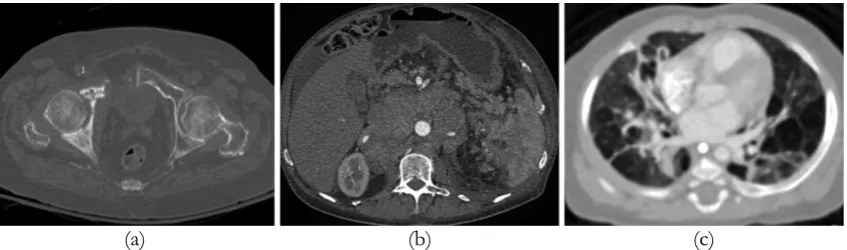
A CT scan obtains a large amount of information about the human body to produce its related cross-sectional images that show valuable medical information. However, because of degradations, the obtained images are considered as a degraded observation of the original scene. Unfortunately, such CT images have poor quality [7]. Usually, degradations such as blurring, noise and low contrast occur due to many reasons such as the use of improper image enhancement and restoration algorithms and low radiation dose [3,8]. Likewise, the obtained medical images through sensors may have a high possibility to be degraded by blur and noise [5]. The existence of noise not only results in unclear images, but also reduces the visibility of low-contrast details in CT images [9]. Figure 1 shows the above-mentioned degradations of CT images. As mentioned earlier, CT scans are useful in producing high-resolution images which are helpful in diagnosing many diseases especially in their primary stages which contribute in decreasing the death toll among patients. However, CT images for some areas of the body, such as the liver, have low contrast which eventually results in an imprecise diagnosis. Certain contrast agents can be inserted to improve the contrast of CT images, but they are harmful or even deadly sometimes for certain patients due to the occurrence of anaphylaxis [6]. Finally, it is necessary to discuss some of the available methods that handle these degradations to provide a better understanding about this topic.
(a) (b) (c)
Figure 1. The common degradations of CT images: (a) a low-contrast image; (b) a noisy image; (c) a blurry image
Related Works
It is important to provide a proper literature review that covers a survey of the existing studies, books and other sources related to the research subject. Hence, some of the available methods that are related to the topics of CT image enhancement and restoration are investigated by writing a constructive summary about each method. In this article, six of the recent methods are discussed for each of contrast enhancement, image denoising and deblurring areas. The review of such studies can assist to provide readers with the required background information about these topics. The relevant methods are discussed and summarized in the following sub-sections.
Contrast Enhancement Methods
concepts have been introduced for the purpose of providing more accurate analysis for various types of images [12,13]. Furthermore, different methods have been provided to improve the low-contrast of CT images. In [14], the author used a gamma correction procedure to process a selected image region of interest (ROI), while in [15] the authors proposed a global histogram equalization technique based on a probability function computed from a defined image ROI. Likewise, the authors of [16] introduced a dynamic histogram equalization technique, which maintains the mean brightness of the input image to produce acceptable results, while the authors of [17] proposed an extreme level eliminating adaptive histogram equalization technique, which implements locally on an image so that its output is adaptive to the local brightness. Moreover, the authors of [18] employed a method which combines a local adaptive Gaussian scale mixture model with a median filter to exploit both clipped and nonlinear binning approaches, while in [19] the authors proposed a modified histogram based fast enhancement technique to increase its efficiency in processing CT images. Each method is discussed in the next sub-sections.
Gamma Correction
In [14], the author suggested a gamma correction technique to enhance the contrast for a defined ROI. This technique functions as follows:
First, an ROI is defined for a CT image which is chosen and saved as a known file format using an interactive procedure.
Second, the contrast of the ROI is improved using a gamma correction procedure. This process is applied to the Y module of a chosen image that is being processed by a YUV colour system.
Third, morphological operators of hat filtering are included, such as opening-closing and top-bottom.
Fourth, a comparison between the morphological operators is achieved to select the most effective operator to be used with the proposed method.
From these comparisons, it is ascertained that the top-bottom is most suitable as it increases the object contrast by means of increasing the details in the murky regions and nearby contours. Finally, the images formats are unified to bmp and then are processed using a Matlab platform.
Global Histogram Equalization
In [15], the authors introduced a global histogram equalization technique to enhance the low CT contrast. In short words, this technique is implemented as follows: initially, the image is filtered using a normalization process. Next, the probability is computed from a defined ROI of every grey-level. Lastly, the histogram is equalized based on the computed probability. Hence, the contrast of the filtered image is enhanced without amplifying the latent image noise. The steps of this technique can be described as follows:
i. Normalize the processed image by reducing the smallest pixel value to zero. ii. Compute the probability for every pixel using the defined ROI values. iii. Compute the cumulative distribution function as follows:
(1)
where L is the highest grey-level value, (nj/ n) is the probability of the jth grey-level in the ROI. iv. Update each pixel in the ROI using the following formula:
(2)
Dynamic Histogram Equalization
In [16], the authors presented a contrast enhancement dynamic histogram equalization (CEDHE) technique, which can produce better results by maintaining the input mean brightness without introducing the unwanted artefacts of the standard histogram equalization. The CEDHE technique can be described as follows:
First, the initial histogram is normalized.
Second, a smoothing procedure is applied to perform a smooth detection for the break point procedure.
Third, a process of break-point detection is applied on the histogram to find the local minimum values.
Fourth, the histogram equalization is applied as every sub-histogram is equalized within its new grey-level distribution depending on a range attained in the previous step during the filtering process.
Although the break point process divides the input histogram properly, it does not assure that any minor segment of the grey-level controls the whole divided histogram segment. Moreover, the examination of every sub-histogram is achieved individually to check if there is any grey-level domination. Indeed, the key motive of dividing the histogram is to reduce the risk of having a dominating low frequency segment of the histogram. Lastly, the histogram equalization is only applied to the sub-histograms obtained from the previous steps.
Adaptive Histogram Equalization
In [17], the authors proposed an extreme level eliminating adaptive histogram equalization (ELEAHE) technique to enhance the contrast of CT images without producing any undesirable processing flaws. This technique runs locally on an image so that its output is adaptive to the local brightness alteration for the input image. This technique functions as follows: it starts by separating
an A×B image into C×D equal-size, square and non-overlapped sub-blocks. Next, processing starts
from the first sub-block to calculate the cumulative distribution function (CDF) and as a result, the histogram of each sub-block is created. After determining the CDF, it is saved in a Z matrix. However, the threshold level of the pixels is different when this technique is locally applied to the image. If all the pixels are considered as extreme levels, the threshold value becomes zero. Therefore, a new threshold setting is utilized to consider this situation. This setting uses the average of sub-block pixels per redistributed level as a reference. Finally, the output image is obtained by a bilinear interpolation technique to estimate the best value for all image pixels and eliminate the blocking artefact. Every sub-block is further separated into four small tiles. Using the CDF value, the mapping function for every sub-block is defined. The rest of the pixels are allocated using four mapping functions of the sub-blocks considering the midpoint pixels nearest to them. Lastly, mapping occurs using the interpolated values.
Local Adaptive Gaussian Scale Mixture Model with a Median Filter
In [18], the authors combined a local adaptive Gaussian scale mixture model with a median filter to improve the low contrast of CT images. This method can achieve the nonlinear requirement using both clipped and nonlinear binning methods. Moreover, CT images were scanned using an adequate digital scanner and then filtered by this method using Matlab. Likewise, the scanned CT images were saved as a tiff file format to obtain a high quality image files. Lastly, the experiments were applied to a dataset of 50 CT images of chest and abdomen organs.
Modified Histogram Based Fast Enhancement
histogram equalization technique, a goal which was successfully achieved. Likewise, the HBFE method is fast and simple, and therefore attracted many developers to increase its filtering efficiency in the medical field. The modified histogram based fast enhancement technique simply calculates the constant k more accurately than the previous algorithm by involving either mean or median or mode of the histogram in its calculation process. Lastly, including this modification allows a better enhancement process for CT images.
Denoising Methods
Image denoising is considered as a vital step in processing different types of medical images including computed tomography. As a result, many denoising methods have been widely used and different innovative concepts have been proposed, including smoothing filters, Wavelets, Curvelets and Ridgelets based approaches, sparse representation, bilateral processing, non-local means and total variation based methods [9]. In recent years, different CT denoising techniques have been proposed all with the same goal of retaining the important information while attenuating the unwanted noise. In 2010, a study was proposed to reduce the CT image noise using a multi-scale Wiener filtering technique [20]. In 2011, a new scheme which combines a Curvelet transform with a Monte-Carlo algorithm was introduced [21]. In 2012, an optimal weight denoising method was proposed as the filtering was achieved on every image block with the aid of a defined database of standard image blocks [22]. In 2013, a new technique was introduced which fuses three versions of the original image processed by three different methods. The first image was denoised by a total variation (TV) algorithm, while the second image was denoised by a Curvelet based method and the third image was the extracted edge information from the noise residual TV by using a Curvelet transform method [23]. In the same year, a new method was proposed to improve the spatial adaptivity of the non-local means method by employing a modified version of pointwise fractal dimension function [24]. In 2014, optimal partial differential equations based method with stopping criteria was proposed, which used an efficient explicit technique to solve the unstable anisotropic diffusion filter and introduced automatic stopping criteria [25]. Each method is discussed in the following sub-sections.
Multiscale Wiener Filtering
In [20], the authors proposed a multi-scale Wiener filtering technique, which analyses the noise of a given CT image. Briefly, this technique employs a two-stage Wavelet transform to decompose an image. Then, for each sub-image, a Wiener filter is implemented. Decomposing the image is achieved using a Wavelet transform to provide the required time-frequency and multi-scale features. Likewise, Wavelets can provide acceptable visual effect in image denoising. A Wiener filter is applied to the three high-frequency sub-images in the first decomposition to reduce their noise. Then, the obtained low-frequency sub-image is decomposed once more and a Wiener filter is applied again to the three high-frequency images. Lastly, an inverse Wavelet transformation is applied to obtain the final result.
Curvelet Transformation
In [21], the authors provided a new processing scheme, which combines a Curvelet transform with a Monte-Carlo algorithm. In short words, this scheme functions as follows: initially, the CT image is decomposed using a Curvelet transform. Then, a Monte-Carlo algorithm is utilized to approximate the high-frequency coefficients. Lastly, the coefficients are processed using a defined threshold. This scheme is applied to CT images that are degraded by Gaussian noise. The detailed steps of this scheme are as follows:
i. Apply a Curvelet transform to the image to obtain the low and high frequency coefficients at every scale.
ii. Estimate the noise variance by utilizing a Monte-Carlo algorithm.
(3)
where T(x) is the threshold and λ is a scalar parameter.
iv. Apply an inverse Curvelet transform to obtain the denoised image.
Optimal Weight Technique
In [22], the authors proposed a new noise reduction technique, in which the denoising process is performed on every block depending on a certain database of standard image blocks. Besides, the authors have considered the noise reduction problem as a constrained optimization problem. In this technique, a given noisy CT image is divided into a number of organized groups of small blocks, wherein the proposed technique is applied to every block as the amount of noise in every block is assumed to be different. The input for this technique is a noisy block and the output is a denoised block, which is defined as a sparse positive linear mixture of the typical blocks in a definite database in that the result is the nearest to the input. The key contributions of this technique are:
First, it can reduce the Gaussian noise efficiently.
Second, it provides an easy formulation of the noise reduction problem in the sparse representation structure into a known quadratic programming problem.
Lastly, the experimental results obtained by applying this technique were better than NLM, K-SVD and BM3D denoising methods, where this technique scored the highest results among the comparable methods.
Adaptive Fusion of Curvelet Transform and Total Variation
In [23], the authors introduced a denoising technique which is suitable for CT images by fusing three versions of the original image which are processed by three different methods. The first image is denoised by a total variation (TV) algorithm, while the second image is denoised by a Curvelet based method and the third image is the extracted edge information from the noise residual of the TV method using Curvelet transform. Moreover, a TV model proposed by [26] was used as the initial denoising step, which is a projection based model that can detect noise in a more accurate way and denoise an image better than the standard TV version. For the Curvelet transform denoising method, a Curvelet that depends on multiscale Ridgelets mixed with a spatial band-pass filtering process was used. This denoising technique can be abridged as:
i. Denoise the CT image by TV and Curvelet methods independently.
ii. Extract the edge information by filtering the noise residual of the TV method with a Curvelet method.
iii. Estimate the variance for every denoised image by TV and Curvelet. iv. Compute the overall variance difference of both denoised images. v. Estimate the edge variance from the edge information image. vi. Perform the fusion process based on the above information.
Non-local Means with Pointwise Fractal Dimension
In [24], the authors introduced a new way to improve the spatial adaptivity of Non-local Means (NLM) by employing a modified version of the pointwise fractal dimension (PWFD) function. The previous PWFD functions are calculated either for image blocks or the entire image. However, the modified PWFD, named as pointwise box-counting dimension (PWBCD), is calculated for each pixel of the processed image. Likewise, the PWBCD function is utilized as a fixed size window to fit various image structures, which ameliorates the spatial adaptivity of NLM algorithm. Moreover, the PWBCD function was mixed with the weight of the difference between the comparable windows of NLM. The steps of this technique are summarized as:
iii. Calculate the variance between the two comparable windows. iv. Calculate the required weights.
v. Approximate the real grey-levels.
Optimal Partial Differential Equations
In [25], the authors proposed a novel denoising technique using an anisotropic diffusion method which has a suitable stopping criterion, wherein it solves the anisotropic diffusion method which is mathematically unstable. Moreover, it offers an automatic stopping standard by using the input image only. This study uses a discrete Perona-Malik equation and solves it using an explicit method. Then, a Euclidean norm L2 is computed in the frequency domain at every iteration to
measure the high frequency energy of the input image. Afterwards, a high frequency relative forward difference function is calculated to evaluate the grey value variations inside the nearby areas of an image. The previous step requires the derivatives information to understand the image structure. Lastly, this technique increases the restoration process speed by introducing additional steps to improve the image quality.
Deblurring Methods
Image deblurring is the process of recovering a latent image from its blurry observation, which leads to the obtaining of a sharper image that helps in extracting and/or observing more information from the degraded image [27]. In image processing context, the deblurring procedure is considered as a vital step that allows access to better quality images. Many innovative methods have been introduced to reduce the blurring of CT images. In 2009, a study was proposed by [8] to deblur CT images in the case of noise existence. This study is adaptive and filters a given image using two phases. In the first phase, a pseudo inverse filter is employed to reduce the blurring artefact, while in the second phase, a multi-level thresholding of complex wavelet coefficients is utilized to reduce the existing noise. In 2010, a study was proposed by [28] to restore blurry-noisy CT images using a combination of Tikhonov and total variation regularizations with non-local means. At the same year, a study was introduced by [29] to deblur CT images using the standard Wiener filter. In 2011, another study was proposed by [30] to deblur CT images using Morlet and Curvelet frames with a Lagrangian pursuit function. In 2012, a study was proposed by [31] to restore blurry-noisy CT images utilizing a constant modulus blind equalization cost function with a kurtosis of error signal. In 2013, another study was proposed by [32] to deblur CT images using a hyperbolic secant square feature with a least-square feature to provide a two-dimensional isotropic hyperbolic secant square filter. Each method is discussed in the following sub-sections.
Daubechies Complex Wavelet Transform Based Multilevel Shrinkage
In [8], the authors proposed a new technique to deblur CT images in case of noise existence. This technique is adaptive due to the use of a definite shrinking function in addition to the median and the mean of the Wavelet coefficients at a specific level. It uses two phases to filter a given image, where in the first phase the pseudo inverse filter is used to reduce the blurring artefact, while in the second phase the noise is reduced by multilevel threshold of the complex Wavelet coefficients. Moreover, the used noise reduction method is a multilevel adaptive threshold with a soft-shrinkage function, which utilizes three parameters of complex Wavelet coefficients that are: the variance of Wavelet coefficients, the mean and the median of absolute Wavelet coefficients at a specific level. This technique retains the translation invariance caused by the complex Wavelet transform. Likewise, this technique can restore the blurred images degraded by Gaussian or non-Gaussian noise. This method can be summarized as:
i. Calculate the symmetric Daubechies complex Wavelet transform of pseudo inverse filtered estimate x', named as w.
(4)
where j is the resolution level, σis the standard deviation of Wavelet coefficients, μ and M are the mean and the median of absolute Wavelet coefficients at j th level for a particular subband.
iii. Calculate the soft-shrinkage function as:
(5)
apply this soft-shrinkage function on the magnitude of complex Wavelet coefficients w to get w'= f
(σ).w, by calculating the threshold of the second step at different resolution levels.
iv. Apply an inverse Wavelet transform on the shrunk Wavelet coefficients to obtain the denoised image.
Wiener Filter
In [29], the authors used the standard Wiener filter to deblur CT images in case of noise existence using a Gaussian point spread function (PSF). It is known that this filter reduces the noise and inverts the blur at the same time. Degradations in a CT image can be described as:
(6) where g(x, y) is the observed image, l(x, y) is the latent image, h(x, y) is the PSF, n(x, y) is the additive noise, (x, y) are spatial coordinates and ⨂ is a convolution operator. Also, the Gaussian PSF is calculated as:
(7)
where σ is the variance of the PSF, in which a higher value leads to more blurring. Next, the Wiener
filter is calculated as:
(8)
where, Ĝ(u,v) is the recovered image, H(u,v) is the optical transfer function (OTF) which is the Fourier transform of the PSF h(x, y), Ḧ(u,v) is the complex conjugate of the OTF, G(u,v) is the Fourier transform of the degraded image g(x, y), (u,v) are frequency coordinates, * is an element-wise multiplication process and W is a constant that represents the ratio of noise power spectrum to the original image power spectrum. Lastly, the images filtered using this technique show a better visual quality than its degraded versions.
Nonlocal Operators
In [28], the authors proposed a combination of two regularizations techniques and a nonlocal method to restore blurry-noisy CT images. Moreover, linear and non-liner methods are used to recover an acceptable quality CT image. The linear regularization methods are Tikhonov and total variation methods, while the non-local method is a non-local means denoising algorithm. This technique can be summarized:
i. Input: a true image f and a distortion factor K.
ii. Pre-processing: calculate u0 via Tikhonov regularization for image deconvolution or filtered back projection for tomographic reconstruction.
iii. Weight computation: For every pixel x, the weight w(x, y) is calculated for each y to fit a
searching window of size 21×21 centred at x.
(9)
where a fixed value of λ must be selected. Then, use e = 1e– 5, dt = 0.1, k = 0, maxIter = 500, in which u0 must be employed as the primary estimation of the iteration.
v. While dogradient descent:
(10)
Then, k = k +1. vi. End while
vii. Output: final solution uk.
Lagrangian Pursuit in Frame Dictionaries
In [30], the authors introduced a new technique to deblur CT images in case of noise existence, where it disintegrates a given image into its basic waveforms chosen in a redundant dictionary that consists of Morlet and Curvelet frames. Then, a Lagrangian pursuit function is applied to determine the optimum set of the dictionary vectors, which leads to attain clearer results. Due to the redundancy feature of the dictionary, a Lagrangian pursuit is used to determine the best set of the dictionary vectors that indicates the few coefficients which hold the required information to deliver clearer images. However, using linear filters only in the denoising process is inefficient, especially when the handled functions have discontinuities. Therefore, additional coefficients are required to show areas close to sharp transitions or edges. Hence, the use of Wavelet transforms overcame this problem by allowing an adaptive demonstration of signal discontinuities with a suitable thresholding of their estimated coefficients. The techniques which used linear filtering failed to produce acceptable results. Therefore, non-linear filtering methods for deblurring and denoising are used to avoid the common errors of the linear techniques. The aforesaid is achieved by creating an optimum estimation of the degraded image, in which only few coefficients can have the energy of the image. The dictionary is created by merging Curvelets to obtain better edges, and Wavelets to detect the low frequency components of an image. Lastly, the structure of this dictionary provides essential spatial adaptability for an image and allows for a good performance in terms of image demonstration sparsity.
Variable Step-Size Blind Restoration Algorithm
In [31], the authors provided a new approach to recover CT images which are degraded by blur and noise using an adaptive variable step-size blind equalization technique. In order to recover a given CT image using this technique, the image must be converted to a one-dimensional row or column sequence. However, this conversion reduces the ability of overcoming the point spread function effects. This technique can be summarized as follows:
First, a dimension reduction method is used to convert the image into a one-dimensional signal.
Second, a constant modulus cost function is implemented followed by an application of a kurtosis of error signal to avoid the imperfections of the constant modulus procedure and the slow convergence problem.
Lastly, due to the use of the above-mentioned steps, a faster and accurate convergence is attained, which provides a better restoration process.
Hyperbolic Secant Square Filter
between smoothing and edge sharpness. In addition, the least square feature can help in providing an adequate regularization. As a result, this filter has a good noise treatment without generating the undesirable frequency boosting near the margins. Therefore, it is applied to improve the important medical details of CT images by emphasizing their edges and fine details. To get the improved image, the high pass output is superimposed to the original images, in which the enhanced images are equivalent to their original ones but added with scaled versions of the edge and line structures. The edge and line structures are attained by a two dimensional hyperbolic secant square filter. Lastly, the accurate detection of the essential edge and line structures produced a successful sharpening process.
Summary
Different studies were explained to support the studied problems of this article, in which six studies for each of contrast enhancement, image denoising and image deblurring were selected and clarified. Regarding the contrast enhancement topic, a collection of methods such as, gamma correction, global histogram equalization, dynamic histogram equalization, adaptive histogram equalization, a combination of a local adaptive Gaussian scale mixture model with a median filter and a modified histogram based fast enhancement techniques have been used by different researchers to improve the poor contrast of CT images. Among the reviewed contrast enhancement methods, the adaptive histogram equalization performed the best as it reduced the unsought murkiness significantly and produced clear results. Regarding the denoising topic, a collection of methods such as, multiscale Wiener filtering, Curvelet transformation, optimal weight denoising, fusion of Curvelet transformation and total variation, pointwise box-counting dimension non-local means and optimal partial differential equations methods have been used by different researchers to reduce the noise from CT images. Among the studied denoising methods, the optimal weight technique performed the best as it reduced the unwanted noise, preserved the small image information and produced sharp details. Regarding the deblurring topic, a collection of methods such as, Daubechies complex Wavelet transform based multilevel shrinkage, Wiener filter, nonlocal operators, Lagrangian pursuit in frame dictionaries, variable step-size blind restoration and hyperbolic secant square filter have been used by different researchers to reduce the blur from CT images. Among the surveyed deblurring methods, the hyperbolic secant square filter performed the best as it reduced the undesirable blur without amplifying the latent image noise. Finally, the performance of the aforesaid methods varies from one to the other leaving promising opportunities for proposing new methods that can achieve better quality results with less processing time.
Conflict of Interest
The authors declare that they have no conflict of interest regarding this work.
References
1. Zarb F, Rainford L, McEntee M. Developing optimized CT scan protocols: Phantom measurements of image quality. Radiography 2011;17(2):109-114.
2. Enjilela E, Hussein E. Refining a region-of-interest within an available CT image. Appl Radiat Isotopes 2013;75:77-84.
3. Khare C, Nagwanshi K. Implementation and Analysis of Image Restoration Techniques. Int J Comput Tr Technol 2011;1(2):1-6.
4. Dakua S, Abi-Nahed J. Patient oriented graph-based image segmentation. Biomed Signal Proces 2013;8(3):325-332.
6. Liang Y, Yanga L, Fanb H. Image enhancement for liver CT images. Proceedings of the SPIE International Conference on Optical Instruments and Technology: Optoelectronic Imaging and Process Technology 2009:75130K1-75130K8.
7. Ciurte A, Nedevschi S. Texture Analysis within Contrast Enhanced Abdominal CT Images. 5th International Conference on Intelligent Computer Communication and Processing, Cluj-Napoca, Romania 2009: 73-78.
10. Sankar P, Rao B. Parallel Architecture for Implementation of Contrast Limited Adaptive Histogram Equalization. Int J Adv Eng Sci Technol 2011;10(1):047-051.
11. Sathya S, Manavalan R. Analysis of Background Detection and Contrast Enhancement of MRI Images. Int J Comput Appl 2011;36(12):16-21.
12. Al-Wadud M. Modified Histogram Equalization for Contrast Enhancement Preserving the Small Parts in Images. Int J Comput Sci Network Sec 2012;12(2):1-4.
13. Seema S. Region Based Contrast Limited Adaptive HE with Additive Gradient for Contrast Enhancement of Medical Images (MRI). Int J Soft Comput Eng 2011;1(4):154-157.
14. Georgieva V. An Approach for Computed Tomography Images Enhancement. Elektron Elektrotech 2010;98(2):71-74.
15. Yousuf M, Rakib M. An Effective Image Contrast Enhancement Method Using Global Histogram Equalization. J Sci Res 2011;3(1):43-50.
16. Ismail W, Sim K. Contrast enhancement dynamic histogram equalization for medical image processing application. Int J Imaging Sys Technol 2011;21(3):280-289.
17. Tan T, Sim K, Tso C, Chong A. Contrast enhancement of computed tomography images by adaptive histogram equalization-application for improved ischemic stroke detection. Int J Imaging Sys Technol 2012;22(3):153-160.
18. Abdallah Y, Siddig M. Contrast Improvement of Chest Organs in Computed Tomography Images using Image Processing Technique. Asian J Med Radiol Res 2013;1(1):39-44.
19. Kandeel A, Abbas A, Hadhoud M, Saghir Z. A Study of a Modified Histogram Based Fast Enhancement Algorithm (MHBFE). Sign Image Proc Int J 2014;5(1):55-67.
20. Ke L, Zhang R. Multiscale Wiener Filtering Method for Low-Dose CT Images. IEEE third International Conference on Biomedical Engineering and Informatics. Yantai, China. 2010:428-431.
21. Deng J, Li H, Wu H. A CT Image Denoise Method Using Curvelet Transform. In: In Ma M, editor. Communication Systems and Information Technology. 1st ed. Berlin: Springer-Verlag Heidelberg; 2011. p. 681-687.
22. Trinh D, Luong M, Rocchisani J, Pham C, Pham H, Dibos F. An optimal weight method for CT image denoising. J El Sci Technol 2012;10(2):124-129.
23. Bhadauria H, Dewal M. Medical image denoising using adaptive fusion of curvelet transform and total variation. Comput El Eng 2013;39(5):1451-1460.
24. Zheng X, Liao Z, Hu S, Li M, Zhou J. Improving Spatial Adaptivity of Nonlocal Means in Low-Dosed CT Imaging Using Pointwise Fractal Dimension. Comput Math Methods Med 2013;2013:1-8.
25. Khanian M, Feizi A, Davari A. An Optimal Partial Differential Equations-based Stopping Criterion for Medical Image Denoising. J Med Sig Sens 2014;4(1):72-83.
26. Chambolle A. An algorithm for total variation minimization and applications. J Math Imaging Vision 2004;20(1-2):89-97.
27. Subashini P, Krishnaveni M, Singh V. Image Deblurring Using Back Propagation Neural Network. World Comp Sci Inf Technol J 2011;1(6):277-282.
12 Appl Med Inform 36(1) March/2015
29. Hussien M, Saripan M. Computed Tomography Soft Tissue Restoration using Wiener Filter. IEEE Student Conference on Research and Development. Putrajaya, Malaysia. 2010:415-420. 30. Zifan A, Liatsis P. Medical Image Deblurring via Lagrangian Pursuit in Frame Dictionaries.
Developments in E-Systems Engineering. Dubai, UAE. 2011:86-91.
31. Sun Y, Zhang L, Duan J. Research of Medical Image Variable Step-Size Blind Restoration Algorithm. In: Hu W (Ed). Advances in Electric and Electronics. 1st ed. Berlin: Springer Heidelberg; 2012. p. 583-590.