The Role of Cytokines in Activation of Coagulation
and Fibrinolysis in Dengue Shock Syndrome*
Catharina Suharti
1, Eric C.M. van Gorp
2, Tatty E. Setiati
3, Wil M.V. Dolmans
4,
Robert J. Djokomoeljanto
1, C. Erik Hack
5, Hugo ten Cate
6, Jos W.M. van der Meer
4Departments of 1Medicine and 3Paediatrics, Faculty of Medicine, Diponegoro University and Dr. Kariadi Hospital, Semarang, Indonesia, 2Department of Medicine, Slotervaart Hospital, Amsterdam, 4Department of Medicine,
University Medical Centre St. Radboud, Nijmegen, 5Central Laboratory of The Netherlands Red Cross Blood Transfusion Service and Department Clinical Chemistry, Academic Hospital Vrije Universiteit, Amsterdam, 6Laboratory for Experimental Internal Medicine, Academic Medical Centre, Amsterdam, The Netherlands
Keywords
Cytokines, coagulation, fibrinolysis, dengue shock syndrome
Summary
In a prospective clinical study of 50 patients with Dengue Shock
Syndrome (DSS), we investigated the association of tumor necrosis
factor-
␣
(TNF-
␣
), interleukin-1

(IL-1

), IL-1 receptor antagonist
(IL-1Ra), and IL-6 with activation markers of coagulation (F1+2 and
TATc) and fibrinolysis (t-PA, PAPc, and D-dimer). We found that
TNF-
␣
, IL-1

and Il-1Ra, but not IL-6, concentrations were elevated in
the circulation during the early stage of infection and at discharge from
hospital. TNF-
␣
was significantly associated with D-dimer, an activation
marker of fibrinolysis (p < 0.003), but not with activation markers of
coagulation. IL-1

was significantly associated with t-PA (p < 0.03).
IL-1Ra was significantly associated with F1+2, TATc (p < 0.04 and
p < 0.02, respectively), whereas IL-6 was significantly associated with
both, activation markers of coagulation (F1+2; p < 0.03) and fibrinolysis
(PAPc; p = 0.002). Our data are in line with studies in bacterial sepsis.
In severe dengue virus infection the same cytokines are involved in the
onset and regulation of hemostasis.
Introduction
The association between infection and activation of coagulation and
fibrinolysis has been studied extensively in Gram negative-sepsis and
endotoxemia (1). Activation of coagulation, as reflected by increasing
plasma levels of prothrombin fragment 1 and fragment 2 (F1+2) and
thrombin-antithrombin complexes (TATc), and activation of fibrinolysis,
as reflected by increasing plasma levels of tissue plasminogen activator
(t-PA) and plasminogen activator inhibitor type 1 (PAI-1), have been
detected in sepsis (2-4) and in experimental endotoxemia in humans (5,
6). Cytokines appear to play a crucial role in the initiation of coagulation
Dengue hemorrhagic fever (DHF) is a severe form of the infection
caused by any of four serotypes of the dengue virus. It is characterized
by fever, thrombocytopenia, bleeding manifestations ranging from mild
to severe, and evidence of plasma leakage. According to the 1997
WHO criteria, dengue shock syndrome (DSS) is defined as DHF with
signs of circulatory failure, including narrow pulse pressure (20 mm
Hg) and hypotension (DHF grade III) or frank shock (DHF grade IV)
(11, 12). Plasma leakage and bleeding are the two major
pathophysio-logical changes in DHF/DSS, determining the severity of disease
(11-14). Little is known about the pathogenesis of bleeding in DHF/
DSS and in an analogy to sepsis it is attractive to speculate that
cyto-kines are key mediators. Preexisting heterotypic dengue antibodies
augment dengue virus infection of monocytes or macrophages (15). An
increased number of dengue virus-infected monocytes may result in
T-lymphocyte activation; the activation of both types of cell may result in
the production of elevated levels of the aforementioned cytokines (16).
We designed a prospective clinical study to investigate the association
of cytokines with activation markers of coagulation and fibrinolysis in
DSS. We investigated those cytokines which are known to play a role
in the activation of coagulation and fibrinolysis in bacterial sepsis.
Patients and Methods
Study Setting
The study was executed in Dr. Kariadi Hospital, Semarang, Indonesia, the university hospital of Diponegoro University. The research protocol was reviewed and approved by the institutional Review Board of the Dr. Kariadi Hospital. Written informed consent was obtained from children’s parents or legal guardians.
Patients
Specimen Collection
Blood specimens were collected in vacutainer tubes (Becton Dickinson, Rutherford, NJ 07417). Specimens for cytokine assays were collected in the acute phase of disease (on day of admission: day 0, on day 1 and 2), and on day 7 or the day of discharge if the patient was hospitalized for less than 7 days. Four ml of blood were collected into sterile tubes containing EDTA in each patient. To this tube 125 µL of aprotinin (Trasylol, Bayer, Leverkusen, Germany; final concentration 625 kallikreine inactivating units/mL) was added through the stopper by a tuberculin needle and syringe. The tube was centrifuged directly at 1250 g for 10 min, and thereafter at 15000 g for 1 min to
remove platelets. The plasma was stored in aliquots at –80° C until assayed for cytokines.
Blood samples for analysis of coagulation and fibrinolysis were collected on the same days as the samples for cytokines. Venous blood (9 vol.) for measurement of F1+2, TATc and D-dimers was drawn into vacutainer tubes containing 0.105 M sodium citrate (1 vol.). For measurement of PAPc, t-PA, blood was collected in siliconized vacutainer tubes containing Polybrene (Janssen Chimica, Belgium) and EDTA (0.05% w/v, and 10 mM, respectively, final concentrations) to prevent in-vitro complex formation. All blood samples were immediately immersed in melting ice and subsequently centrifuged at 4° C for 20 min at 1600⫻g. Plasma samples were stored at –80° C until assayed.
To collect appropriately timed specimens for serological assays (≥day 6 after onset of fever) (18-21), 2 ml of blood was collected on day of admission and at discharge. Blood was centrifuged 1000-1500 rpm for 10 min, afterwards, serum was transferred to screw cap Eppendorf tubes and stored at –80° C, until assayed. For the transport from Indonesia to The Netherlands (which lasts longer than 15 h) the samples for cytokines, coagulation, fibrinolysis, and serological assays were kept on dry ice.
Cytokine Assays
TNF-␣, IL-and IL-1Ra were measured in duplicate by nonequilibrium radioimmunoassay (RIA) (22). Recombinant human TNF-␣, IL-1and IL-1Ra were calibrated against standards provided by the National Institute of Biolo-gical Standards and Control (Potters Bar, UK), with the sensitivity of the assay with 100 µl sample of 40 pg/ml, 40 pg/ml, and 80 pg/ml, respectively. IL-6 was measured with ELISA obtained from the Central Laboratory of the Netherlands Red Cross Blood Transfusion Service, Amsterdam, The Netherlands, according to manufacturer’s instruction; circulating concentrations exceeding 10 pg/ml were considered to be elevated.
Coagulation and Fibrinolysis Assays
F1+2 and TATc levels were determined with the use of commercially available ELISA kits, according to the manufacturer’s instructions (Enzygnost F1+2 micro and TAT micro, Dade Behring, Marburg, Germany). For the measurement of D-dimer levels, the TintElize D dimer ELISA from Biopool (Sweden) was used, according to the manufacturer’s instructions. t-PA was measured with sandwich ELISA kits using specific monoclonal antibodies as described before (23). PAPc was measured with radioimmuno-assay as described previously (24).
Serological Assays
The diagnosis of dengue infection was confirmed by serological assays. A capture and indirect enzyme-linked immunosorbent assay (ELISA) detected Table 1 Characteristics in 50 patients with dengue shock syndrome
dengue specific IgM and IgG antibodies in serum samples, according to a previously described procedure (25).
Statistical Analysis
Continuous data were described as mean with SD or median with range. Nominal data were described as percentage (%). Multiple linear regression analysis was done to determine the association between cytokines and activation markers of coagulation and fibrinolysis on the day of admission. P values of less than 0.05 were considered to indicate statistical significance. All statistical analyses were performed with SPSS for Windows, version 9.0.
Results
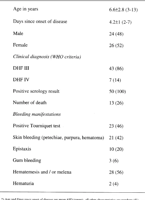
Between June and November 1996, 50 children with a clinical
diagnosis of DSS, were enrolled in the study. Baseline characteristics of
the patients are listed in Table 1. Thirteen patients (26%) died during
follow up on the intensive care unit. The clinical diagnosis was confirmed
by serological assay in all patients, either by an IgM response or a
fourfold rise in IgG titres. Antibody profiles were typical for secondary
dengue infection.
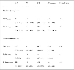
Activation of Coagulation and Fibrinolysis
As shown in Table 3, F1+2 and TATc were persistently high from
admission to discharge, reflecting activation of coagulation. Also,
persistently elevated levels of t-PA, PAPc, and D-dimer were found
during the study period, reflecting activation of the fibrinolytic system.
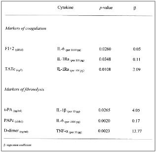
Association of Plasma Concentrations of Cytokines with Activation
Markers of Coagulation and Fibrinolysis
TNF-
␣
was significantly associated with D-dimer, a marker of
fibrinolysis, but not with activation markers of coagulation. Studies in
bacterial infection have clearly indicated the role of TNF-
␣
in the
activation of the fibrinolytic system, but the role of TNF-
␣
in the
activation of the coagulation system seems to be less direct. A single
intravenous injection of recombinant TNF into human volunteers
induced activation of the common pathway of the coagulation cascade,
resulting in thrombin formation, as reflected by elevated plasma levels
of F1+2 (3), and activation of the fibrinolytic system characterized by
increased levels of urokinase-type PA (u-PA) and t-PA, followed by
PAI-1 (4). An injection of E. coli endotoxin induced increased levels of
TNF-
␣
and IL-6, and resulted in activation of fibrinolysis as reflected
by increased levels of t-PA, PAPc, which was subsequently offset by
release of PAI-1, and a long-lasting onset of coagulation activation
characterized by increase of prothrombin F1+2 and TATc (9).
However, further studies have demonstrated that anti-TNF treatment
did not affect endotoxin-induced coagulation activation (26-28), but did
inhibit the activation of the fibrinolytic system in animal and human
models with low-grade endotoxaemia (26, 27, 29). Thus our findings in
DHF are in line with these findings in endotoxemia.
We demonstrated that IL-1

was significantly associated with tPA,
a marker for activation of fibrinolysis. The role of IL-1

in the activation
of coagulation as well as fibrinolysis has been demonstrated by injection
of IL-1

, or, indirectly by injection of IL-1Ra (5, 6). Infusion of IL-1
into baboons induced activation of the fibrinolytic system, reflected by
increased levels of t-PA, and subsequently the offset of fibrinolysis by
PAI-1, followed by the activation of coagulation, characterized by
increased levels of TATc (5). Administration of IL-1Ra in baboons
with lethal bacteremia, and in patients with sepsis have significantly
attenuated the activation of coagulation and fibrinolysis (6).
We also found that IL-1Ra was significantly associated with the
activation markers of coagulation F1+2 and TATc, leading to the
conclusion that the regulation of coagulation and fibrinolysis by IL-1

and IL-1Ra in this severe viral infection is similar to that in sepsis.
We demonstrated that IL-6 is significantly associated with activation
markers of both coagulation and fibrinolysis. The role of this cytokine
in the activation of coagulation has been demonstrated by injection of
recombinant IL-6, as well as by administration of anti-IL-6 antibody.
Infusion of recombinant IL-6 into patients with renal cell carcinoma
resulted in increased levels of F1+2 and TATc, while fibrinolysis was
not clearly affected (30).
In the present study we found that the median levels of IL-6 were
normal, but with extremely high values in 3 patients (5823, 86000 and
199000 pg/ml). Two of these 3 patients died and in these two patients,
very high levels of TNF-
␣
(2500 and 2500 pg/ml), IL-1Ra (2500 and
2500 pg/ml), and F1+2 (7.51 and 14.2 nMol/l, normal <1.1 nMol/l)
were found. The very high levels of F1+2 reflect the degree of coagulation
activation, which may result in a considerable fibrin formation, leading
to multiorgan failure and ultimately to death.
We conclude that in dengue shock syndrome the same cytokines are
involved in the onset and regulation of coagulation and fibrinolysis as
in bacterial infections.
Acknowledgements
Participants in this project, besides the authors, were Dr. D.P.M. Brandjes from Department of Medicine, Slotervaart Hospital, Amsterdam, Prof. Moeljono S. Trastotenojo, Dr. Herawati Juslam, from the Deparment of Paediatrics and Prof. Soenarto, Dr. Widiono L. from the Department of Internal Medicine, Dr. Kariadi Hospital, Diponegoro University, Semarang, Indonesia. We thank Y.T. van der Heide from the Clinical Chemistry and Hematology Laboratory, Slotervaart Hospital, Amsterdam, The Netherlands, for managing the blood samples, and Dr. J. Groen and Prof. A. Osterhaus from the Institute of Virology, Erasmus Medical Centre, Rotterdam, for performing the diagnostic assays. We thank also Johanna van der Ven-Jongekrijg from the laboratory of the Table 4 Results of multiple linear regression analysis
Department of General Internal Medicine, University Medical Centre St. Radboud, Nijmegen, The Netherlands and GJ van Mierloo and AJM Eerenberg from the Central Laboratory of The Netherlands Red Cross Blood Transfusion Service, Amsterdam, The Netherlands, for performing the cytokine assays.
References
1. Van Gorp ECM, Suharti C, ten Cate H, Dolmans WMV, van der Meer JWM, ten Cate JW, Brandjes DPM. Infectious Diseases and Coagulation Disorders. J Infect Dis 1999; 180: 176-86.
2. Osterud B, Flaegstad T. Increased tissue thromboplastin activity in monocytes of patients with meningococcal infections related to unfavourable prognosis. Thromb Haemost 1983; 49: 5-7.
3. Voss R, Matthias FR, Borkowski G, Reitz D. Activation and inhibition of fibrinolysis in septic patients in an internal intensive care unit. B J Haematol 1990; 75: 99-105.
4. Brandtzaeg P, Joo GB, Brusletto B, Kierulf P. Plasminogen activator inhibitor 1 and 2, and ␣2-antiplasmin, and endotoxin levels in systemic meningococcal disease. Thromb Res 1990; 57: 271-78.
5. Van Deventer SJH, Büller HR, Ten Cate JW, Aarden LA, Hack CE, Sturk A. Experimental endotoxaemia in humans: analysis of cytokine release and coagulation, fibrinolytic and complement pathways. Blood 1990; 76: 2520-26.
6. Suffredini AF, Harpel PC, Parrillo JE. Promotion and subsequent inhibition of plasminogen activator after administration of intravenous endotoxin to normal subjects. N Engl J Med 1989; 320: 1165-72.
7. Van der Poll T, Büller HR, Ten Cate H, Wortel CH, Bauer KA, van Deventer SJH, Hack CE, Sauerwein HP, Rosenberg RD, ten Cate JW. Activation of coagulation after administration of tumor necrosis factor in healthy subjects. N Engl J Med 1990; 322: 1622-27.
8. Van der Poll T, Levi M, Büller HR, van Deventer SJH, de Boer JP, Hack CE, ten Cate JW. Fibrinolytic response to tumor necrosis factor in healthy subjects. J Exp Med 1991; 174: 729-32.
9. Jansen PM, Boermeester MA, Fischer E, de Jong IW, van der Poll T, Moldawer LL, Hack CE, Lowry SF. Contribution of interleukin-1 to activation of coagulation and fibrinolysis, to neutrophil degranulation and the release of sPLA2 in sepsis. Studies in non-human primates following interleukin-1␣administration and during lethal bacteremia. Blood 1995; 86: 1027-34.
10. Boermeester MA, van Leeuwen PAM, Coyle SM, Houdijk AJP, Eerenberg AM, Wolbink GJ, Pribble JP, Stiles DM, Wesdorp RIC, Hack CE, Lowry SF. Interleukin-1 receptor blockade in patients with sepsis syndrome: evidence that interleukin-1 contributes to the release of interleukin-6, elastase and phospholipase A2, and to the activation of the complement, coagulation and fibrinolytic system. Arch Surg 1995; 130: 739-48. 11. Dengue haemorrhagic fever: diagnosis, treatment, prevention and control.
2d ed. Geneva: World Health Organization 1997; 12-47.
12. Dengue and dengue hemorrhagic fever in the Americas: guidlines for prevention and control.Washington, DC: Pan American Health Organization Sci Publ 1994; 3-21.
13. Bhamarapravati N. Pathology of dengue infections. In: Dengue and dengue hemorrhagic fever. Gubler DJ, Kuno G, eds. Wallingford, UK: Cab International 1997; 115-32.
16. Kurane I, Rothman AL, Livingstone PG, Green S, Gagnon SJ, Janus J, Innis BL, Nimmannitya S, Nisalak A, Ennis FA. Immunopathologic mechanisms of dengue hemorrhagic fever and dengue shock syndrome. Arch Virol (Suppl) 1994; 9: 59-64.
17. Jarvis C. Physical examination and health assessment. Philadelphia: WB Saunders 1993; 178.
18. Rigau-Perez JG, Clark GG, Gubler DJ, Reiter P, Sanders EJ, Vorndam AV. Dengue and dengue hemorrhagic fever. Lancet 1998; 352: 971-77. 19. Sharp TW, Wallace MR, Hayes CG, Sanchez JL, DeFraites RF, Arthur RR,
Thornton SA, Batchelor RA, Rozmajzl PJ, Hanson RK, Wu SJ, Iriye C, Burans JP. Dengue fever in U.S. troops during operation restore hope, Somalia, 1992-1993. Am J Trop Med Hyg 1995; 53: 89-94.
20. Innis BL, Nisalak A, Nimmannitya S, Kusalerdcharya S, Chongswasdi V, Suntayakorn S, Puttisri P, Hoke CH. An enzyme-linked immunosorbent assay to characterize dengue infections where dengue and Japanese Encephalitis co-circulate. Am J Trop Med Hyg 1989; 40: 418-27. 21. Ruechusatsawat K, Morita K, Tanaka M, Vongcheree S, Rojanasuphot S,
Warachit P, Kanai K, Thongtradol P, Nimnakorn M, Kanungkid S, Igarashi A. Daily observation of antibody levels among dengue patients detected by enzyme-linked immunosorbent assay (ELISA). Jpn J Trop Med Hyg 1984; 22: 9-12.
22. Drenth JP, van Uum SH, van Deuren M, Pesman GJ, van der Ven-Jongekrijg J, van der Meer JWM. Endurance run increases circulating IL-6 and IL-1Ra but downregulates ex-vivo TNF-alpha and IL-1beta production. J Appl Physiol 1995; 79: 1497-03.
23. De Boer JP, Creasy AA, Chang A, Roem D, Brouwer MC, Eerenberg AJ, Hack CE, Taylor FB Jr. Activation patterns of coagulation and fibrinolysis in baboons following infusion with lethal or sublethal dose of Escherichia coli. Circ Shock 1993; 39: 59-67.
24. Levi M, de Boer JP, Roem D, ten Cate JW, Hack CE. Plasminogen activation in vivo upon intravenous infusion of DDAVP. Quantitative assessment of plasmin-alpha-2 antiplasmin complex with a novel monoclonal antibody based radioimmunoassay. Thromb Haemost 1992; 67: 111-16. 25. Groen J, Velzing J, Copra C, Balentien E, Deubel V, Vorndam V, Osterhaus
ADME. Diagnostic value of dengue virus-specific IgA and IgM serum antibody detection. Microbes and infection 1999; 1: 1-6.
26. Van der Poll T, Levi M, Van Deventer SJH, Ten Cate H, Haagmans BL, Biemond BJ, Büller HR, Hack CE, Ten Cate JW. Differential effects of anti-tumor necrosis factor monoclonal antibodies on systemic inflammatory responses in experimental endotoxaemia in chimpanzees. Blood 1994; 83: 446-51.
27. Van der Poll T, Coyle SM, Levi M, Jansen PM, Dentener M, Barbosa K, Buurman WA, Hack CE, ten Cate JW, Agosti JM, Lowry SF. Effect of recombinant dimeric tumor necrosis factor receptor on inflammatory responses to intravenous endotoxin in normal humans. Blood 1997; 89: 3727-34.
28. Hinshaw LB, Tekamp-Olson P, Chang ACK, Lee PA, Taylor Jr FB, Murray CK, Peer GT, Emerson Jr TE, Passey B, Kuo GC. Survival of primates in LD100 septic shock following therapy with antibody to tumor necrosis factor (TNF). Circ Shock 1990; 30: 279-92.