Disturbed ratio of erythrocyte and plasma
S
-adenosylmethionine/
S
-adenosylhomocysteine in peripheral
arterial occlusive disease
Franziska M.T. Loehrer
a,1, Martin Tscho¨pl
b, Christian P. Angst
a, Piotr Litynski
a,
Kurt Ja¨ger
b, Brian Fowler
a,*, Walter E. Haefeli
c,d,2aMetabolic Unit,Uni6ersity Children’s Hospital Basel,(UKBB),Romergasse8,CH-4005Basel,Switzerland bDepartment of Medicine,Di6ision of Angiology,Uni6ersity Hospital Basel,CH-4031Basel,Switzerland cDepartment of Medicine,Di6ision of Clinical Pharmacology,Uni6ersity Hospital Basel,CH-4031Basel,Switzerland
dDepartment of Pharmacy,Uni6ersity of Basel,CH-4051Basel,Switzerland Received 28 May 1999; received in revised form 21 February 2000; accepted 1 March 2000
Abstract
Altered homocysteine metabolism associated with peripheral arterial occlusive disease (PAOD) may lead to impairment of vital methylation reactions through accumulation of S-adenosylhomocysteine (AdoHcy) as well as through alteration of the ratio
S-adenosylmethionine (AdoMet)/AdoHcy. We determined AdoMet, AdoHcy, their ratio, and homocysteine in plasma as well as AdoMet, AdoHcy, and their ratio in erythrocytes of 61 patients with PAOD (age 49 – 93) and 50 healthy controls (age 41 – 87). Geometric mean values of plasma homocysteine, AdoMet, and AdoHcy were significantly increased in patients compared with controls (15.5 vs. 10.4mmol/l**; 107 vs. 52.3* nmol/l; 55.0 vs. 23.1** nmol/l, respectively; *PB0.01, **PB0.001), while the ratio of AdoMet/AdoHcy was decreased in patients (1.92 vs. 2.52*). In erythrocytes patients exhibited increased levels of AdoHcy compared with controls (309 vs. 205 nmol/l**) whereas AdoMet (3351 vs. 3732 nmol/l*) and the ratio of AdoMet/AdoHcy (11.8 vs. 19.1**) were decreased. The odds ratio (OR) for developing PAOD with decreased AdoMet/AdoHcy ratio after adjustment for kidney function was significant for erythrocyte levels 514.2 (OR, 7.1 (6.9 – 7.2, 95% CI). In addition, hematocrit levels were found to be significantly decreased in patients versus controls (0.35 vs. 0.42 l/l**) and were significantly correlated with the ratio of AdoMet/AdoHcy in erythrocytes of the patients. Since the ratio of AdoMet/AdoHcy is closely linked with the activity of numerous enzymatic methylation reactions, these results suggest that methylation may be impaired in these patients. © 2001 Elsevier Science Ireland Ltd. All rights reserved.
Keywords:Peripheral arterial occlusive disease;S-Adenosylmethionine;S-Adenosylhomocysteine; Homocysteine; Erythrocytes; Methylation; High performance liquid chromatography
www.elsevier.com/locate/atherosclerosis
1. Introduction
In the last two decades several retrospective and prospective studies have shown that mild
hyperhomo-cysteinemia is associated with peripheral arterial occlu-sive (PAOD), coronary artery, and cerebrovascular disease, as well as with venous thrombosis independent of other risk factors such as smoking, arterial hyperten-sion, hypercholesterolemia, and diabetes mellitus [1 –
12]. The pathophysiological implications of
hyperhomocysteinemia, however, are not fully under-stood. Homocysteine is produced during methionine metabolism via the adenosylated compounds S -adeno-sylmethionine (AdoMet) and S-adenosylhomocysteine (AdoHcy) [13]. The main physiological function of this metabolic pathway (Fig. 1) is probably the maintenance of adequate amounts of AdoMet, which not only plays
* Corresponding author. Tel.: +41-61-6856275; fax: + 41-61-6856566.
E-mail address:[email protected] (B. Fowler).
1Present address: James Lance Glaxo Wellcome Medicines Re-search Unit, Prince of Wales Hospital, Parkes 10 East, Randwick 2031, NSW, Australia.
2Present address: Department of Internal Medicine VI, Clinical Pharmacology and Pharmacoepidemiology, University of Heidelberg, D-69115 Heidelberg, Germany.
a key role in the enzymatic regulation of homocysteine metabolising reactions, but is also the main methyl donor in numerous enzymatic trans-methylation reac-tions, which lead to the formation of AdoHcy [14]. AdoHcy acts as a competitive inhibitor of these AdoMet-dependent trans-methylation reactions and its affinity to most methyltransferases is greater than the affinity of the substrate AdoMet [15]. Increases in Ado-Hcy concentration relative to AdoMet will therefore result in inhibition of trans-methylation reactions. In-deed, the ratio of AdoMet/AdoHcy is crucial in the regulation of enzymatic trans-methylation reactions [15,16] and a decrease of the ratio may inhibit trans -methylation reactions in various tissues [16] thereby potentially affecting biosynthesis of a wide range of endogenous compounds such as proteins, hormones, phospholipids, neurotransmitters, DNA and RNA [17] (Fig. 1). Even though thermodynamics favour the biosynthesis of AdoHcy, AdoHcy normally remains low in vivo provided that homocysteine is removed immediately [15]. Hence, elevations of homocysteine levels may lead to an increase in AdoHcy and therefore to the potentially detrimental alterations of the ratio AdoMet/AdoHcy described above.
Indeed, in a previous study [18], we detected low whole blood AdoMet concentrations in patients with coronary artery disease, pointing to possible alterations of methylation reactions in these patients compared with healthy controls. Moreover, recent studies showed a decreased ratio of AdoMet/AdoHcy in erythrocytes [19,20] and in plasma [21] of end-stage renal failure patients in whom the incidence of moderate hyperho-mocysteinemia is high as is the risk for vascular disease. So far no information about AdoMet/AdoHcy ratios is
available in patients with PAOD. The aim of this study was to investigate AdoMet and AdoHcy in erythrocytes and plasma as a metabolic consequence of altered homocysteine metabolism or of related factors such as renal function in patients with PAOD.
2. Subjects and methods
2.1. Patients and control subjects
Sixty one patients (aged 49 – 93; 32 women and 29 men, Table 1) with PAOD were investigated at the University Hospital in Basel (between 1994 and 1997) after giving informed consent. Six patients had claudi-catio (Fontaine, stage II), and 59 suffered from critical limb ischemia (stage III/IV). Thirteen patients had di-agnosed chronic renal insufficiency, 24 patients had diabetes; 27 were hypertensive; 13 were current, 16 previous and 20 were non-smokers. Twenty-six patients had a history of one or more vascular events other than PAOD (in total 24 coronary artery and five cerebrovas-cular events). Fifty healthy control subjects (aged 41 – 87; 28 women and 22 men, three smokers, Table 1) were randomly selected within the same city. At least 4 h after the last food intake, blood samples for the determination of homocysteine and its metabolites, he-matological parameters and for routine clinical chem-istry (Hitachi 911 Automatic Analyser) were drawn.
2.2. Sample preparation
Ethylene diamine tetra acetic acid (EDTA) blood samples for homocysteine, 5-methyltetrahydrofolate (MeTHF), AdoMet, and AdoHcy were placed on ice after collection and processed within half an hour. Plasma for MeTHF measurement, processed under light protection, was added to ascorbic acid (10 mg/ml). For AdoMet and AdoHcy, samples were deproteinised immediately by adding 0.625 ml of a 10% perchloric acid solution to 1 ml of plasma or an equal amount of 5% perchloric acid solution to whole blood, respec-tively, followed by thorough mixing. All samples were stored at −70°C until analysis [18,22].
2.3. High performance liquid chromatography (HPLC) determination of homocysteine, MeTHF (in plasma),
AdoMet and AdoHcy (in plasma and erythrocytes)
Plasma samples for total homocysteine, MeTHF, AdoMet, and AdoHcy determination in plasma were processed and subjected to reversed phase chromatog-raphy with fluorescence detection as previously de-scribed [22], with the following modifications of the HPLC conditions for AdoHcy and AdoMet. Etheno-derivatives of both metabolites were analysed
Table 1
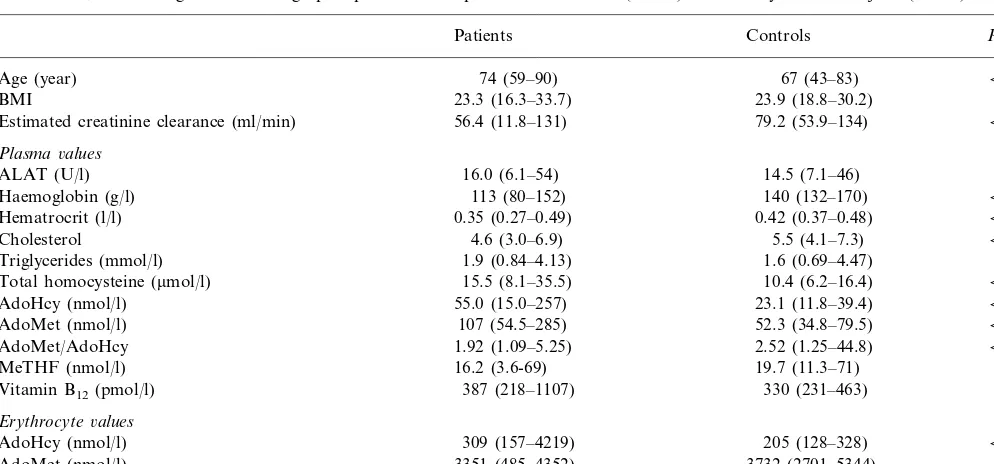
Biochemical, haematological and demographic parameters of patients with PAOD (n=61) and healthy control subjects (n=50)a
Controls P-values
Estimated creatinine clearance (ml/min) 79.2 (53.9–134) B0.001
Plasma6alues
ALAT (U/l) 16.0 (6.1–54) 14.5 (7.1–46) NS
Haemoglobin (g/l) 113 (80–152) 140 (132–170) B0.001
0.42 (0.37–0.48)
0.35 (0.27–0.49) B0.001
Hematrocrit (l/l)
4.6 (3.0–6.9)
Cholesterol 5.5 (4.1–7.3) B0.001
1.9 (0.84–4.13)
Triglycerides (mmol/l) 1.6 (0.69–4.47) NS
10.4 (6.2–16.4)
15.5 (8.1–35.5) B0.001
Total homocysteine (mmol/l)
55.0 (15.0–257)
AdoHcy (nmol/l) 23.1 (11.8–39.4) B0.001
52.3 (34.8–79.5)
107 (54.5–285) B0.001
AdoMet (nmol/l)
2.52 (1.25–44.8)
AdoMet/AdoHcy 1.92 (1.09–5.25) B0.01
19.7 (11.3–71)
16.2 (3.6-69) 0.06
MeTHF (nmol/l)
Vitamin B12(pmol/l) 387 (218–1107) 330 (231–463) 0.06
Erythrocyte6alues
AdoHcy (nmol/l) 309 (157–4219) 205 (128–328) B0.001
3732 (2701–5344) B0.01
AdoMet (nmol/l) 3351 (485–4352)
19.1 (9.73–34.2)
11.8 (0.69–22.3) B0.001
AdoMet/AdoHcy
aValues are expressed as median (95% range). Note that the values of AdoMet/AdoHcy ratios are the median ratios of individual subjects, not ratios of the median concentrations of the whole group. The estimated creatinine clearance was calculated using the following equation [150-age (year)]×weight (kg)×0.9 for females,×1.1 for males/serum creatinine (mmol/l) [23].
rately on a 4.0×200 mm Hypersil RP-18 (3 mm)
column with a guard column 4.0×20 mm filled with the same packing material. Etheno – AdoHcy was eluted at a flow rate of 0.8 ml/min with a 0.1 mol/l sodium acetate buffer containing 4.3% acetonitrile adjusted to pH 4.45 with acetic acid. Separation of the Etheno – AdoMet was performed at a flow rate of 0.7 ml/min with the same buffer containing 10 mmol/l heptanesul-fonic acid and 3.95% acetonitrile. After 35 min the column was flushed with 100% acetonitrile for 10 min followed by equilibration at initial conditions for 10 min. The retention time for AdoHcy was 10 – 12 min, and for AdoMet 29 – 31 min. Standards in the range 15 – 1 000 nmol/l for AdoMet and 5 – 500 nmol/l for AdoHcy were prepared in 0.4 mol/l perchloric acid and aliquots stored at −20°C. The inter-assay coefficients of variation (CV) for AdoMet and AdoHcy were 8.2 and 9.1%, respectively (n=15); the intra-assay CVs 9.8 and 9.9%, respectively (n=15), and the detection limits 10 and 5 nmol/l, respectively, with a signal to noise ratio ]5.
AdoMet and AdoHcy in erythrocytes were deter-mined in deproteinised whole blood samples after thaw-ing and centrifugthaw-ing for 3 min at 4500×g. The supernatant was filtered through a 0.45 mm HV
Mil-lipore filter and analysed immediately by a slightly modified method of Perna et al. [20]. Briefly 100 ml of
sample was injected on a 4.6×250 mm Zorbax C-8 column. Compounds were detected with a Milton Roy Spectro Monitor 3100 UV detector at 254 nm. The
column was equilibrated with a 50 mmol/l NaH2PO4/10
mmol/l heptanesulfonic acid buffer containing 5% ace-tonitrile adjusted to pH 3.21. The compounds were eluted at a flow rate of 1.5 ml/min with a linear gradient increasing from 5 to 20% acetonitrile over 20 min, and then with 20% acetonitrile for a further 10 min. The retention time for AdoHcy was 14 min, for AdoMet 17 min. Repeated analysis in 20 selected sam-ples confirmed stability for at least 10 h at a tempera-ture below 10°C.
Standards in the range 0.25 – 5 mmol/l for AdoMet
and 0.1 – 15 mmol/l for AdoHcy were prepared in 0.4
mol/l perchloric acid and aliquots stored at −20°C. The inter-assay CV for AdoMet and AdoHcy were 6.8 and 9.7%, respectively (n=10); the intra-assay CVs 4.6 and 7.6%, respectively (n=10), and the detection limits 0.1 and 0.05 mmol/l, respectively. The recovery of
AdoMet and AdoHcy determined by adding trace amounts of both compounds to whole blood prior to deproteinisation was 9892.6 and 9792.9% (mean9
S.D.), respectively. Erythrocyte concentrations were calculated by multiplying the difference of plasma and whole blood values by 100 divided by hematocrit.
2.4. Vitamin B12 determination
Total vitamin B12 determination was performed by a
3. Statistical analysis
All patients and control subjects were included in the computations regardless of renal function, which was considered by adjustment for estimated creatinine clear-ance [23] in the statistical analysis. Unpaired compari-sons were tested by the Mann – Whitney U-test. The relationship between pairs of variables was tested by linear regression analysis and/or multiple regression analysis. Adjustments for age, gender, estimated crea-tinine clearance and/or homocysteine as well as the influence of diseases, gender or smoking on methionine metabolites were tested by multifactorial analysis (ANOVA). P-values B0.05 were considered signifi-cant. These tests were performed with the software package Student SYSTAT 1.0 for windows (SYSTAT Inc., Evanston, IL, USA). Logistic regression analysis (odds ratio (OR)) was performed using the PROC PROBIT procedure with the SAS software package. Unless indicated otherwise values are expressed as the medians (95% range).
4. Results
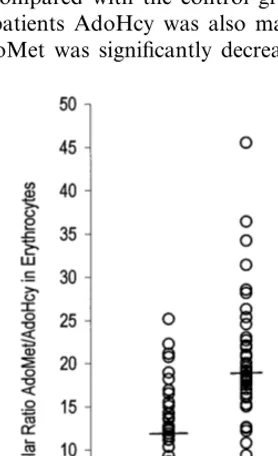
Table 1 shows the biochemical, hematological and demographic parameters of patients and control sub-jects. In plasma, AdoMet and AdoHcy levels were both significantly higher and the ratio significantly lower in patients compared with the control group. In erythro-cytes of patients AdoHcy was also markedly elevated, while AdoMet was significantly decreased, resulting in
an even greater decrease of this ratio in erythrocytes of PAOD patients compared with controls (Fig. 2).
Homocysteine concentrations were above 13.5mmol/l
in 11 (21%) of the control subjects, which is the upper limit of our own healthy control population normal range [18]. This limit, however, was determined in a younger control group with subjects aged 20 – 70 years. The 95th percentile in the present control group was 15.8 mmol/l. In patients mean total plasma
homocys-teine concentrations were significantly elevated com-pared with healthy subjects. Thirty-nine (64%) patients had homocysteine levels above 13.5 mmol/l and 29
(47.5%) above 15.8 mmol/l (95th percentile of the
con-trol subjects).
Bivariate linear regression analysis revealed no rela-tionship between the adenosylated metabolites and ho-mocysteine in patients or controls, whereas renal function (expressed as estimated creatinine clearance) was correlated with plasma AdoMet (R= −0.4, PB
0.01, in patients), plasma AdoHcy (R= −0.5, PB
0.001, in patients) and homocysteine (R= −0.34,
PB0.01 and R= −0.39, PB0.01, in patients and controls, respectively). Multiple regression analysis was performed to further evaluate the relationship of plasma AdoHcy and/or homocysteine with AdoMet. This revealed a strong influence of AdoHcy on AdoMet (R=0.836, PB0.001), but no correlation with homo-cysteine (R=0.03,P=NS).
Patients (42%) had impaired renal function (crea-tinine clearance B50 ml/min). After multifactorial ANOVA with estimated creatinine clearance, age, and gender as covariates, the differences between patients and controls of the standardised mean values of homo-cysteine and the adenosylated metabolites remained significant, but not the difference in the ratio of AdoMet/AdoHcy in plasma, which was mainly deter-mined by renal function (standardised mean values (9S.E.M.), 2.4290.2 and 2.6090.2 for patients and controls, respectively, P=NS, and P=0.02 for the influence of renal function in the model). Further inclu-sion of homocysteine in the above model had no addi-tional influence on the difference of the adenosylated metabolites between the two groups.
The OR for cases and control subjects per quartile increase of the AdoMet/AdoHcy ratios in erythrocytes and plasma, after adjustment for the estimated crea-tinine clearance, showed a high prevalence of PAOD within the lowest quartile of the ratio for erythrocytes (514.2), but not for plasma values (Table 2).
No relationship between the vitamins MeTHF or vitamin B12 and homocysteine or between the vitamins
and the adenosylated metabolites was observed. Gender differences were evident for body mass index (BMI) in patients (21.6 (16.1 – 32.4) kg/m2
and 25.4 (19.4 – 33.7), for women and men, respectively, PB
0.05) and for plasma total homocysteine in patients
Table 2
OR for patients to develop PAOD by quartile decrease of the ratio of AdoMet/AdoHcy in plasma and in erythrocytes (quartile 1 vs. quartile 4) after adjustment for the estimated creatinine clearance
Ratio in erythrocytesb
Quartilea Ratio in plasma
Adjusted ORc(95% CId) Adjusted ORc(95% CId)
514.2
1 7.07 (6.92, 7.22) 51.86 1.48 (0.29, 2.67)
2.06 (−1.90, 2.23) 1.87–2.52
14.2–19.1 1.33 (0.14, 2.52)
2
]19.2
3 1 2.53–3.3 0.58 (−0.77, 1.93)
4 ]3.31 1
aBased on the control values only.
bBased on control values but the last two quartiles were made into one as there were no patients in the last quartile. cOdds ratio adjusted for the estimated creatinine clearance by means of a logistic model.
dConfidence interval.
(15.3 (7.9 – 27.2)mmol/l and 18.3 (8.7 – 40.3), for women
and men, respectively, PB0.001). The gender differ-ence in homocysteine persisted also when renal function or BMI was considered.
In addition no influence of smoking, diabetes mellitus or arterial hypertension on any of the measured metabolites was observed in the two groups.
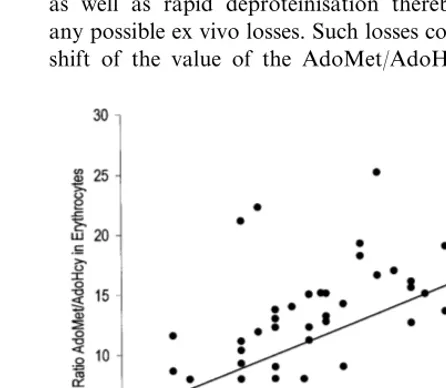
Hematocrit and haemoglobin levels were significantly decreased in patients compared with the controls (Table 1). No difference was found for other haematological parameters such as MCH, MCHC, MCV (values not shown). After multifactorial ANOVA analysis with the ratio of AdoMet/AdoHcy in erythrocytes and the esti-mated creatinine clearance as covariates the difference of the standardised mean values of hematocrit and haemoglobin between patients and controls was less strong but still significant (PB0.05 for both parame-ters), mainly influenced by the AdoMet/AdoHcy ratio in erythrocytes (PB0.05). Linear regression analysis in the whole patient group revealed a significant correla-tion between hematocrit and AdoMet/AdoHcy ratio in erythrocytes (R=0.421, P=0.01, Fig. 3), which was absent in the control group. This correlation remained significant when adjusted for the estimated creatinine clearance. No correlation between the haematological parameters and any of the plasma parameters measured was found.
5. Discussion
This study on homocysteine and related metabolites such as AdoMet and AdoHcy determined in different compartments (plasma, erythrocyte) of PAOD patients revealed a number of notable findings.
First, in accordance with other studies [8,24 – 26] a substantial proportion of our PAOD patients showed elevated plasma homocysteine, one of whose suggested toxic effects is disturbed endothelial function [27]. The percentage of patients with homocysteine levels above
the 95th percentile of the control group was higher than in previous reports [8,25], probably because patients with impaired kidney function, a strong predictor of homocysteine concentrations [28,29] and frequent in PAOD [30], were included in our study and because patients were on average older than in many previous studies.
Second, in erythrocytes the ratio of AdoMet/AdoHcy was markedly reduced in the patient group with more minor changes in the concentrations of these metabo-lites. Our control values are somewhat higher than those of AdoMet, 2.790.1 mmol/l and AdoHcy,
0.7790.09, resulting in a much lower ratio of 3.5 reported by Perna et al. [31]. This may be due to the determination of the metabolites in whole blood and plasma separately and calculation of the erythrocyte concentrations based on hematocrit levels in our study, as well as rapid deproteinisation thereby minimising any possible ex vivo losses. Such losses could result in a shift of the value of the AdoMet/AdoHcy ratio. The
lower ratios in patients could be a consequence of increased extracellular levels of homocysteine which can freely enter erythrocytes [19] and be converted to Ado-Hcy due to the thermodynamics of the AdoAdo-Hcy
hydro-lase reaction [15], whereas remethylation of
homocysteine to methionine does not occur in erythro-cytes [19,31] preventing de novo synthesis of AdoMet. Alternatively an accelerated requirement of AdoMet for
trans-methylation reactions e.g. methylation of erythro-cyte membrane proteins [19], could lead to increased formation of AdoHcy, which subsequently leads to a release of the resulting homocysteine into plasma as demonstrated by Dudman and co-workers 1998 [32], and, due to the lack of remethylation in red cells, to a depletion of AdoMet. In both cases the ratio of AdoMet/AdoHcy in erythrocytes may be expected to decrease to a much larger extent than in plasma as indeed found in this study.
The ratio of AdoMet/AdoHcy and the concentration of AdoHcy are reported to be crucial in the regulation oftrans-methylation reactions, for example as reported in brain [16]. Thus the decreased ratios in erythrocytes, which are independent of kidney function and the association between PAOD and low AdoMet/AdoHcy values demonstrated by the OR may point to disturbed
trans-methylation reactions in vascular disease patients. Indeed disturbed erythrocyte membrane protein methyl esterification, atrans-methylation reaction, was associ-ated with increased intra-cellular AdoHcy levels and a decreased AdoMet/AdoHcy ratio [20], and suggested to cause hemolysis and anaemia in patients with chronic renal failure. This observation is supported by the correlation between hematocrit and the AdoMet/ Ado-Hcy ratio in our patients, which remained when esti-mated creatinine clearance was included as a co-variate in the statistical analysis. Whether the hematocrit and the AdoMet/AdoHcy ratio are causally linked requires further studies focussing on occurrence of anaemia, which has also been observed in cardiovascular disease patients [33] and including additional red cell parame-ters such as reticulocyte counts.
Third, plasma levels of both AdoMet and AdoHcy were increased in the patient group with relatively higher increases of the latter resulting in a reduced ratio of AdoMet/AdoHcy. The compartmental origin of plasma AdoMet and AdoHcy is not fully understood but increases of these compounds, seen post methionine loading in control subjects [22], and evidence of Ado-Hcy export in tissue culture [34] suggests plasma levels of these two compounds may reflect tissue metabolism. Regression analysis was performed in attempting to explain these changes in plasma levels of AdoMet and AdoHcy. Although these levels correlate with each other, possibly reflecting inhibition of enzymatic AdoMet dependent trans-methylation reactions [35], they do not correlate with homocysteine, even though
the thermodynamics of the AdoHcy hydrolase reaction favour the synthesis of AdoHcy [15] when homocys-teine accumulates.
The lack of a statistical correlation between homo-cysteine and AdoMet or AdoHcy may reflect the mainly intra-cellular nature of the adenosyl derivatives whereas homocysteine is readily exported into the ex-tracellular compartment. Also regulation of AdoMet and AdoHcy is multifactorial and other factors than the accumulation of homocysteine will influence AdoMet and AdoHcy levels and their ratio in plasma. For example there was a striking influence of kidney function on the plasma ratios of AdoMet/AdoHcy as demonstrated by the multifactorial ANOVA as well as by the OR in plasma after adjustment for renal dys-function. In contrast to the AdoMet/AdoHcy ratios in erythrocytes which were related to PAOD indepen-dently from kidney function, decreased plasma ratios seem not to be directly related to the disease itself, but are rather predicted by the estimated creatinine clear-ance. These findings confirm the modulation of me-thionine metabolism by renal function, which has been shown to be a good predictor for homocysteine espe-cially when kidney function is impaired [28,29] as in a substantial proportion of our patients.
Fourth, mean MeTHF values were slightly lower in our patients compared with controls, consistent with our previous findings in patients with coronary artery disease (5.2 – 54.8 and 8.5 – 74.7 nmol/l in patients and controls, respectively; PB0.05 [18]). However, there was no statistically significant correlation between ho-mocysteine and folate (measured as MeTHF) in the present study, in contrast to our [18], and another previous study [36] the latter measuring total folate. This may be due to the higher MeTHF in the present study, with MeTHF levels below the 5th percentile (11 nmol/l) of our own healthy population range in only 16% of our patients, since there is evidence that this association is mainly found when folate levels are low [37,38].
concentrations, combined treatment with vitamin B6,
which stimulates trans-sulphuration and should lower AdoHcy, together with folate, which will increase the AdoMet pool, may be more effective than either vita-min given singly.
Acknowledgements
These studies were supported by grants from the
Swiss National Science Foundation (No.
3200-045988.95/98); the Treubel Foundation, Basel and F. Hoffmann-La Roche Ltd., Vitamins and Fine Chemi-cals Division Exploratory Research, Basel, Switzerland. We are indebted to P.D., Dr P. Huber of the Depart-ment of Central Laboratories, University Hospital Basel, Switzerland for the analysis of vitamin B12.
References
[1] Taylor LM, Moneta GL, Sexton GJ, Schuff RA, Porter JM. Prospective blinded study of the relationship between plasma homocysteine and progression of symptomatic peripheral arterial disease. J Vasc Surg 1999;29:8 – 19.
[2] Mercie´ P, Seigneur M, Conri C, Boisseau MR. Hyperhomocys-teinaemia and endothelial dysfunction in peripheral arterial dis-ease. Thromb Res 1999;93:97 – 9.
[3] Refsum H, Ueland PM, Nygard O, Vollset SE. Homocysteine and cardiovascular disease. Annu Rev Med 1998;49:31 – 62. [4] Perry IJ, Refsum H, Morris RW, Ebrahim SB, Ueland PM,
Shaper AG. Prospective study of serum total homocysteine concentration and risk of stroke in middle-aged British men. Lancet 1995;346:1395 – 8.
[5] Malinow MR. Plasma homocyst(e)ine: a risk factor for arterial occlusive diseases. J Nutr 1996;126:S1238 – 43.
[6] Stampfer MJ, Malinow MR, Willett WC, et al. A prospective study of plasma homocyst(e)ine and risk of myocardial infarc-tion in US physicians. J Am Med Assoc 1992;268:877 – 81. [7] van den Berg M, Stehouwer CD, Bierdrager E, Rauwerda JA.
Plasma homocysteine and severity of atherosclerosis in young patients with lower-limb atherosclerotic disease. Arterioscler Thromb Vasc Biol 1996;16:165 – 71.
[8] Brattstro¨m L, Israelsson B, Norrving B, Bergqvist D, Tho¨rne J, Hultberg B, Hamfelt A. Impaired homocysteine metabolism in early-onset cerebral and peripheral occlusive arterial disease. Effects of pyridoxine and folic acid treatment. Atherosclerosis 1990;81:51 – 60.
[9] Boushey CJ, Beresford SAA, Omenn GS, Motulsky AG. A quantitative assessment of plasma homocysteine as a risk factor for vascular disease. J Am Med Assoc 1995;274:1049 – 57. [10] Graham IM, Daly LE, Refsum HM, et al. Plasma homocysteine
as a risk factor for vascular disease: the European concerted action project. J Am Med Assoc 1997;277:1775 – 81.
[11] den Heijer M, Koster T, Blom HJ, et al. Hyperhomocysteinemia as a risk factor for deep-vein thrombosis. New Engl J Med 1996;334:759 – 62.
[12] Selhub J, Dangelo A. A relationship between homocysteine and thrombotic disease. Am J Med Sci 1998;316:129 – 41.
[13] Ueland PM, Refsum H, Brattstro¨m L. Plasma homocysteine and cardiovascular disease. In: Robert BF, Jr, editor. Atherosclerotic Cardiovascular Disease, Hemostasis, and Endothelial Function. New York: Marcel Dekker, 1992:183 – 236.
[14] Chiang PK, Gordon RK, Tal J, Zeng GC, Doctor BP, Pard-hasaradhi K, Mccann PP.S-adenosylmethionine and methyla-tion. FASEB J 1996;10:471 – 80.
[15] Finkelstein JD. Methionine metabolism in mammals. J Nutr Biochem 1990;1:228 – 37.
[16] McKeever M, Molloy A, Weir DG, Young PB, Kennedy DG, Kennedy S, Scott JM. An abnormal methylation ratio induces hypomethylation in vitro in the brain of pig and man, but not in rat. Clin Sci 1995;88:73 – 9.
[17] Mathews CK, van Holde KE. Biochemistry. Redwood City, CA, USA: Benjamin/Cummings, 1990:713.
[18] Loehrer FMT, Angst CP, Jordan PP, Ritz R, Haefeli WE, Fowler B. Low whole bloodS-adenosylmethionine and correla-tion between 5-methyltetrahydrofolate and homocysteine in coronary artery disease. Arterioscler Thromb Vasc Biol 1996;16:727 – 33.
[19] Perna AF, Ingrosso D, Galletti P, Zappia V, De Santo NG. Membrane protein damage and methylation reactions in chronic renal failure. Kidney Int 1996;50:358 – 66.
[20] Perna AF, Ingrosso D, Zappia V, Galletti P, Capasso G, De Santo NG. Enzymatic methyl esterification of erythrocyte mem-brane proteins is impaired in chronic renal failure. Evidence for high levels of the natural inhibitor S-adenosylhomocysteine. J Clin Invest 1993;91:2497 – 503.
[21] Loehrer FMT, Angst CP, Brunner FP, Haefeli WE, Fowler B. Evidence for disturbedS-adenosylmethionine/S -adenosylhomo-cysteine ratio in patients with end-stage renal failure: a cause for disturbed methylation reactions. Nephrol Dial Transplant 1998;13:656 – 61.
[22] Loehrer FMT, Angst CP, Browne G, Frick G, Haefeli WE, Fowler B. The effect of methionine loading on 5-methyltetrahy-drofolate,S-adenosylmethionine andS-adenosylhomocysteine in plasma of healthy humans. Clin Sci 1996;91:79 – 86.
[23] Dettli L. The kidney in pre-clinical and clinical pharmacokinet-ics. Jpn J Clin Pharmacol Ther 1984;15:234 – 41.
[24] Taylor LM, Jr, DeFrang RD, Harris EJ, Jr, Porter JM. The association of elevated plasma homocyst(e)ine with progression of symptomatic peripheral arterial disease. J Vasc Surg 1991;13:128 – 36.
[25] Molgaard J, Malinow MR, Lassvik C, Holm AC, Upson B, Olsson AG. Hyperhomocyst(e)inaemia: an independent risk fac-tor for intermittent claudication. J Intern Med 1992;231:273 – 9. [26] Malinow MR, Kang SS, Taylor LM, et al. Prevalence of hyper-homocyst(e)inemia in patients with peripheral arterial occlusive disease. Circulation 1989;79:1180 – 8.
[27] Bellamy MF, McDowell IFW. Putative mechanisms for vascular damage by homocysteine. J Inherit Metab Dis 1997;20:307 – 15. [28] Chauveau P, Chadefaux B, Coude M, Aupetit J, Hannedouche T, Kamoun P, Jungers P. Increased plasma homocysteine con-centration in patients with chronic renal failure. Miner Elec-trolyte Metabol 1992;18:196 – 8.
[29] Bostom AG, Lathrop L. Hyperhomocysteinemia in end-stage renal disease: prevalence, etiology, and potential relationship to arteriosclerotic outcomes. Kidney Int 1997;52:10 – 20.
[30] Tscho¨pl M, Tsakiris DA, Marbet GA, Labs KH, Ja¨ger K. Role of hemostatic risk factors for restenosis in peripheral arterial occlusive disease after transluminal angioplasty. Arterioscler Thromb Vasc Biol 1997;17:3208 – 14.
[31] Perna AF, Ingrosso D, De Santo NG, Galletti P, Zappia V. Mechanism of erythrocyte accumulation of methylation inhibitor
S-adenosylhomocysteine in uremia. Kidney Int 1995;47:247 – 53. [32] Dudman NPB, Fu W, Yang K, Silberberg JS, Crooks R. Homo-cysteine production in chronic renal failure. Neth J Med 1998;52(suppl.):S22.
[34] Svardal AM, Djurhuus R, Ueland PM. Disposition of homocys-teine and S-3-deazaadenosyl-homocysteine in cells exposed to 3-deazaadenosine. Mol Pharmacol 1986;30:154 – 8.
[35] Niederwieser A. Inborn errors of pterin metabolism. In: Botez MI, Reynolds EH, editors. Folic Acid in Neurology, Psychiatry and Internal Medicine. New York: Raven, 1979:349 – 84. [36] Mansoor MA, Bergmark C, Svardal AM, Lonning PE, Ueland
PM. Redox status and protein binding of plasma homocysteine and other aminothiols in patients with early-onset peripheral vascular disease. Homocysteine and peripheral vascular disease. Arterioscler Thromb Vasc Biol 1995;15:232 – 40.
[37] Boushey CJ, Beresford SA, Omenn GS, Motulsky AG. A quan-titative assessment of plasma homocysteine as a risk factor for vascular disease. Probable benefits of increasing folic acid in-takes. J Am Med Assoc 1995;274:1049 – 57.
[38] Selhub J, Jacques PF, Wilson PW, Rush D, Rosenberg IH. Vitamin status and intake as primary determinants of homocys-teinemia in an elderly population. J Am Med Assoc 1993;270:2693 – 8.
[39] Ward M, McNulty H, McPartlin J, Strain JJ, Weir DG, Scott JM. Plasma homocysteine, a risk factor for cardiovascular dis-ease is lowered by physiological doses of folic acid. Q J Med 1997;90:519 – 24.
[40] Brattstrom L. Vitamins as homocysteine-lowering agents. J Nutr 1996;126:S1276 – 80.
[41] den Heijer M, Brouwer IA, Bos GMJ, et al. Vitamin supplemen-tation reduces blood homocysteine levels. A controlled trial in patients with venous thrombosis and healthy volunteers. Arte-rioscler Thromb Vasc Biol 1998;18:356 – 61.
![Fig. 1. Important AdoMet-dependent trans-methylation reactions inmammals [17].](https://thumb-ap.123doks.com/thumbv2/123dok/3147660.1384323/2.612.35.257.481.684/fig-important-adomet-dependent-trans-methylation-reactions-inmammals.webp)