Thioguanine-based DENV-2 NS2B/NS3
protease inhibitors: Virtual screening,
synthesis, biological evaluation and molecular
modelling
Maywan Hariono1,2, Sy Bing Choi1,3☯, Ros Fatihah Roslim1☯, Mohamed Sufian Nawi1,4☯, Mei Lan Tan5, Ezatul Ezleen Kamarulzaman1, Nornisah Mohamed1, Rohana Yusof6, Shatrah Othman7, Noorsaadah Abd Rahman7, Rozana OthmanID8, Habibah A. WahabID1,9*
1School of Pharmaceutical Sciences, Universiti Sains Malaysia, Minden, Pulau Pinang, Malaysia,2Faculty of Pharmacy, Sanata Dharma University, Maguwoharjo, Sleman, Yogyakarta, Indonesia,3School of Data Sciences, Perdana University, Blok B and d1, MARDI Complex, Jalan MAEPS Perdana, Serdang, Selangor,
4Department of Pharmaceutical Chemistry, Kulliyah of Pharmacy, International Islamic University Malaysia, Kuantan, Pahang, Malaysia,5Advanced Medical and Dental Institute, Universiti Sains Malaysia, Bertam, Pulau Pinang, Malaysia,6Department of Molecular Medicine, Faculty of Medicine, Universiti Malaya, Kuala Lumpur, Malaysia,7Department of Chemistry, Faculty of Science, Universiti Malaya, Kuala Lumpur, Malaysia,8Department of Pharmacy, Faculty of Medicine, Universiti Malaya, Kuala Lumpur, Malaysia,
9Malaysian Institute of Pharmaceuticals and Nutraceuticals, Ministry of Science, Technology and Innovation, Halaman Bukit Gambir, Bayan Lepas, Pulau Pinang, Malaysia
☯These authors contributed equally to this work. *[email protected],[email protected]
Abstract
Dengue virus Type 2 (DENV-2) is predominant serotype causing major dengue epidemics. There are a number of studies carried out to find its effective antiviral, however to date, there is still no molecule either from peptide or small molecules released as a drug. The present study aims to identify small molecules inhibitor from National Cancer Institute database through virtual screening. One of the hits,D0713(IC50= 62μM) bearing thioguanine
scaf-fold was derivatised into 21 compounds and evaluated for DENV-2 NS2B/NS3 protease inhibitory activity. Compounds18and21demonstrated the most potent activity with IC50of
0.38μM and 16μM, respectively. Molecular dynamics and MM/PBSA free energy of binding calculation were conducted to study the interaction mechanism of these compounds with the protease. The free energy of binding of18calculated by MM/PBSA is -16.10 kcal/mol compared to the known inhibitor, panduratin A (-11.27 kcal/mol), which corroborates well with the experimental observation. Results from molecular dynamics simulations also showed that both18and21bind in the active site and stabilised by the formation of hydro-gen bonds with Asn174.
Citation:Hariono M, Choi SB, Roslim RF, Nawi MS, Tan ML, Kamarulzaman EE, et al. (2019) Thioguanine-based DENV-2 NS2B/NS3 protease inhibitors: Virtual screening, synthesis, biological evaluation and molecular modelling. PLoS ONE 14 (1): e0210869.https://doi.org/10.1371/journal.
Copyright:©2019 Hariono et al. This is an open access article distributed under the terms of the Creative Commons Attribution License, which permits unrestricted use, distribution, and reproduction in any medium, provided the original author and source are credited.
Data Availability Statement:All relevant data are within the paper and its Supporting Information files.
Introduction
Dengue, caused by Dengue Virus (DENV), is the most important mosquito-borne viral disease affecting the tropics and subtropics [1]. Endemic in more than 100 countries [2,3], the virus is estimated to cause 390 million infections each year [4]. DENV infections can result in serious diseases including dengue fever, dengue hemorrhagic fever (DHF), dengue shock syndrome (DSS) and even death [5]. There are no approved antiviral drugs for these diseases and cur-rently, patients are treated with supportive care to relieve fever, pain, and dehydration [6]. A tetravalent dengue vaccine (CYD-TDENV or Dengvaxia), the first dengue vaccine has recently been registered in several countries [7]. Despite being a leading cause of hospitalisation and death among children in some Asian and Latin American countries [8], this vaccine is not rec-ommended for use in children under 9 years of age due to safety concerns [7]. Therefore, there exists an urgent need for antiviral therapies to treat dengue.
DENV carries a positive single strand RNA in its genome and five serotypes (DENV-1 to 5) have been identified. DENV-2 is the most prevalent type in dengue epidemic, especially in the South East Asian region and has been associated with severe dengue cases [9]. The new sero-type (DENV-5) [10] was discovered in 2013 in Sarawak, Malaysia further complicates the pre-vention and treatment of the disease. The virus genome is encoded by three structural proteins (C, prM, E) as well as seven non-structural proteins (NS1, NS2A, NS2B, NS3, NS4A, NS4B and NS5) [11]. Of these proteins, NS2B/NS3protease (NS2B/NS3pro) has been well studied and regarded as a promising target in anti-dengue discovery [12–15].
NS3 is a trypsin like serine protease which plays a role in post-translation in the virus matu-ration. This domain has a catalytic triad made up of His51, Asp75 and Ser135 and its activity is enhanced by NS2B as the cofactor [16,17]. This cofactor contributes to the NS3 activity through its hydrophilic region which is responsible for holding and promoting the activation of NS3 while the hydrophobic region takes part in membrane association upon the cleavage process [18,19].
Previous discoveries of dengue inhibitors by targeting NS2B/NS3pro activity were mainly based on the non-prime substrates which were identified by profiling dengue virus using tetra-peptides [20–22]. Significant challenges arise as the protein possesses a solvent-exposed, topo-logically shallow active site and dependent on the selectivity for substrates containing basic amino acids (arginine and lysine) at P1 and P2 positions [23]. Nevertheless, many peptidic or modified peptidic molecules have been discovered to have good NS2B/NS3pro inhibition activities [15,24–30]. In addition, there are also reports of potent small molecule NS2B/ NS3pro inhibitors from natural products (panduratin[31], agathisflavone and quercetin [32]), from synthetic medicinal chemistry (dehydronaphthalene [33], benzimidazole [34], and thia-diazoloacrylamide [35]) or from the utilisation of computational methods [36–39].
Here, we report the discovery of potential NS2B/NS3pro inhibitors designed based on thio-guanine scaffold identified through the virtual screening of compounds library from National Cancer Institute (NCI) diversity set II. Twenty-four compounds were found asin silicohits based on the free energy of binding (ΔGbind) ranking from a total of 1990 compounds. Twenty
of them were obtained from NCI forin vitroassay, out of which four demonstrated moderate inhibition towards DENV-2 NS2B/NS3pro (IC50= 29–77μM). Although Diversity0713
(D0713) is not the most potent compound identified in the virtual screening, the structure contains thioguanine (TGor 6-thioguanine) scaffold can be used as a template to develop a series of analogues as its simple chemical structure benefits feasible synthetic steps.TGshowed 56% inhibition at 200μg/mL (1.2 mM) indicating that even without any modification to the structure, the scaffold itself is able to inhibit the protease activity. This compound is a drug classed as anti-neoplastic agent and used with other compounds in treating leukemia [40] and
ScienceFund Grant No. 02-05-23-SF0019 (2014-2015). We thank National Cancer Institute, US for the complementary samples of the 20 NCI hit compounds.
has been investigated in many pharmacological activities such as immunomodulators in auto-immune diseases [41] and transplant graft rejection [42]. From this initial study, we are inter-ested to utilise thioguanine scaffold in the design of DENV-2 NS2B/NS3pro inhibitor(s). Thus, in this study, we designed, synthesised thioguanine analogues and investigated their possible DENV-2 NS2B/NS3pro inhibition activity. We hope that this study could contribute to the efforts in discovering novel and potent anti-dengue agents.
Materials and methods
Virtual screening
Virtual screening was carried out using AutoDockVina [43] (www.autodock.scripps.edu). The DENV-2 NS2B/NS3pro model was taken from published article [44], where the model was built based on the DENV-2 complex cofactor-protease using the crystal structure of NS2B/ NS3pro West Nile Virus (WNV) as the template. The docking procedure was initiated by the preparation of NS2B/NS3pro as a macromolecule using AutoDock Tools (version 1.5.6) with default parameters for docking with AutoDock Vina. The exhaustiveness was set to 8 and other parameters were unchanged. The centre of the grid box was set at 30.71, 50.48 and 4.10Åin x, y, z coordinates, respectively, with a box size of 25 x 25 x 25 points. The internal validation was done by re-docking the tetrapeptide inhibitor (Bz-Nle-Lys-Arg-Arg-H) with the RMSD value not greater than 2Å. The external validation was done using panduratin A (a competitive inhibi-tor), that dock at the same binding site of tetrapeptide inhibitor. Thein silicoscreening of 1990 ligands from NCI diversity set (II) was carried out using the docking parameters above. The hit compounds were ranked according to the free energy of binding (ΔGbind) and analysis of their
binding modes were performed using Discovery Studio 3.5 (www.accelrys.com).
DENV-2 NS2B/NS3pro expression and purification, optimum activity and
inhibition assay
The DENV-2 NS2B/NS3pro expression was carried out according to the established method by Yusofet al., (2000) [45] with minor modifications according to published articles [46–50]. The plasmid encoding the NS2B/NS3 protease sequence from DENV-2 was transformed into
Escherichia colistrain XL1-Blue transformed with pQE30.CF40.gly(T).NS3pro expression plasmid were grown in LB medium containing 10μg/ml ampicillin at 37˚C until the OD600
reached 0.6. First, the cells were incubated at 37˚C, 200 rpm until OD600reached ~0.6. One ml
of isopropyl-β-D-thiogalactopyranoside (0.5 mM in LB medium) was added to the bacterial cells for 2 hours to induce protein expression. Expression of the recombinant protein was induced by the addition of 0.5 M IPTG and the culture was incubated for 2 hours.
Bradford protein assay. The purified protein was then analysed with 12% SDS-PAGE, pooled and stored at -80˚C for further use in the dengue protease activity and inhibition studies. The dengue protease activity assay was developed as previously described [51] with a slight modifi-cation [46,47,50]. Briefly, the assay system comprised of 200 mM Tris-HCl (pH 8.5) buffer, DENV-2 NS2B/NS3pro and Boc-GRR-MCA as the substrate. Protease optimum assay was executed to ascertain maximum protease activity at constant concentration of the substrate (25μM). The protease concentrations were varied within 0–10μM. The 7-Amino-4-methyl-coumarin (AMC) [51] produced was measured as fluorescence intensity atλexcitationof 340 nm
andλemissionof 440 nm by using ELISA modulus microplate reader.
The compounds were also checked for its requirement to pass the pan assay interference compounds (PAINS) (http://cbligand.org/PAINS/) before tested for their inhibition activities against DENV-2 NS2B/NS3pro at a range of concentrations (0 to 300μg/mL). The result shows that all the tested compounds, except forD1855passed the PAINS filter. The concentra-tion of the protease being used was 3μM initially incubated with the compound for 10 minutes at 37˚C with 200 rpm of rotation. Subsequently, 25μM of the substrate was added and the incubation was further prolonged for 60 minutes. The fluorescence intensity at 340/ 440 nm was measured as the AMC byproduct was released upon the peptide substrate cleavage by the protease. The experiments were triplicated and Panduratin A was used as the positive control.
Synthesis of thioguanine derivatives
All reactions were carried out using standard techniques for the exclusion of moisture [52,53] except those in aqueous media. The progress of reaction was monitored using TLC on 0.25 mm silica F254and detected under UV light, or iodine vapor.1H-NMR and13C-NMR spectra
were determined using Bruker Avance 500 spectrometer with TMS as an internal standard and the mass spectra were determined using XEVO-G2TOF #YCA153. Melting points were obtained using a STUART SMP electro-thermal apparatus and were uncorrected. Anhydrous reactions were carried out in over-dried glassware under a nitrogen atmosphere. The detail method of synthesis, the compounds’ structures as well as the numbering due to NMR assigna-tion can be seen inS1 Text.
Molecular docking simulation
Molecular dynamics simulation
To gain insight into the binding interaction of compounds18, 21and panduratin A with NS2B/NS3pro, molecular dynamics simulations were carried out using Amber 14 [57]. The following is the description of the setups of NS2B/NS3pro-18(compound18),
NS2B/NS3pro-21(compound21) and NS2B/NS3panduratin. All systems were prepared using LEaP pro-gram. Amber generalised force filed (GAFF) were assigned to the three compounds whilst amber ff14SB to NS2B/NS3pro. Each system was neutralised with sodium ions and solvated using TIP3P water in a 53.57, 46.51, 44.05Åtruncated octahedral water box. To eliminate the steric clashes, each system was subjected to a total of 500 stepwise minimisation using steepest descent followed by conjugated gradient. The solvent of the system was first heated to 100 K with an NVT ensemble, followed by heating of the whole system to 300 K with an NPT ensem-ble. Throughout the MD simulation, a 0.2 fs time step, SHAKE algorithm, periodic boundary, and 10Åcutoffs were applied. The analysis for RMSD and gyration was taken from the begin-ning. The equilibration phase was 1 ns, and the production phase of the simulation was 69 ns, with a total simulation time of 70 ns for each system. Analysis was conducted using CPPTRAJ modules in AmberTools 14 and visualised through VMD.
MM/PBSA calculation
A total of 100 frames were extracted from the last nanosecond of the 70 ns simulation trajec-tory files. MM/PBSA calculations were performed on each frame using MMPBSA.py module in AMBER 14. Energy compositions of compounds18,21as well as panduratin A with NS2B/ NS3pro was dissected accordingly.
Results and discussion
Virtual screening and the confirmation of activity of the
in silico
hits
In this study, 1990 compounds from NCI Diversity Set II were docked into the active site pocket of DENV-2 NS2B/NS3pro. The active site is made up of important amino acid residues in S1-S4 subpockets such as Asp129_NS3, Ser135_NS3, Tyr150_NS3 and Tyr161_NS3 (S1 pocket); Asp81_NS2B, Gly82_NS2B, Ser83_NS2B, Asp75_NS3 and Asn152_NS3 (S2); Ser85_NS2B, Ile86_NS2B and Lys87_NS2B (S3); Val154_NS3 and Ile155_NS3 (S4). The 24 top hit compounds were investigated for their interaction with the active site’s residues and ranked in term of the free energy of binding (ΔGbind= -10 kcal/mol to -5 kcal/mol, seeS1 Table. We identified that there are more than 50 ligands docked into the active site with that
ΔGbindrange, but only 24 of them shows interesting interactions with the essential amino acid
residues. The selection ofΔGbindrange is adopted from the study reported by Shityakov (2014)
that Gibbs free energy of binding<6.0 kcal/mol is clustered as active when this prediction is
highly correlated with the experimental results with R2= 0.880; F = 692.4 standard error of estimate = 0.775 and p-value = 0.0001 [58]. This virtual screening study has a limitation in which no decoy database is being used to validate the docking protocol. The non-binding mol-ecule could be selected over the true ligand called as false positive. Decoy database is the false positive hits which may improve the validity of the test by generating the true positive rate as well as enrichment factor. Unfortunately, the decoy for this DENV-2 NS2B/NS3pro has not been available in a database of useful decoy (DUD dude.docking.org), therefore, the validation of this virtual screening only relies on internal control docking and byin vitroassay [59].Fig 1
Dengue protease inhibition assay
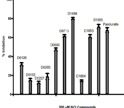
From the 24 hits identified in the virtual screening, only 20 were available for the inhibition assay. Of these 20 compounds, only 10 compounds exhibited significant inhibition activity towards DENV-2 NS2B/NS3pro activity (Fig 2). The other 10 NCI compounds exhibited neg-ligible inhibition (<5% inhibition at 200μM) towards the protease activity.
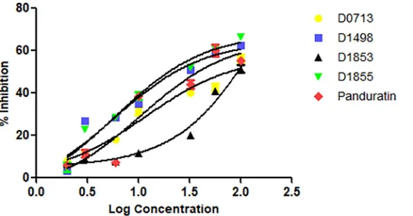
Four NCI compounds with the percentage of inhibition greater than 50% were selected for further assay to determine their IC50(Fig 3).D1855showed the strongest inhibition towards
the protease activity with IC50= 29μM followed byD1498(48μM),D0713(62μM) and
D1853(77μM) (Table 1). Panduratin A was used as a control in this experiment. Previously, panduratin A was isolated from finger root (Boesenbergia rotunda(L.)) demonstrating com-petitive inhibition toward DENV-2 NS2B/NS3pro with Ki= 25μM [31].
Fig 1. Docked conformation of top 24 NCI compounds in the active pocket of DENV-2 NS2B/NS3pro.The NS2B and NS3 domains are presented as surface form (blue area = NS2B, red area = NS3pro). Insets are the two ligands that bound to the NS2B, instead of NS3pro.
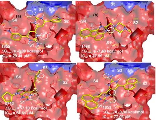
D1855demonstrated the best inhibition towards the protease activity at concentration less than 50μM as the relative protease activity dramatically decreased from 100 to 30% (IC50=
29μM). This inhibition is probably provided by the hydrogen bonding with Gly153 and His51 and the phenyl ring of the compound formedπ-π- interactions with Tyr161 and His51 (Fig 4 (A)).D1498(IC50= 48μM) is the second most potent of these four NCI compounds where
the activity might be contributed by H-bond interaction with Gly151 of NS3,π—πinteraction between its anthracene ring with Tyr161 andπ-cation interaction with His51 (Fig 4(B)).
D0713also demonstrated significant inhibition towards the protease with IC50= 62μM. The
docked pose shows the formation of H-bonds between its amino groups with Asn152, Gly151, and Ser135 andπ—πinteraction between its guanine ring and Tyr161 (Fig 4(C)). Although the IC50ofD1853is approximately 77μM, this compound showed nearly 70% inhibition of
the protease activity at 200μM. As with the above NCI compounds, this ligand also formedπ
-πas well asπ-cation interactions with Tyr161 and His51, respectively, in addition to the H-bond interaction between its amino groups with Gly151, Ser135 and Gly153 (Fig 4(D)). Fig 2.In vitroDENV-2 NS2B/NS3pro inhibition assay of Panduratin A and selected NCI compounds; NCI code D0265, D0685, D0227, D0152, D0126, D1804, D1855, D1498, D0713 and D1853 with D in the code stands for Diversity.The assays contained 0μM NCI compounds were taken as 0% protease inhibition.
In our quest to identify suitable scaffold for designing potential novel and potent dengue protease inhibitors, we took into consideration the structure ofD0713. Although this is not the most potent compound identified in the virtual screening, the structure contains thiogua-nine (TGor 6-thioguanine) scaffold.TG,a known drug used with other compounds in treat-ing leukemia [40] has also been investigated in other pharmacological activities such as in autoimmune diseases [41] and transplant graft rejection [42]. In addition, its simple chemical structure also benefits feasible synthetic steps, thus,TGis selected as a template to develop a series of analogues. Therefore, in order to test our hypothesis,TGwas subjected to the protease inhibition assay and it was found that the molecule showed 56% inhibition at 200μg/mL (1 mM) indicating that even without any modification to the structure, the scaffold itself is able to inhibit the protease activity. Thus,TGwas chosen as the lead structure in the design and synthesis of potential anti-dengue compounds.
Design, synthesis and protease inhibition activity of thioguanine derivatives
The various thioguanine derivatives designed are shown inTable 2with the scaffold being illustrated inFig 5. The design was initiated by connecting the aromatic hydrophilic group as hydrogen bond acceptor (HBA) (1–3)with the amino group; and aromatic hydrophobic group with sulfonyl in4–6at R1. In7–13,acyl groups such as acetyl, butanoyl, isobutanoyl,
pentanoyl, isopentanoyl and hexanoyl groups were placed at the same position, while benzoyl group was instead placed in14. Replacing aliphatic hydrophobic group at R3with cyclopentyl
group, as present inD0713, yielded15.
Compounds1–3are imine derivatives ofTG. Imine itself has an electron withdrawing nature, albeit weakly. Attaching another EWG such as NO2(1) and COOH (2) to the
Fig 3. Plot of % DENV-2 NS2B/NS3pro inhibition vs log concentration of the four NCI compounds.Panduratin A was used as a control in this experiment.
https://doi.org/10.1371/journal.pone.0210869.g003
Table 1. Topin vitrohits from NCI diversity set compounds towards both S1 and S2 pockets of DENV-2 NS2B/ NS3pro.
Ligands ΔGbind(Kcal/ mol) Experimental IC50(μM)
D1498 -7.40 48
D1853 -9.90 77
D1855 -8.90 29
D0713 -7.10 62
Panduratin A -6.30 56
benzimine (in theparaposition) as R1rendered the compounds to be inactive (IC50>
1000μM). Interestingly, attaching an electron donating group (EDG) to benzimine at R1(3)
showed high protease inhibition (IC50= 28μM). These raised the idea to maintain the EWG
group as a linker between the two aromatic rings ofTGand the phenyl group which was modi-fied to have EDG character.
Compounds4and5were designed to have sulfonyl (EWG) with CH3and/ or OCH3
(EDG) on benzenesulfonyl as R1. These compounds demonstrated lower activities than3with
IC50of 68 and 55μM, respectively. In6,the same group attached to R1was also placed at R2
during the synthesis but changes in activity were insignificant. Placing at R1with acetyl or
extended alkyl chain (7–13), in general resulted in marked reduction in protease inhibition, with the exception noted for11which showed IC50= 80μM.
Placing a simple aromatic hydrophobic group (benzoyl) as seen in14did not increase the activity. Exploring placement at R3with an aliphatic hydrophobic group (15) also did not Fig 4. Docked poses of (a)D1855, (b)D1498, (c)D0713and (d)D1853. The NS2B and NS3pro domains are presented as surface form (blue area = NS2B, red area = NS3pro). The H-bond interactions are assigned as yellow dots inside the white circles.
produce a compound with any improvement in the protease inhibition activity. At this point, it was decided to synthesize six more compounds with expected higher activities.
Table 2. The list of TG derivatives with their experimental IC50against DENV -2 NS2B/NS3pro.
Ligands R1 R2 R3 IC50(μM)
1 (4-nitrophenyl)methanimine H H 1995
2 4-(iminomethyl)benzoic acid H H 1367
3 4-(iminomethyl)-2-methoxy-6-nitrophenol H H 28
4 3-methoxybenzenesulfinic acid H H 68
5 4-methoxybenzenesulfinic acid H H 55
6 3-methylbenzenesulfinic acid 3-methylbenzenesulfinic acid H 63
7 acetyl H H 151
8 butanoyl H H 57
9 isobutanoyl H H 3893
10 pentanoyl H H 1037
11 3-methylbutanoyl H H 80
12 hexanoyl H H 168
13 palmitoyl H H 132
14 benzoyl H H 370
15 acetyl H cyclopentyl 556
D0713 H 2-methylpyridine cyclopentyl 62
TG H H H 753
Panduratin A - - - 56
https://doi.org/10.1371/journal.pone.0210869.t002
Fig 5. The structure of thioguanine scaffold.
Inspired by7,8and11where the compounds have amide group with promising inhibitory activity, six compounds (16–21,seeTable 3) having amide group with diverse alkyl chains were designed. As the natural substrate of the protease is peptide, it made sense to incorporate amide groups into the inhibitor structure in order to mimic the peptide character. High activ-ity of18, 19 and 21towards DENV-2 NS2B/NS3pro (IC50of 0.38, 54 and 16μM, respectively)
could be due to this mimic of peptide character. Compound18with pentanamide chains at both R1and R2possessed the highest activity against the protease. However, attaching benzyl
group at R2(19) dramatically decreased the activity from 0.38 to 54μM. Interestingly,
attach-ing benzyl group at R3significantly increased the activity to 16μM. Compounds16and17
have amide group extended by shorter alkyl chains showed moderate activity with IC50= 97
and 80μM, respectively. To confirm the essentiality of amide group, in compound20, the amide group at R1was omitted and at R2,it was replaced with methylnaphthalene whilst at R3
with isopropyl chain. As predicted, the activity of20significantly decreased with IC50=
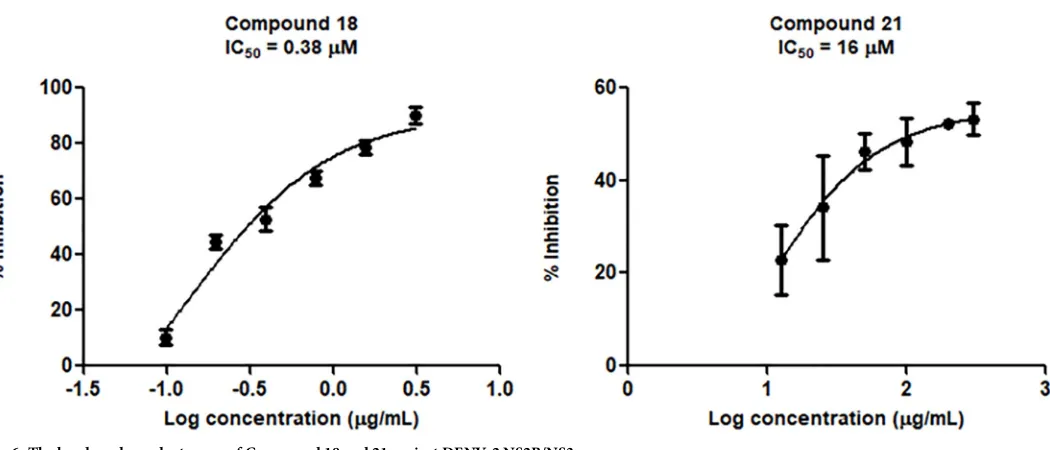
258μM. The activity-dose curve of compounds18and21are presented inFig 6.
Molecular docking simulation
From the docking results, several interactions were identified between NS2B/NS3pro with18
and21. For18, interacting residues such as Ser157(135), Tyr183(161) and Gly175(153) indi-cated potential hydrogen bonding interactions. The numbering in the bracket indicates the Table 3. The structures of six newly designed compounds and their activity against DENV-2 NS2B/NS3pro.
Ligands R1 R2 R3 IC50(μM)
16 propanoyl propanoyl H 97
17 isopropanoyl isopropanoyl H 80
18 pentanoyl pentanoyl H 0.38
19 pentanoyl benzyl H 54
20 H 2-methylnaphtyl isopropyl 258
21 pentanoyl benzyl benzyl 16
https://doi.org/10.1371/journal.pone.0210869.t003
Fig 6. The log dose dependent curve of Compound 18 and 21 against DENV-2 NS2B/NS3pro.
numbering in Wichapong model. Binding stability is contributed by all the hydrophobic inter-actions surrounding the entire binding site with the free energy of binding– 7.49 kcal/mol (Fig 7). In addition,21demonstrated binding affinity (ΔG = -8.10 kcal/mol) with the hydrogen bonding also observed with Asn174.
Stability and conformational changes in MD complexes
To further substantiate the stability and conformational changes, MD simulation was carried out to understand the dynamic features of the two best compounds with respect to time at nanosecond scale. Overall the backbone RMSD values for Apo, NS2B/NS3pro-18, NS2B/ NS3pro-21are<2Åthroughout the 70 ns simulation time reflecting the stability of the
sys-tems. NS2B/NS3pro-Panduratin adopted a higher RMSD with the range of 2.0 to 2.5Åat 20 to 50 ns simulation time, however, it stabilised at<2Åfrom 50 to 70 ns of simulation time
(Fig 8).
The gyration value indicates the compactness of the protein which in turn reflects directly on the folding and unfolding of the protein where unfolding of the protein would affect the kinetic of the protein activity. The gyrations of all the four systems (apoenzyme,18,21and Panduratin A) are fluctuating in less than 1Åat 15.5 to 16.5Å. No unfolding is observed with a low fluctuation on the gyration.
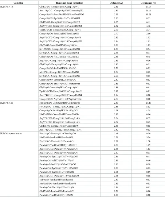
Hydrogen bonding analysis
Protein-ligand interaction in general is stabilised by various types of weak interactions with hydrogen bonding interaction is potentially one of the most important interactions. Hydrogen Fig 7. Binding orientation and interaction mode of compound with compound18(A) and compound21(B) in molecular docking simulation.
bond formation is observed with several important interacting residues such as His73(51), Asp151(129), Ser157(135), Asn174(152) and Tyr183(161) in both systems (Table 4) (The num-ber in parenthesis is according to Wichapong model). Asn174 in NS2B/NS3pro played an important role in forming hydrogen bonds which occupied more than 60% of the simulation time with18. Asp97(75) was also found to form hydrogen bonds with both nitrogen groups of
18with the total occupancy of 8.22% simulation time. It is interesting to note that Tyr183(161) and Ser157(135) which are important in the protease activity also demonstrated weak hydro-gen bonds formation with the occupancy of 2.85% and 2.73%, respectively. It is thus postulated that these hydrogen network together with the hydrophobic clusters contributed to the bind-ing affinity of18.
The number of hydrogen bond formation observed with21is lesser as compared to18. His73 formed a hydrogen bond with nitrogen group in21with 27.4% occupancy of the total 70ns simulation time. Two hydrogen bonds between Ser157 and the nitrogen and oxygen groups of21with 5.12% and 4.58% occupancy while very weak hydrogen bonds were observed with other interacting residues such as Asp97, Asn174 and Tyr183 with less than 1% occupancies.
Fig 8. (A) Time evolution of RMSD of NS2B/NS3pro backbone CA unbounded (Apo) (black), bounded with panduratin A (red),18(green) and21(blue). (B) Time evolution of radius of gyration of NS2B/NS3pro backbone CA unbounded (Apo) (black), bounded with panduratin A (red),18(green) and21(blue).
Table 4. Hydrogen bonds between compound 18 and compound 21 with NS2B/NS3pro that found with at least 0.1% occupancy throughout 70ns simulation time.
Complex Hydrogen bond formation Distance (Å) Occupancy (%)
In the case of panduratin A, only weak hydrogen bonds with less than 10% occupancies throughout 70ns were found with residues such as Phe152, His73, Asp151 and Tyr183. The formation of hydrogen bonding is significantly weaker as compared to18and21.
Comparison of free energy of binding between panduratin A, 18 and 21
The interaction and decomposition energies of the interactions between panduratin A,18and
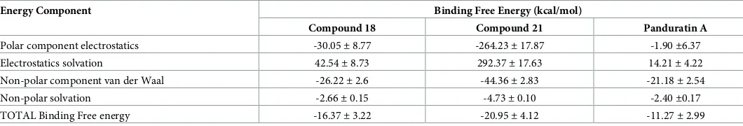
21were calculated using MM/PBSA. A total of 100 snapshots were extracted every 10 ps inter-val from the last nanosecond. The absolute binding free energy (Table 5) for panduratin A,18
and21are-11.27±2.99, -16.10±2.70 and -18.24±4.66 kcal/mol, respectively. Non-polar interaction between the binding site residues as well as the aliphatic chain (pentanoyl) of18
and21provides major contribution to the binding energy. The binding of panduratin A against NS2B/NS3pro is less favourable as compared to18and21. Similarly, major contribu-tion of panduratin A binding is mainly from the non-polar interaccontribu-tion. This can be inferred from the fact that only weak hydrogen bonds were found between panduratin A and NS2B/ NS3pro with low occupancies. This reflects that the binding of panduratin A is stabilised mainly by van der Waals and hydrophobic interactions.
The binding free energy estimated by MMPBSA calculation indicated that21has better binding as compared to18but the IC50for18is better.18and21have identical pentanoyl R1
group but21has two aromatic benzyl groups at R2and R3instead of a carbonyl chain and
hydrogen for18at R2and R3, respectively. Higher van der Waals interaction was observed
from MM/PBSA calculation for21due to the presence of these two benzyl groups at R2and
R3. However, these interactions might be overestimated using MM/PBSA approach [60].
Further discussion
Thioguanine was identified as a potential scaffold for DENV-2 NS2B/NS3pro inhibitor based on virtual screening,in vitroassay and molecular modelling. The thioguanine scaffold is com-posed of pyramidine and imidazole rings attached to an amine and a thiol group which may contribute to the compound’s activity. As far as we are aware, no report of pyrimidine inhibi-tion to DENV-2 NS2B/NS3pro, however, 2-(benzylthio)-6-oxo-4-phenyl-1,6-dihydropyrimi-dine has been reported to demonstrate an activity against SARS-CoV-3C like protease [61]. In addition, pyrimidine has also been shown to actively stop the growth of DENV-2 but it relies on the capability of pyrimidine to inhibit dihydroorotate dehydrogenase (DHODH), an enzyme required for viral pyrimidine biosynthesis [62]. Imidazoles have also been investigated against dengue virus [63] however the exact molecular mechanism or which protein they tar-get is still unknown. The 6-thioguanine scaffold has been reported to non-competitively inhibit ubiquitin specific protease in various cancers [64]. With this background, we postu-lated that thioguanine derivatives might potentially be good inhibitors against DENV-2 NS2B/ NS3pro.
Table 5. Binding free energy predicted using MM/PBSA calculation for 18, 21 and panduratin A.
Energy Component Binding Free Energy (kcal/mol)
Compound 18 Compound 21 Panduratin A
Polar component electrostatics -30.05±8.77 -264.23±17.87 -1.90±6.37 Electrostatics solvation 42.54±8.73 292.37±17.63 14.21±4.22 Non-polar component van der Waal -26.22±2.6 -44.36±2.83 -21.18±2.54
Non-polar solvation -2.66±0.15 -4.73±0.10 -2.40±0.17
TOTAL Binding Free energy -16.37±3.22 -20.95±4.12 -11.27±2.99
Two most active compounds (18and21) against DENV2-NS2B/NS3pro have an amide functional group (instead of an amino group). Amide is a part of peptide which is recognised as the key point for the protease substrate. In the docking study, the amide group is marked as a HBD with N-amide interacts with O-phenol of Tyr161 (in18) and Asn152 (in21) via H-bond. Previously, amide, such asα-ketoamide [65], arylcyanoacrylamide [66], and aminoben-zamide [67] has been incorporated in the design of DENV NS2B/NS3pro inhibitors. The alkyl chain of18and phenyl-benzyl group of21demonstrate hydrophobic features and agreeable to docking results which show that these groups bound at the hydrophobic pocket surrounded by Val154, Ile86, Met84 and Val155. These also correlate with the decomposition energies as computed by MM/PBSA which highlight that the major contribution to the interaction with DENV-2 NS2B/NS3pro is from non-polar interaction.
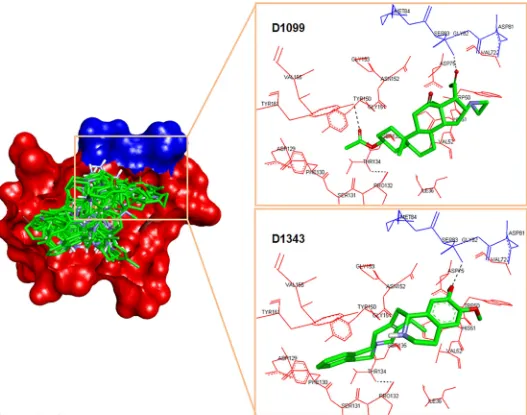
It is interesting to note that the binding orientations from the MD simulations for both compounds are different despite sharing similar thioguanine (Fig 9). The thioguanine moiety is facing toward His73 and Tyr183 for18and21, respectively. Compound18has an acyl group at R2which provides additional electron lone pair enabling the formation of hydrogen
bonds that can strengthen its binding towards NS2B/NS3pro. This reflected clearly in the hydrogen bond analysis where18formed more hydrogen bonds as compared to21. Regardless of the slightly lower absolute binding energy from the MM/PBSA prediction, the binding interaction analysis of18with NS2B/NS3pro agrees well with experimental results. The Fig 9. Binding orientation and interaction mode of compound with18(A),21(B) and panduratin A (C) in molecular dynamics simulation.
absolute free energies of binding for both two active compounds are also more negative than panduratin A supporting thein vitroresults in which compound18and21better IC50value
(0.38 and 16μM, respectively) than panduratin A (56μM).
Conclusions
We presented here the computational design of potential DENV-2 NS2B/NS3pro inhibitors. From the virtual screening of National Cancer Institute Database, four compounds including
D0713were observed to have moderate inhibition activity on the protease.D0713has a thio-guanine scaffold in its structure prompting us to consider the scaffold for designing the new thioguanine derivatives as potential DENV-2 NS2B/NS3pro inhibitors. Fifteen compounds were synthesised and the bioactivity against dengue protease showed variation in inhibition activity from inactive to moderately active (1000>IC50>18μM). Based on this information, a
further design of six new compounds was conducted by concentrating on the attachment of amide group. All the compounds showed inhibition activity against the dengue protease with compound18being the most potent (IC50of 0.38μM). This result agrees well with the MM/
PBSA calculation which showed that the interactions are mainly contributed by polar and non-polar interactions. Hydrogen bonding analysis demonstrates the importance of amino acid residues Asn174 (occupancy 60%), Asp75 (8.22%), Tyr183 and Ser157 (2.85 and 2.73%). This is further supported by the experimental results which showed that18could be further developed as DENV-2 NS2B/NS3pro inhibitor. It is hoped that the results obtained from this study could be used in designing more active compounds as potential dengue protease inhibitors.
Supporting information
S1 Text. Synthesis of thioguanine derivatives.General procedure for synthesis of Schiff base-thioguanine compounds, acetamide-base-thioguanine compounds,N-alkylation of acetamide-thio-guanine, andS/N-benzylation of acetamide-thioguanine.
(PDF)
S1 Fig. Structure of 1–21.The numbering system is according to the NMR characterisation. (PDF)
S2 Fig. The molecular interaction between Panduratin A with DENV-2 NS2B/NS3pro.The NS2B-NS3pro is presented in a surface form. The ligands are presented in a stick form in which carbons are grey while oxygens are red. The ligand is docked at the active site of the pro-tease with the hydrophilic moieties surrounded by catalytic site(His51, Asp75 and Ser135) whereas the nonpolar part of the ligand closes nearly to hydrophobic area of the protease wherein phenyl ring posseses H-bond as well asπ-πinteractions with Tyr161.
(PDF)
S1 Table. The 24 NCI compounds selected from virtual screening.The NCI compounds are named in two codes and the free energy of binding upon DENV-2 NS2B/NS3pro binding site is expressed in kcal/mol.
(PDF)
Author Contributions
Conceptualization:Habibah A. Wahab.
Investigation:Maywan Hariono, Sy Bing Choi, Ros Fatihah Roslim, Mohamed Sufian Nawi.
Methodology:Maywan Hariono, Sy Bing Choi, Ros Fatihah Roslim, Mohamed Sufian Nawi, Habibah A. Wahab.
Project administration:Habibah A. Wahab.
Resources:Habibah A. Wahab.
Supervision:Mei Lan Tan, Rohana Yusof, Shatrah Othman, Noorsaadah Abd Rahman, Rozana Othman, Habibah A. Wahab.
Validation:Maywan Hariono, Sy Bing Choi, Habibah A. Wahab.
Visualization:Maywan Hariono, Sy Bing Choi.
Writing – original draft:Maywan Hariono, Sy Bing Choi.
Writing – review & editing:Nornisah Mohamed, Noorsaadah Abd Rahman, Rozana Oth-man, Habibah A. Wahab.
References
1. Murray NE, Quam MB, Wilder-Smith A. Epidemiology of dengue: past, present and future prospects. Clin Epidemiol. 2013; 5: 299–309.https://doi.org/10.2147/CLEP.S34440PMID:23990732
2. Brady OJ, Gething PW, Bhatt S, Messina JP, Brownstein JS, Hoen AG, et al. Refining the global spatial limits of dengue virus transmission by evidence-based consensus. PLoSNegl Trop Dis. 2012; 6: e1760.
3. Guzman MG, Harris E. Dengue. Lancet. 2015; 385: 453–465.https://doi.org/10.1016/S0140-6736(14) 60572-9PMID:25230594
4. Bhatt S, Gething PW, Brady OJ, Messina JP, Farlow AW, Moyes CL, et al. The global distribution and burden of dengue. Nature. 2013; 496: 504–507.https://doi.org/10.1038/nature12060PMID:23563266
5. Pang T, Cardosa MJ, Guzman MG. Of cascades and perfect storms: the immunopathogenesis of den-gue haemorrhagic fever-denden-gue shock syndrome (DHF/DSS). Immunol Cell Biol. 2007; 85: 43–45. https://doi.org/10.1038/sj.icb.7100008PMID:17130899
6. Simmons CP, McPherson K, Van Vinh Chau N, Hoai Tam DT, Young P, Mackenzie J, et al. Recent advances in dengue pathogenesis and clinical management. Vaccine. 2015; 33: 7061–7068.https:// doi.org/10.1016/j.vaccine.2015.09.103PMID:26458808
7. WHO, Dengue vaccine: WHO position paper—July 2016.Wkly Epidemiol Rec. 2016; 91: 349–64. PMID:27476189
8. WHO, Dengue: Guidelines for diagnosis, treatment, prevention and control: New Edition. 2013/06/14 ed. World Health Organisation: Geneva 2009.
9. Galula JU, Shen WF, Chuang ST, Chang GJ, Chao DY. Virus-like particle secretion and genotype-dependent immunogenicity of dengue virus serotype 2 DNA vaccine. J Virol. 2014; 88: 10813–10830. https://doi.org/10.1128/JVI.00810-14PMID:25008922
10. Normile D. First new dengue virus type in 50 years. Health. 2013.
11. Henchal EA, Putnak JR. The dengue viruses. Clin Microbiol Rev. 1990; 3: 376–396. PMID:2224837
12. Natarajan S. NS3 protease from flavivirus as a target for designing antiviral inhibitors against dengue virus. Genet Mol Biol. 2010; 33: 214–219.https://doi.org/10.1590/S1415-47572010000200002PMID: 21637471
13. Wahab HA, Yusof R, Rahman NA. A search for vaccines and therapeutic for dengue: A review. Curr Comput-Aid Drug. 2007; 3:, 101–112.
14. Behnam MA, Nitsche C, Boldescu V, Klein CD. The medicinal chemistry of dengue virus. J Med Chem. 2016; 59: 5622–5649.https://doi.org/10.1021/acs.jmedchem.5b01653PMID:26771861
15. Behnam MA, Graf D, Bartenschlager R, Zlotos DP, Klein CD. Discovery of nanomolar dengue and west nile virus protease inhibitors containing a 4-benzyloxyphenylglycine residue. J Med Chem. 2015; 58: 9354–9370.https://doi.org/10.1021/acs.jmedchem.5b01441PMID:26562070
17. Yin Z, Patel SJ, Wang WL, Chan WL, Ranga Rao KR, Wang G, et al. Peptide inhibitors of dengue virus NS3 protease. Part 2: SAR study of tetrapeptide aldehyde inhibitors. Bioorg Med Chem Lett. 2006; 16: 40–43.https://doi.org/10.1016/j.bmcl.2005.09.049PMID:16246563
18. Chanprapaph S, Saparpakorn P, Sangma C, Niyomrattanakit P, Hannongbua S, Angsuthanasombat C, et al. Competitive inhibition of the dengue virus NS3 serine protease by synthetic peptides representing polyprotein cleavage sites. Biochem Biophys Res Commun. 2005; 330: 1237–1246.https://doi.org/10. 1016/j.bbrc.2005.03.107PMID:15823576
19. Steuer C, Heinonen KH, Kattner L, Klein CD. Optimization of assay conditions for dengue virus prote-ase: Effect of various polyols and nonionic detergents. J Biomol Screen. 2009; 14: 1102–1108.https:// doi.org/10.1177/1087057109344115PMID:19726784
20. Frecer V, Miertus S. Design, structure-based focusing and in silico screening of combinatorial library of peptidomimetic inhibitors of Dengue virus NS2B-NS3 protease. J Comput Aided Mol Des. 2010; 24: 195–212.https://doi.org/10.1007/s10822-010-9326-8PMID:20306283
21. Yang CC, Hsieh YC, Lee SJ, Wu SH, Liao CL, Tsao CH, et al. Novel dengue virus-specific NS2B/NS3 protease inhibitor, BP2109, discovered by a high-throughput screening assay. Antimicrob Agents Che-mother. 2011; 55: 229–238.https://doi.org/10.1128/AAC.00855-10PMID:20937790
22. Katzenmeier G. Inhibition of the NS2B–NS3 protease: towards a causative therapy for dengue virus dis-eases. Dengue Bull. 2004; 28: 58–67.
23. Yotmanee P, Rungrotmongkol T, Wichapong K, Choi SB, Wahab HA, Kungwan N, et al. Binding spesifi-city of polypeptide substrates in NS2B/NS3pro serine protease of dengue virus type 2: A molecular dynamics study. J Mol Graph Model 2015, 60, 24–33.https://doi.org/10.1016/j.jmgm.2015.05.008 PMID:26086900
24. Nitsche C, Behnam MA, Steuer C, Klein CD. Retro peptide-hybrids as selective inhibitors of the dengue virus NS2B-NS3 protease. Antiviral Res. 2012; 94: 72–79.https://doi.org/10.1016/j.antiviral.2012.02. 008PMID:22391061
25. Behnam MA, Nitsche C, Vechi SM, Klein CD. C-terminal residue optimization and fragment merging: discovery of a potent peptide-hybrid inhibitor of dengue protease. ACS Med Chem Lett. 2014;, 5: 1037–1042.https://doi.org/10.1021/ml500245vPMID:25221663
26. Nitsche C, Schreier VN, Behnam MA, Kumar A, Bartenschlager R, Klein CD. Thiazolidinone–peptide hybrids as dengue virus protease inhibitors with antiviral activity in cell culture. J Med Chem. 2013; 56: 8389–8403.https://doi.org/10.1021/jm400828uPMID:24083834
27. Rothan HA, Han HC, Ramasamy TS, Othman S, Rahman NA, Yusof R. Inhibition of dengue NS2B-NS3 protease and viral replication in Vero cells by recombinant retrocyclin-1. BMC Infect Dis. 2012; 12:314. https://doi.org/10.1186/1471-2334-12-314PMID:23171075
28. Rothan HA, Abdulrahman AY, Sasikumer PG, Othman S, Rahman NA, Yusof R. Protegrin-1 inhibits dengue NS2B-NS3 serine protease and viral replication in MK2 cells. J Biomed Biotechnol. 2012; 2514–2582.
29. Prusis P, Junaid M, Petrovska R, Yahorava S, Yahorau A, Katzenmeier G, et al. Design and evaluation of substrate-based octapeptide and non substrate-based tetrapeptide inhibitors of dengue virus NS2B-NS3 proteases. Biochem Biophys Res Commun. 2013; 434: 767–772.https://doi.org/10.1016/j.bbrc. 2013.03.139PMID:23587903
30. Xu S, Li H, Shao X, Fan C, Ericksen B, Liu J, et al. Critical effect of peptide cyclization on the potency of peptide inhibitors against Dengue virus NS2B-NS3 protease. J Med Chem. 2012; 55: 6881–6887. https://doi.org/10.1021/jm300655hPMID:22780881
31. Kiat TS, Pippen R, Yusof R, Ibrahim H, Khalid N, Rahman NA. Inhibitory activity of cyclohexenyl chal-cone derivatives and flavonoids of fingerroot, Boesenbergia rotunda (L.), towards dengue-2 virus NS3 protease. Bioorg Med Chem Lett. 2006; 16: 3337–40.https://doi.org/10.1016/j.bmcl.2005.12.075 PMID:16621533
32. de Sousa LRF, Wu H, Nebo L, Fernandes JB, Kiefer W, Kanitz M, et al. Flavonoids as noncompetitive inhibitors of dengue virus NS2B-NS3 protease: Inhibition kinetics and docking studies. Bioorg Med Chem Lett. 2015; 23: 466–470.
33. Viswanathan U, Tomlinson SM, Fonner JM, Mock SA, Watowich SJ. Identification of a novel inhibitor of dengue virus protease through use of a virtual screening drug discovery Web portal. J Chem Inf Model. 2014; 54: 2816–2825.https://doi.org/10.1021/ci500531rPMID:25263519
34. Raut R, Beesetti H, Tyagi P, Khanna I, Jain SK, Jeankumar VU, et al. A small molecule inhibitor of den-gue virus type 2 protease inhibits the replication of all four denden-gue virus serotypes in cell culture. Virol J. 2015; 12: 1.https://doi.org/10.1186/s12985-014-0235-7
36. Yusof R, Rahman NA, Wahab HA, Othman R, Padmanabhan R. Design of Chemotherapeutic Agent for Dengue Virus Protease. 2008.
37. Lee YK, Tan SK, Wahab HA, Yusof R, Rahman NA. Nonsubstrate based inhibitors of dengue virus ser-ine protease: a molecular docking approach to study binding interactions between protease and inhibi-tors. Asia Pac J Mol Biol Biotechnol. 2007; 15: 53–59.
38. Cabarcas-Montalvo M, Maldonado-Rojas W, Montes-Grajales D, Bertel-Sevilla A, Wagner-Dobler I, Sztajer H, et al. Discovery of antiviral molecules for dengue: In silico search and biological evaluation. Eur J Med Chem. 2016; 110: 87–97.https://doi.org/10.1016/j.ejmech.2015.12.030PMID:26807547
39. Othman R, Wahab HA, Yusof R, Rahman NA. Analysis of secondary structure predictions of dengue virus type 2 NS2B/NS3 against crystal structure to evaluate the predictive power of the in silico meth-ods. In Silico Biol. 2007; 7: 215–224. PMID:17688447
40. Adamson PC, Poplack DG, Balis FM. The cytotoxicity of thioguanine vs mercaptopurine in acute lym-phoblastic leukemia. Leuk Res. 1994; 18: 805–810. PMID:7967706
41. Osterman MT, Kundu R, Lichtenstein GR, Lewis JD. Association of 6-thioguanine nucleotide levels and inflammatory bowel disease activity: a meta-analysis. Gastroenterology. 2006; 130: 1047–53.https:// doi.org/10.1053/j.gastro.2006.01.046PMID:16618398
42. Elion GB. The purine path to chemotherapy. Biosci Rep. 1989; 9: 509–529. PMID:2679902
43. Trott O, Olson AJ. AutoDock Vina: improving the speed and accuracy of docking with a new scoring function, efficient optimization, and multithreading. J Comput Chem. 2010; 31: 455–461.https://doi. org/10.1002/jcc.21334PMID:19499576
44. Wichapong K, Pianwanit S, Sippl W, Kokpol S. Homology modeling and molecular dynamics simula-tions of Dengue virus NS2B/NS3 protease: insight into molecular. J Mol Recognit. 2010; 23: 283–300. https://doi.org/10.1002/jmr.977PMID:19693793
45. Yusof R, Clum S, Wetzel M, Murthy HM, Padmanabhan R. Purified NS2B/NS3 serine protease of den-gue virus type 2 exhibits cofactor NS2B dependence for cleavage of substrates with dibasic amino acids in vitro. J Biol Chem. 2000; 275: 9963–9969. PMID:10744671
46. Heh CH, Othman R, Buckle MJ, Sharifuddin Y, Yusof R, Rahman NA. Rational discovery of dengue type 2 non-competitive inhibitors. Chem Biol Drug Des. 2013; 82: 1–11.https://doi.org/10.1111/cbdd. 12122PMID:23421589
47. Rahman NA, Hadinur MS, Rashid N, Muhamad M, Yusof R. Studies on quercuslusitanica extracts on DENV-2 replication. Dengue Bull. 2006; 30: 260–269.
48. Qiagen, The QIA expressionist—A handbook for high-level expression and purification of 6xHis-tagged proteins. 5th ed ed.; 2003.
49. Leung D, Schroder K, White H, Fang NX, Stoermer MJ, Abbenante G, et al. Activity of recombinant den-gue 2 virus NS3 protease in the presence of a truncated NS2B co-factor, small peptide substrates, and inhibitors. J Biol Chem. 2001; 276: 45762–45771.https://doi.org/10.1074/jbc.M107360200PMID: 11581268
50. Zandi K, Lim TH, Rahim NA, Shu MH, Teoh BT, Sam SS, et al. Extract of Scutellariabaicalensis inhibits dengue virus replication. BMC Complement Altern Med. 2013; 13: 91. https://doi.org/10.1186/1472-6882-13-91PMID:23627436
51. Prakash C, Vijay IK. A new fluorescent tag for labeling of saccharides. Anal Biochem. 1983; 128: 41–6. PMID:6846798
52. Hariono M, Wahab HA, Tan ML, Rosli MM, Razak IA. 9-Benzyl-6-benzylsulfanyl-9H-purin-2-amine. Acta Cryst E. 2014; 70: o288.
53. Hariono M, Ngah N, Wahab HA, Abdul Rahim AS. 2-Bromo-4-(3, 4-dimethyl-5-phenyl-1, 3-oxazolidin-2-yl)-6-methoxyphenol. Acta Cryst E. 2012; 68: o35–o36.
54. HyperChem (TM) Profesional 8.0, Hypercube, Inc.
55. Morris GM, Lindstrom RHW, Sanner MF, Belew RK, Goodsell DS, Olson AJ, AutoDock4 and AutoDock-Tools4: Automated docking with selective receptor flexibility. J Comput Chem. 2009; 28: 73–86.
56. Laskowski RA, Swindells MB. LigPlot+: multiple ligand-protein interaction diagrams for drug discovery. J Chem Inf Model. 2011; 51: 2778–2786.https://doi.org/10.1021/ci200227uPMID:21919503
57. Le Grand S, Go¨tz AW, Walker RC. SPFP: Speed without compromise—A mixed precision model for GPU accelerated molecular dynamics simulations. Comput Phys Commun. 2013; 184: 374–380.
59. Chaput L, Martinez-Sanz J, Saettel N, Mouawad L. Benchmark of four popular virtual screening pro-grams: construction of the active/decoy dataset remains a major determinant of measured perfor-mance. J Cheminform. 2016; 8: 56.https://doi.org/10.1186/s13321-016-0167-xPMID:27803745
60. Genheden S, Ryde U. The MM/PBSA and MM/GBSA methods to estimate ligand-binding affinities. Expert Opin Drug Discov. 2015; 10: 449–461.https://doi.org/10.1517/17460441.2015.1032936PMID: 25835573
61. Ramajayam R, Tan K-P, Liu H-G, Liang P-H. Synthesis, docking studies, and evaluation of pyrimidines as inhibitors of SARS-CoV 3CL protease, Bioorg Med Chem Lett. 2010; 20: 3569–3572.https://doi.org/ 10.1016/j.bmcl.2010.04.118PMID:20494577
62. Wang QY, Bushell S, Qing M, Xu HY, Bonavia A, Nunes S, et al. Inhibition of dengue virus through sup-pression of host pyrimidine biosynthesis. J Virol. 2011; 85: 6548–6556.https://doi.org/10.1128/JVI. 02510-10PMID:21507975
63. Saudi M, Zmurko J, Kaptein S, Rozenski J, Neyts J, Van Aerschot A. Synthesis and evaluation of imid-azole-4,5- and pyrazine-2,3-dicarboxamides targeting dengue and yellow fever virus. Eur J Med Chem. 2014; 87: 529–539.https://doi.org/10.1016/j.ejmech.2014.09.062PMID:25285371
64. Chuang S-J, Cheng S-C, Tang H-C, Sun C-Y, Chou C-Y. 6-Thioguanine is noncompetitive and slow binding inhibitor of human deubiquitinating protease USP2. Sci Rep. 2018; 8: 1–9.https://doi.org/10. 1038/s41598-017-17765-5
65. Steuer C, GeGe C, Fischl W, Heinonen KH, Bartenschlager R, Klein CD. Synthesis and biological eval-uation ofα-ketoamides as inhibitors of the Dengue virus protease with antiviral activity in cell-culture. Bioorg Med Chem. 2011; 19: 4067–4074.https://doi.org/10.1016/j.bmc.2011.05.015PMID:21641807
66. Nitsche C, Steuer C, Klein CD. Arylcyanoacrylamides as inhibitors of the dengue and west nile virus proteases. Bioorg Med Chem. 2011; 19: 7318–7337.https://doi.org/10.1016/j.bmc.2011.10.061PMID: 22094280