Introduction
!The usual outcome of acute inflammation is suc-cessful resolution and repair of tissue damage. However, it has also been recognized as the cen-tral cause of many prevalent diseases, including arthritis, periodontal disease, cardiovascular dis-eases, cancer, and Alzheimerʼs disease [1]. Cyto-kines are primarily involved in host responses to infection, immune responses, inflammation, and trauma. Several cytokines, called proinflamma-tory cytokines, tend to increase disease symp-toms and produce fever, inflammation, tissue de-struction, shock, and even death when they are administered to humans. Therefore, compounds that could block or inhibit the production of proinflammatory cytokines might be beneficial anti-inflammatory agents [2].
The genus Piper(Piperaceae) is well known for producing a large number of physiologically ac-tive compounds and is widely used in folklore medicine in the West Indies and Latin America. The Piperaceae family, in which the genusPiper
belongs, comprises approximately 2000 species distributed in the tropical regions of the world [3, 4]. Piper aduncum L. is widely used in folk medicine to treat stomachaches (Jamaica) and as an inflammatory, diarrhea (Peru), and anti-steptic to heal wounds (Brazil and Papua New Guinea) [3–5]. Previous phytochemical studies of P. aduncumreported the isolation of dihydrochal-cones, chaldihydrochal-cones, flavanones, chromene, phenyl-propanoids, and benzoic acid derivatives [6–8]. Several pharmacological effects ofP. aduncum ex-tracts have been demonstrated, including anti-leishmanial, antibacterial, cytotoxic, and antifun-gal activities [6, 9]. Furthermore, some of the iso-lated compounds have been shown to be active against promastigote and intracellular amasti-gotes, causing damaging effects on DNA, and leishmanial, antimicrobial, molluscicidal, anti-tumor, and antifungal activities [8, 10, 11]. How-ever, anti-inflammatory studies on P. aduncum are limited. To date, only an essential oil (dilla-piole) of this plant was investigated for anti-inflammatory activity in the carrageenan-induced rat paw edema model [12].
As a part of our ongoing investigations on the chemical constituents of medicinal plants with
Abstract
!
Four new compounds, acacetin 8-C-[β -D-apiofur-anosyl-(1→2)-β-D-glucopyranoside] (1), 7-me-thoxyacacetin 8-C-[β-D-apiofuranosyl-(1→3)-β -D-glucopyranoside] (2), 7-methoxyacacetin 8-C-[β-D-glucopyranosyl-(1→2)-β -D-glucopyrano-side] (3), and 4′′′-O-acetylacacetin 8-C-[α -L-rhamnopyranosyl-(1→2)-β-D-glucopyranoside] (4), along with ten known compounds (5–14), were isolated fromPiper aduncumleaves. The ef-fects of these compounds on lipopolysaccharide-induced expression of the proinflammatory cyto-kines IL-12 p40, IL-6, and TNF-αin bone
marrow-derived dendritic cells were evaluated. Com-pounds2, 3, 6, 8, 9, and11–13inhibited the pro-duction of both IL-12 p40 and IL-6, with IC50 val-ues ranging from 0.35 ± 0.01 to 1.40 ± 0.04 µM and 1.22 ± 0.02 to 3.79 ± 0.10 µM, respectively. Com-pounds5and10only showed strong inhibition effects on the production of IL-12 p40, with IC50 values of 2.76 ± 0.08 and 0.39 ± 0.05 µM, respec-tively. However, all compounds showed weak ac-tivity or no acac-tivity on TNF-αproduction at the tested concentrations.
Supporting informationavailable online at http://www.thieme-connect.de/products
* These authors contributed equally to this work.
Anti-inflammatory Flavonoid C-Glycosides from
Piper aduncum
Leaves
Authors Nguyen Phuong Thao1, 2*, Bui Thi Thuy Luyen1*, Wahyu Widowati3, Nurul Fauziah4, Maesaro Maesaroh4, Tati Herlina5,
Zahid Manzoor6, Irshad Ali6, Young Sang Koh6, Young Ho Kim1
Affiliations The affiliations are listed at the end of the article
Key words
l" Piper aduncum l" Piperaceae
l" Flavonoid C‑glycoside
l" IL‑12 p40
l" IL‑6
l" TNF‑α
l" LPS
received February 25, 2016
revised May 1, 2016
accepted May 5, 2016
Bibliography
DOIhttp://dx.doi.org/
10.1055/s-0042-108737 Published online
Planta Med © Georg Thieme Verlag KG Stuttgart · New York ·
ISSN 0032‑0943
Correspondence
Prof. Dr. Young Ho Kim
College of Pharmacy Chungnam National University 99 Yuseong-gu
Daejeon 305–764
Korea
Phone: + 82 4 28 21 59 33 Fax: + 82 4 28 23 65 66 [email protected]
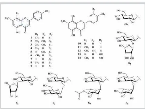
anti-inflammatory activities, we found that the methanolic ex-tract ofP. aduncumshowed significant anti-inflammatory effects in vitrowith an inhibition value of 65.9 % (20.0 µg/mL). In the present study, we report the isolation and structural elucidation of four new flavonoids (1–4) and ten known compounds (5–14) fromP. aduncumleaves (l"Fig. 1). Also, the anti-inflammatory ef-fects of these compounds on lipopolysaccharide (LPS)-induced expression of the proinflammatory cytokines IL-12 p40, IL-6, and TNF-αin bone marrow-derived dendritic cells (BMDCs) were evaluated.
Results and Discussion
!A MeOH extract of the leaves ofP. aduncumwas suspended in H2O and successively partitioned with CH2Cl2 and EtOAc. The EtOAc fraction was separated by various chromatographic ex-periments to afford 14 C-glycosyl flavones. Ten known com-pounds including isospinosin (5) [13], 2-O-β-D-glucosyl-8-C-β -D-glucosyl-4′-O-methylapigenin (6) [14], apigenin-8-C-neohes-peridoside (7) [15], 2″-O-α-rhamnosyl-4′-O-methyl vitexin (8) [16], ficuflavoside (8-C-(2″-O-β-D-apiofuranosyl)-β -D-glucopyr-anosyl apigenin) (9) [17], apigenin 8-C-β-glucopyranoside (10) [18], cytisoside (11) [19], isoembigenin (12) [20], orientin (13) [21], and 4′-O-methylorientin (14) [22] were identified by NMR analysis as well as comparison with previous reports (l"Fig. 1). The C-glycosyl flavones were isolated for the first time from the leaves of this plant, indicating that these compounds could be considered chemotaxonomic markers for the identification of P. aduncum.
Compound1was obtained as a yellow amorphous powder. The HR‑ESI‑MS of1was consistent with the molecular formula of C27H30O14. The13C NMR and DEPT spectra (l"Table 1) revealed 27 carbon signals, including 15 signals that were assigned to a flavone aglycon, as well as two anomeric and nine other oxygen-ated carbon signals, of which three methylenes were ascribable to C-4′′′of an apiofuranosyl unit, and C-6′′of a glucopyranosyl unit, respectively. Assignment of the protons and carbons in the aglycone moiety was done based on 2D spectra. All of the sugar proton resonances were assigned by a COSY experiment, and the corresponding13C resonances were then identified by the HMQC.
The COSY spectra of compound1indicated two individual corre-lation systems of sugar units [H-1′′(δH4.99)/H-2′′(δH4.24)/H‑3′′ (δH3.65)/H‑4′′(δH3.70)/H‑5′′(δH3.46)/H-6′′(δH3.84 and 4.01)] and [H-1′′′(δH5.16)/H-2′′′(δH3.78)]. The connectivity of these fragments was determined through HMBC. The downfield shift was observed for C-2′′(δC77.4), as well as HMBC correlations of H-1′′′(δH5.16) with C-2′′(δC77.4), and H-2′′(δH4.24) with C-1′′′ (δC111.1), C-1′′(δC73.7), and C-8 (δC105.4), demonstrating the (1→2) linkage between the apiosyl and glucosyl units in1. Fur-thermore, acid hydrolysis of1, followed by TLC, GC analysis, and a comparison with authentic D-apiose, confirmed the presence of a D-apiose moiety in that compound (see Materials and Meth-ods). A C-8 substituted acacetin structure was suggested by the 1H (δ
H6.25, s, H-6) and13C NMR (δC105.4 for 8 and 99.3 for C-6) signals characteristic of C-8-glycosylated flavones [23]. In ad-dition, the HMBC correlations of H-1′′ (δH 4.99) with C-7 (δC 164.1), C-8 (δC105.4), and C-9 (δC158.4) confirmed that the di-saccharide chain was bonded by a C-glycosidic linkage at the 8-position. Accordingly, compound1was identified as acacetin 8-C-[β-D-apiofuranosyl-(1→2)-β-D-glucopyranoside].
Compound2was obtained as a yellow amorphous powder. The HR‑ESI‑MS was consistent with the molecular formula of C28H32O14. The1H and13C NMR spectra (l"Table 1) of2were sim-ilar to those of1. The differences between the spectra of com-pounds1and2were with regard to the presence of two methoxy groups and the linkage between two sugar units, glucose and apiose. The downfield shift of C-3′′(δC84.8) and the upfield shift of C-2′′(δC70.9) of the glucopyranosyl unit, in comparison with those observed in1, were indicative of a glycosidation at C-3′′. In addition, the interglycosidic linkage of the apiofuranosyl moiety in2was established unambiguously by the HMBC spectrum to be at the C-3′′position of the glucopyranosyl unit based on the cross-peak between H-1′′′(δH5.20) with C-3′′(δC84.8) and H-3′′ (δH3.45) with C-1′′′(δC109.8). The position of the disaccharide chain was determined as C-8 based on the HMBC correlations of the anomeric proton H-1′′ (δH 4.76) of glucosyl with C-7 (δC 164.0), C-8 (δC105.2), and C-9 (δC155.8) of an aglycon. Two me-thoxy groups were located at C-7 (δC164.0) and C-4′(δC163.0), as indicated by the HMBC correlations of a methoxy proton atδC 3.89 with C-7 (δC164.0), and atδH3.87 with C-4′(δC163.0), re-spectively. In addition, acid hydrolysis of2, followed by TLC, GC analysis, and a comparison with authentic D-apiose, confirmed the presence of a D-apiose moiety in2(see Materials and Meth-ods). Therefore, the structure of2was identified as 7-methoxy-acacetin 8-C-[β-D-apiofuranosyl-(1→3)-β-D-glucopyranoside]. Compound3was obtained as a yellow powder. The HR‑ESI‑MS spectra of3indicated the molecular formula of C29H34O15. The
1H and13C NMR spectra of3showed the similar aglycon of2as
7-methoxyacacetin (l"Table 1). The presence of a glucopyranosyl unit, instead of a terminal apiofuranosyl unit, was the main dif-ference in the structure of3. Two individual correlations for the spin systems of H-1′′/H-2′′/H-3′′/H-4′′/H-5′′/H-6′′ and H-1′′′/ H-2′′′/H-3′′′/H-4′′′/H-5′′′/H-6′′′were observed in the COSY spectra of3, which supports the backbone proton assignments of the two sugar units. HMBC correlations of the anomeric proton H-1′′′(δH 3.87)/C-2′′(δC81.3) and proton H-2′′(δH4.05)/C‑1′′′(δC105.3), as well as the downfield shift of C-2′′(δC81.3), suggested the struc-ture of aβ-D-glucopyranosyl (1→2)β-D-glucopyranosyl disac-charide. The position of the sugar moiety in3through a C‑C link-age was identified as C-8 based on the HMBC correlations of the anomeric proton H-1′′(δH 4.83) with C-8 (δC 105.3), C-7 (δC 164.0), and C-9 (δC 155.9). Acid hydrolysis experiments
con-Fig. 1 Chemical structures of compounds1–14isolated fromP. aduncum leaves. (Color figure available online only.)
firmed the presence of D-glucose in3. This conclusion was fur-ther supported by TLC, GC analysis, and comparisons with an authentic D-glucose sample. Thus, compound 3was identified as 7-methoxyacacetin 8-C-[β-D-glucopyranosyl-(1→2)-β -D-glu-copyranoside].
Compound4 was obtained as yellow powder. The HR‑ESI‑MS spectra of4indicated the molecular formula of C30H34O15. The
1H NMR spectra of4showed signals of six aromatic protons,
in-cluding two singlet signals, four aromatic protons, one methoxy group, two methyl groups, and two anomeric protons, together with carbinol protons, suggesting the presence of two sugar units. The13C NMR spectra displayed 30 carbon resonance sig-nals, including 15 carbons belonging to an aglycon moiety, 12 car-bons of sugar units, 1 methoxy carbon, and 2 carcar-bons of an acetyl group.
The1H and13C NMR data showed doubling of the signals due to restricted rotation (l"Table 2). The rotameric signals show almost the same1H NMR signal intensity of 3 : 2. Hence, the rotameric partners of4could also be observed in the13C NMR spectrum. Because of the similar1H and13C resonance signals between4 and1, the aglycon of4was identified as acacetin. The presence of glucose and rhamnose units was observed in the1H,13C, and COSY spectra of4. Furthermore, acid hydrolysis of4, followed by TLC, GC analysis, and a comparison with authentic L-rhamnose,
confirmed the presence of an L-rhamnose moiety in4(see Mate-rials and Methods). The HMBC correlations of the anomeric pro-ton of rhamnose H-1′′′(δH5.42/5.30) and C-2′′(δC76.2/76.1) of glucose, proton H-2′′(δH4.20) of glucose and C-1′′′(δC101.3) of rhamnose, and C-8 (δC105.9/105.7) of the A ring suggested the structure of anα-L-rhamnopyranosyl (1→2)β-D-glucopyranosyl disaccharide. The position of an acetyl group at C-4′′′was con-firmed by the cross-peak observed between H-4′′′(δH4.53/4.61) and C-1′′′′(δC172.5), as well as the downfield shift of C-4′′′(δC 75.2). The C-8 substituted structure was determined by the HMBC correlation of H-1′′(δH5.05) of the glucopyranosyl unit with C-8 (105.9/105.7), C-7 (δC164.4/164.3), and C-9 (δC157.7/ 156.2) of the aglycon. Therefore, compound4was assigned as 4′′′-O-acetylacacetin 8-C-[α-L-rhamnopyranosyl-(1→2)-β -D-glucopyranoside].
The isolated compounds were assessed for anti-inflammatory ac-tivity on LPS-induced expression of the proinflammatory cyto-kines IL-12 p40, IL-6, and TNF-αby BMDCs (see Materials and Methods). Many natural products have been shown to exhibit anti-inflammatory activities by inhibiting the production of proinflammatory cytokines, including IL-12 p40, IL-6, and TNF-α, which play a crucial role in host defenses and inflammation [24]. Thus, agents that block the excessive production of these cy-tokines might be candidates for use in the treatment of
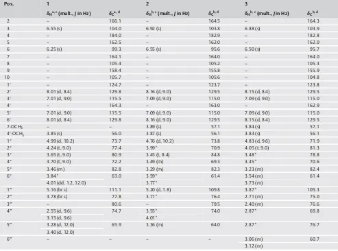
inflam-Table 1 1H and13C NMR spectroscopic data for compounds1–3.
Pos. 1 2 3
δHa, c(mult.,Jin Hz) δCa, d δHb, c(mult.,Jin Hz) δCb, d δHb, c(mult.,Jin Hz) δCb, d
2 – 166.1 – 164.5 – 164.3
3 6.55 (s) 104.0 6.92 (s) 103.8 6.88 (s) 103.9
4 – 184.0 – 182.9 – 182.8
5 – 162.5 – 162.0 – 162.0
6 6.25 (s) 99.3 6.55 (s) 95.6 6.50 (s) 95.7
7 – 164.1 – 164.0 – 164.0
8 – 105.4 – 105.2 – 105.3
9 – 158.4 – 155.8 – 155.9
10 – 105.7 – 105.6 – 104.8
1′ – 124.7 – 123.7 – 123.8
2′ 8.01 (d, 8.4) 129.8 8.16 (d, 9.0) 129.5 8.15 (d, 8.4) 129.5
3′ 7.01 (d, 9.0) 115.5 7.09 (d, 9.0) 115.0 7.09 (d, 9.0) 115.0
4′ – 164.3 – 163.0 – 162.9
5′ 7.01 (d, 9.0) 115.5 7.09 (d, 9.0) 115.0 7.09 (d, 9.0) 115.0
6′ 8.01 (d, 8.4) 129.8 8.16 (d, 9.0) 129.5 8.15 (d, 8.4) 129.5
7-OCH3 – – 3.89 (s) 57.1 3.84 (s) 57.1
4′-OCH3 3.85 (s) 56.0 3.87 (s) 56.1 3.83 (s) 56.1
1″ 4.99 (d, 10.2) 73.7 4.76 (d, 10.2) 73.8 4.83 (d, 9.6) 71.9
2″ 4.24 (t, 9.0) 77.4 3.99* 70.9 4.05 (t, 9.0) 81.3
3″ 3.65 (t, 9.0) 80.9 3.45 (t, 8.4) 84.8 3.48* 78.8
4″ 3.70 (t, 9.0) 72.2 3.49 (m) 69.3 3.45* 70.6
5″ 3.46 (m) 82.8 3.29 (m) 82.3 3.23 (m) 82.4
6″ 3.84* 63.0 3.59* 61.4 3.54 (m) 61.4
4.01 (dd, 1.2, 12.0) 3.77* 3.73 (m)
1‴ 5.16 (br s) 111.1 5.20 (d, 1.8) 109.8 3.87* 105.3
2‴ 3.78 (br s) 77.8 3.71* 76.4 2.71 (m) 75.0
3‴ – 80.6 – 79.5 2.40 (m) 76.6
4‴ 2.55 (d, 9.6) 74.7 3.55* 74.0 2.87* 69.8
3.15 (d, 9.6) 4.01*
5‴ 3.28 (d, 12.0) 65.9 3.36 (m) 64.0 2.87* 76.7
3.40 (d, 12.0)
6‴ – – – – 3.06 (m) 60.7
3.12 (m)
aCD3OD;bDMSO-d6;c600 MHz;d150 MHz. *Overlapped signals. Assignments were done by DEPT, HMQC, HMBC, and COSY
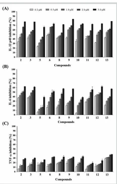
matory diseases. BMDCs were pretreated with the isolated com-pounds for 18 h, and then viability was measured using the MTT assay. At a concentration of 5.0 µM, compound4showed toxicity. Other compounds did not exhibit significant cytotoxic effects (data not shown). Thus, the effects of compounds1–3and5–14 on LPS-stimulated production of proinflammatory cytokines, in-cluding IL-12 p40, IL-6, and TNF-α, in BMDCs were evaluated at concentrations of 0.2, 0.5, 1.0, 2.0, and 5.0 µM. SB203580, a bi-cyclic imidazole compound and an inhibitor of p38 mitogen-acti-vated protein (MAP) kinase [25], was used as a positive control, with IC50values of 5.00 ± 0.16, 3.50 ± 0.12, and 7.20 ± 0.13 µM for IL-12 p40, IL-6, and TNF-α, respectively. Compounds2, 3, 5, 6, and8–13potently inhibited IL-12 p40 production in LPS-stimu-lated BMDCs with IC50 values ranging from 0.35 ± 0.01 to
2.76 ± 0.08 µM (l"Table 3andFig. 2 A). Among them, compound 9showed the greatest inhibitory activity, up to 83.74 % at a con-centration of 5.0 µM and 42.66 % after dilution to 0.2 µM. Other compounds were weak or inactive (l"Fig. 2 A).
To investigate the effects of the tested compounds on other pro-inflammatory cytokines, their effects on IL-6 production were measured. Compounds2, 3, 6, 8, 9, and11–13showed potent in-hibition of IL-6 production, with IC50 values ranging from 1.22 ± 0.02 to 3.79 ± 0.10 µM (l"Table 3). Compound 3showed the greatest inhibition of IL-6 production, with 61.63 % at a con-centration of 5.0 µM and 37.47 % when diluted to 0.2 µM (l"Fig. 2 B). Compounds5and10showed moderate activity, with inhibition percentages ranging from 4.11 % to 39.11 % at the tested concentrations. Other compounds were inactive. In addi-tion, the isolated compounds were also evaluated for TNF-α pro-duction. However, all of them showed weak activity or were in-active at the tested concentrations from 0.2 to 5.0 µM (l"Fig. 2 C). As shown inl"Table 3, compounds2, 3, 6, 8, 9, and11–13 inhib-ited the production of both IL-12 p40 and IL-6. Compounds5and 10showed potent inhibitory effects only on the production of IL-12 p40. Compounds1, 7, and14were inactive in terms of pro-duction of both IL-12 p40 and IL-6.
Flavonoids, one of the most widespread groups of natural prod-ucts, possess a wide variety of pharmacological and biochemical properties, including antioxidant, antimicrobial, antiallergenic, and anti-inflammatory effects. Among them, flavonoid C-glyco-sides such as vitexin, isoorientin, orientin, and isovitexin were shown to have significant anti-inflammatory activity [26, 27]. Therefore, our results were in agreement with those from pre-vious studies. This study has provided evidence of the anti-inflammatory properties of flavonoid C-glycosides that could be promising in the development of anti-inflammatory agents.
Materials and Methods
!General experimental procedures
Optical rotation was recorded on a JASCO DIP-370 automatic dig-ital polarimeter. The NMR spectra were measured using a JEOL ECA 600 spectrometer with TMS as the internal standard. The ESI mass spectra were performed on an AGILENT 1100 LC‑MSD trap spectrometer. The HR‑ESI‑MS were obtained from an Agilent 6530 Accurate-Mass Q‑TOF LC/MS system. GC used an instru-ment (SHIMADZU GC-14B). Silica gel (70–230, 230–400 mesh, Merck) and YMC RP-18 resins (75 µm, Fuji Silysia Chemical Ltd.) were used as absorbents in the CC. TLC plates (silica gel 60 F254 and RP-18 F254, 0.25 µm, Merck) were purchased from Merck KGaA. Spots were detected under UV radiation (254 and 365 nm) and by spraying the plates with 10 % H2SO4, followed by heating with a heat gun. Other chemical reagents and standard compounds were purchased from Sigma-Aldrich.
Plant material
The leaves ofP. aduncumwere collected from Coblong-Bandung, West Java, Indonesia in September 2014 and taxonomically iden-tified by the staff at the Herbarium Laboratory, Department of Bi-ology, School of Life Sciences and TechnBi-ology, Bandung Institute of Technology, Bandung, West Java, Indonesia. A voucher speci-men (BIT-1481) was deposited at the Herbarium of the Depart-ment of Biology, School of Life Sciences and Technology, Bandung Institute of Technology.
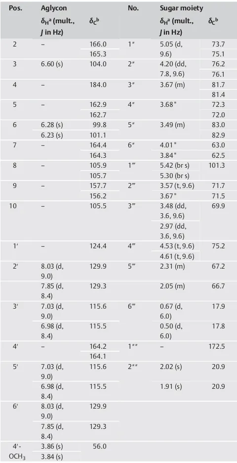
Table 2 1H and13C NMR spectroscopic data for compound4in CD
3OD.
Pos. Aglycon No. Sugar moiety
δHa(mult.,
a600 MHz;b150 MHz. *Overlapped signals. Assignments were done by DEPT, HMQC,
HMBC, and COSY
Extraction and isolation
The dried leaves ofP. aduncum(3.2 kg) were extracted with 70 % ethanol (10 L × 3 times) under reflux condition. Evaporation of the solvent under reduced pressure gave the crude extract (379 g), which was suspended in H2O and successively separated with CH2Cl2and EtOAc to yield CH2Cl2(93 g) and EtOAc extracts (27 g).
The EtOAc extract (27 g) was fractionated on a silica gel CC eluting with gradient solvent systems of CH2Cl2-MeOH (0–100% MeOH, stepwise) to obtain six fractions (C.1 through C.6). Compound12 (20 mg) was isolated from fraction C.3 by YMC reverse-phase (RP)-18 CC using MeOH‑H2O (1/1, v/v) as the eluent, and further purified by YMC RP-18 CC eluting with acetone-H2O (1/2, v/v). Fraction C.4 was separated on silica gel CC eluting with CH2Cl2 -MeOH‑H2O (4/1/0.1, v/v/v) to give six subfractions (C.4.1 through C.4.6). Compounds2(10 mg),3(70 mg), and11(38 mg) were sep-arated from fraction C.4.2 by silica gel CC using CH2Cl2-MeOH (6/ 1, v/v) and further purified by Sephadex® LH-20 CC using MeOH‑H2O (1/1, v/v) as the eluent. Fraction C.4.4 was fraction-ated on YMC RP-18 CC eluting with MeOH‑H2O (1/2, v/v) to ob-tain three fractions (C.4.4.1 through C.4.4.3). Compounds 1 (12 mg) and14(10 mg) were obtained from fraction C.4.4.2 by Sephadex®LH-20 CC eluting with MeOH‑H
2O (1/1, v/v). Fraction C.5 was isolated on YMC RP-18 CC eluting with MeOH‑H2O (1/2, v/v) to obtain four fractions (C.5.1 through C.5.4). Similarly, com-pound 13 (40 mg) was afforded from the fraction C.5.1 by Sephadex®LH-20 CC using MeOH‑H
2O (1/1, v/v) as the eluent. Compounds5(8 mg),9(9 mg), and10(7 mg) were isolated from fraction C.5.2 by Sephadex®LH-20 CC eluting with MeOH‑H
2O (1/1, v/v). And finally, fraction C.5.4 were separated by Sephadex®LH-20 CC using MeOH‑H
2O (1/1, v/v) and further pu-rified by silica gel CC using CH2Cl2-MeOH‑H2O (4/1/0.1, v/v/v) as eluents to afford compounds4(20 mg),6(10 mg),7(15 mg), and 8(100 mg).
Acacetin 8-C-[β‑D-apiofuranosyl-(1→2)-β‑D-glucopyranoside] (1): Yellow amorphous powder; [α]D25:−65.3° (c0.15, MeOH); IR (KBr) νmax: 3367, 2925, 1650, 1602, 1561, 1496, 1070, and 1000 cm−1; UV (MeOH)λmax(logε) 329 (4.39), 270 (4.31) nm; HR‑ESI‑MS m/z: 579.1726 [M + H]+ (Calcd. C
27H31O14 for 579.1714) and 601.1544 [M + Na]+ (Calcd. C
27H30NaO14 for 601.1533);1H NMR (600 MHz, CD
3OD) and13C NMR (150 MHz, CD3OD) are given inl"Table 1.
7-Methoxyacacetin 8-C-[β‑D-apiofuranosyl-(1→3)-β‑ D-gluco-pyranoside] (2): Yellow amorphous powder; [α]D25: −75.8° (c 0.15, MeOH); IR (KBr)νmax: 3375, 2931, 1650, 1602, 1570, 1523, 1459, 1350, and 1055 cm−1; UV (MeOH)λmax(logε) 331 (4.42), 272 (4.32) nm. HR‑ESI‑MS m/z: 615.1685 [M + Na]+ (Calcd. C28H32NaO14for 615.1684);1H NMR (600 MHz, DMSO-d6) and
13C NMR (150 MHz, DMSO-d
6) are given inl"Table 1.
7-Methoxyacacetin 8-C-[β‑D-glucopyranosyl-(1→2)-β‑ D-gluco-pyranoside](3): Yellow powder; [α]D25:−49.7° (c0.1, MeOH); IR (KBr) νmax: 3369, 2925, 1651, 1610, 1570, 1480, 1100, and 1040 cm−1; UV (MeOH)λmax(logε) 332 (4.43), 268 (4.27) nm. HR‑ESI‑MS m/z: 623.1979 [M + H]+ (Calcd. C
29H35O15 for 623.1970), 645.1800 [M + Na]+ (Calcd. C
29H34NaO15 for 645.1790); 1H NMR (600 MHz, DMSO-d
6) and 13C NMR (150 MHz, DMSO-d6) are given inl"Table 1.
4′′′-O-Acetylacacetin 8-C-[α‑L-rhamnopyranosyl-(1→2)-β‑ D-glu-copyranoside](4): Yellow powder; [α]D25:−56.0° (c0.15, MeOH); IR (KBr)νmax: 3414, 2936, 1704, 1650, 1570, 1490, 1446, 1350, and 1045 cm−1; UV (MeOH)λ
max(logε) 334 (4.44), 271 (4.28)
Fig. 2 AEffect of compounds2, 3, 5, 6, and8–13on IL-12 p40
pro-duction by LPS-stimulated BMDCs.BEffect of compounds2, 3, 5, 6, and
8–13on IL-6 production by LPS-stimulated BMDCs.CEffect of compounds
2, 3, 5, 6, and8–13on TNF-αproduction by LPS-stimulated BMDCs. The data are presented as inhibition rate (%). Data represent the mean ± SD of at least three independent experiments performed in triplicate. Statistical significance is indicated as determined by one-way ANOVA followed by
Dunnettʼs multiple comparison test, p < 0.05, using GraphPad Prism 6.
Table 3 Anti-inflammatory effects of selected compounds on LPS-stimulated BMDCs.
Compounds IC50values (µM)
IL-12 p40 IL-6
2 0.87 ± 0.06 1.54 ± 0.09
3 0.56 ± 0.01 1.22 ± 0.02
5 2.76 ± 0.08 > 5.0
6 0.41 ± 0.02 3.17 ± 0.23
8 1.40 ± 0.04 2.95 ± 0.17
9 0.35 ± 0.01 3.79 ± 0.10
10 0.39 ± 0.02 > 5.0
11 1.26 ± 0.02 2.07 ± 0.21
12 0.56 ± 0.03 2.27 ± 0.05
13 0.39 ± 0.05 3.71 ± 0.04
SB203580a 5.00 ± 0.16 3.50 ± 0.12
aSB203580 was used as a positive control
nm. HR‑ESI‑MSm/z: 635.1975 [M + H]+ (Calcd. C
Acid hydrolysis and sugar identification
Each compound (2.0 mg) was dissolved in 1.0 N HCl (dioxane/ H2O, 1 : 1, v/v, 1.0 mL) and then heated to 80 °C in a water bath for 3.5 h. The acidic solution was neutralized with silver carbon-ate and the solvent thoroughly driven out under N2gas over-night. After extraction with ethyl acetate, the aqueous layer was first TLC analyzed [individual and co-analysis with standard sam-ple: glucose (Rf0.29), rhamnose (Rf0.57), and apiose (Rf0.59); CHCl3/MeOH/H2O, 3 : 2 : 0.3] and then concentrated to dryness using N2gas. The residue was dissolved in 0.1 mL of dry pyridine, and then L-cysteine methyl ester hydrochloride in pyridine (0.06 M, 0.1 mL) was added to the solution. The reaction mixture was heated at 60 °C for 2 h, and 0.1 mL of trimethylsilylimidazole solution was added, followed by heating at 60 °C for 1.5 h. The dried product was partitioned withn-hexane and H2O (0.1 mL, each), and the organic layer was analyzed by GC; Column: SPB-1 (0.25 mm × 30 m); detector FID, column temp 210 °C, injector temperature 270 °C, detector temperature 300 °C, carrier gas He. The absolute configuration of the monosaccharide was con-firmed to be D-glucose, L-rhamnose, and D-apiose by comparison of the retention time of the monosaccharide derivative (tR14.11, 4.50, and 4.67 min, respectively) with that of authentic sugar de-rivative samples prepared in the same manner (the retention times of standards D-glucose, L-rhamnose, and D-apiose were 14.12, 4.50, and 4.68 min, respectively.
Cell culture and measurement of cytokine production
BMDCs were grown from wild-type C57BL/6 mice (Orient Bio Inc.) as previously described [28]. All animal procedures were ap-proved and performed according to the guidelines of the Institu-tional Animal Care and Use Committee of Jeju NaInstitu-tional University (#2010–0028). Briefly, the mouse tibia and femur were obtained by flushing with Dulbeccoʼs modified Eagleʼs medium to yield bone marrow cells. The cells were cultured in RPMI 1640 me-dium containing 10% heat-inactivated FBS (Gibco), 50.0 µMβ -mercaptoethanol, and 2 mM glutamine supplemented with 3 % J558 L hybridoma cell culture supernatant containing granulo-cyte-macrophage colony stimulating factor. The culture medium was replaced with fresh medium every second day. On day 6 of the culture, non-adherent cells and loosely adherent DC aggre-gates were harvested, washed, and resuspended in RPMI 1640 supplemented with 5% FBS.The BMDCs were incubated in 48-well plates in 0.5 mL containing 1 × 10⁵cells per well, and then treated with the isolated com-pounds at different concentrations for 1 h before stimulation with 10.0 ng/mL LPS from Salmonella Minnesota (Alexis). Super-natants were harvested 18 h after stimulation. Concentrations of compounds IL-12 p40, IL-6, and TNF-αin the culture supernatant were determined by ELISA (BD PharMingen) according to the manufacturerʼs instructions. SB203580 was the product of Calbiochem.
The inhibitory activity(I)was expressed as the inhibition rate (%), which was calculated from the following formula:
I¼CdcvCdcvCdcc100
Where:Cdcv: Cytokine level (ng/mL) in vehicle-treated DC;Cdcc: Cytokine level (ng/mL) in compound-treated DC.
Statistical analysis
All data represent the mean ± SD of at least three independent experiments performed in triplicate. Statistical significance is in-dicated as determined by one-way ANOVA followed by Dunnettʼs multiple comparison test, p < 0.05, using the GraphPad Prism 6.01 program (GraphPad Software Inc.).
Supporting information
Spectral data of compounds 1–4 are available as Supporting Information.
Acknowledgements
!This study was supported by the Basic Science Research Program through the National Research Foundation of Korea (NRF) funded by the Ministry of Education (2009–0 093 815).
Conflict of Interest
!The authors declare no conflict of interest.
Affiliations
1College of Pharmacy, Chungnam National University, Daejeon, Republic of
Korea
2Institute of Marine Biochemistry (IMBC), Vietnam Academy of Science and
Technology (VAST), Caugiay, Hanoi, Vietnam
3Faculty of Medicine, Maranatha Christian University, Bandung, West Java,
Indonesia
4Biomolecular and Biomedical Research Center, Aretha Medika Utama,
Bandung, West Java, Indonesia
5Department of Chemistry, Mathematic and Natural Sciences, University of
Padjadjaran, Jatinangor Sumedang, West Java, Indonesia
6School of Medicine, Brain Korea 21 PLUS Program, and Institute of Medical
Science, Jeju National University, Jeju, Republic of Korea
References
1Schwab JM, Serhan CN.Lipoxins and new lipid mediators in the
resolu-tion of inflammaresolu-tion. Curr Opin Pharmacol 2006; 6: 414–420
2Dinarello CA.Proinflammatory cytokines. Chest 2000; 118: 503–508
3Okunade AL, Hufford CD, Clark AM, Lentz D.Antimicrobial properties of
the constituents ofPiper aduncum. Phytother Res 1997; 11: 142–144
4Morandim Ade A, Bergamo DCB, Kato MJ, Cavalheiro AJ, Bolzani Vda S,
Furlan M.Circadian rhythm of anti-fungal prenylated chromene in
leaves ofPiper aduncum. Phytochem Anal 2005; 16: 282–286
5Morandim ADA, Kato MJ, Cavalheiro AJ, Furlan M.Intraspecific
variabil-ity of dihydrochalcone, chromenes and benzoic acid derivatives in leaves of Piper aduncumL. (Piperaceae). Afr J Biotechnol 2009; 8: 2157–2162
6Orjala J, Wright AD, Behrends H, Folkers G, Sticher O, Rüegger H, Rali T.
Cytotoxic and antibacterial dihydrochalcones from Piper aduncum. J Nat Prod 1994; 57: 18–26
7Morandim AA, Bergamo DC, Kato MJ, Cavalheiro AJ, Bolzani VS, Furlan M.
Circadian rhythm of anti-fungal prenylated chromene in leaves ofPiper
aduncum. Phytochem Anal 2005; 16: 282–286
8Baldoqui DC, Kato MJ, Cavalheiro AJ, Bolzani Vda S, Young MCM, Furlan
M. A chromene and prenylated benzoic acid from Piper aduncum. Phytochemistry 1999; 51: 899–902
9Okunade AL, Hufford CD, Clark AM, Lentz D.Antimicrobial properties of
the constituents ofPiper aduncum. Phytother Res 1997; 11: 142–144
10Picolo CRD, Bezerra MP, Gomes KS, Passero LFD, Laurenti MD, Martins
EGA, Sartorellia P, Lago JHG. Antileishmanial activity evaluation of
adunchalcone, a new prenylated dihydrochalcone fromPiper aduncum
L. Fitoterapia 2014; 97: 28–33
11Orjala J, Erdelmeier CA, Wright AD, Rali T, Sticher O.Five new prenylated
p-hydroxybenzoic acid derivatives with antimicrobial and
cidal activity fromPiper aduncumleaves. Planta Med 1993; 59: 546–
551
12Parise-Filho R, Pastrello M, Camerlingo CEP, Silva GJ, Agostinho LA, de
Souza T, Magri FMM, Ribeiro RR, Brandt CA, Polli MC.The
anti-inflam-matory activity of dillapiole and some semisynthetic analogues. Pharm Biol 2011; 49: 1173–1179
13Cheng G, Bai Y, Zhao Y, Tao J, Liua Y, Tu G, Ma L, Liao N, Xu X.Flavonoids
fromZiziphus jujubaMill var.spinosa. Tetrahedron 2000; 56: 8915–
8920
14Khallouki F, Hmamouchi M, Younos C, Soulimani R, Essassic EM.A new
flavonoid from the aerial parts of Chrysanthemum viscidehirtum. Fitoterapia 2000; 71: 413–416
15Bjorøy Ø, Rayyan S, Fossen T, Kalberg K, Andersen ØM.
C-glycosylantho-cyanidins synthesized from C-glycosylflavones. Phytochemistry 2009; 70: 278–287
16Larionova M, Spengler I, Nogueira C, Quijano L, Ramírez-Gualito K,
Cortés-Guzmán F, Cuevas G, Calderón JS.A C-glycosylflavone fromPiper
ossanum, a compound conformationally controlled by CH/πand other
weak intramolecular interactions. J Nat Prod 2010; 73: 1623–1627
17Kiem PV, Cuong NX, Nhiem NX, Thu VK, Ban NK, Minh CV, Tai BH, Hai TN,
Lee SH, Jang HD, Kim YH.Antioxidant activity of a new
C-glycosylfla-vone from the leaves of Ficus microcarpa. Bioorg Med Chem Lett 2011; 21: 633–637
18Rayyan S, Fossen T, Nateland HS, Andersen ØM.Isolation and
identifica-tion of flavonoids, including flavone rotamers, from the herbal drug
‘Crataegi Folium Cum Flore’(Hawthorn). Phytochem Anal 2005; 16: 334–341
19Sharaf M, El-Ansari MA, Matlin SA, Saleh NAM.Four flavonoid
glyco-sides fromPeganum harmala. Phytochemistry 1997; 44: 533–536
20Hilsenbeck RA, Mabry TJ.C-glycosylflavones fromSiphonoglossa sessilis.
Phytochemistry 1983; 22: 2215–2217
21Kato T, Morita Y.C-glycosylflavones with acetyl substitution from
Rumex aeetosaL. Chem Pharm Bull 1990; 38: 2277–2280
22Nicholls KW, Bruce AB.Flavonoids ofLupinus arboreus. Phytochemistry
1979; 18: 1078
23Markham KR, Whitehouse LA.Unique flavonoid glycosides from the
New Zealand white pine,Dacrycarpus dacrydioides. Phytochemistry 1984; 23: 1931–1936
24Wei WC, Sung PJ, Duh CY, Chen BW, Sheu JH, Yang NS.
Anti-inflamma-tory activities of natural products isolated from soft corals of Taiwan between 2008 and 2012. Mar Drugs 2013; 11: 4083–4126
25Lee JC, Laydon JT, McDonnell PC, Gallagher TF, Kumar S, Green D, McNulty
D, Blumenthal MJ, Heys JR, Landvatter SW, Strickler JE, Mclaughlin MM,
Siemens IR, Fisher SM, Livi GL, White JR, Adams JL, Young PR.A protein
kinase involved in the regulation of inflammatory cytokine biosynthe-sis. Nature 1994; 372: 739–746
26Xiaoa J, Capanoglu E, Jassbi AR, Miron A.Advance on the flavonoid
C-glycosides and health benefits. Cri Rev Food Sci Nut, advance online publication 13 October 2015; DOI: 10.1080/10408398.2015.1067595
27Borghi SM, Carvalho TT, Staurengo-Ferrari L, Hohmann MSN, Pinge-Filho
P, Casagrande R, Verri jr. WA.Vitexin inhibits inflammatory pain in
mice by targeting TRPV1, oxidative stress, and cytokines. J Nat Prod 2013; 76: 1141–1149
28Koo JE, Hong HJ, Dearth A, Kobayashi KS, Koh YS.Intracellular invasion of
orientia tsutsugamushi activates inflammasome in ASC-dependent manner. PLoS One 2012; 7: e39042