The Development of PC-Based BME-ITB Beads to Evaluate
Antibiotic Release Kinetics from Bone Cement
(A Preliminary Report)
Hermawan Nagar Rasyid1,2, Kuspriyanto2, Tati L Mengko2, Soegijardjo Soegijoko2
1 Dept. of Orthopaedic and Traumatology, Faculty of Medicine Universitas Padjadjaran/Hasan Sadikin Hospital Bandung 2 Biomedical Engineering Program, Department of Electrical Engineering, Institut Teknologi Bandung
ABSTRACT
Inadequate treatment of the acute phase of hematogenous osteomyelitis (bone infection) allows the local pathological process either to persist and become chronic or to become relatively quiescent for a time, only to recur at a later date. Both the persistent chronic form and the recurrent chronic form of bone infection are exceedingly difficult to eradicate. Chronic bone infection can seldom be completely eradicated until all the infected dead bone has separated, or sequestrated, and has either been extruded spontaneously through a sinus tract or been removed surgically (sequesterectomy). Antibacterial therapy is required both systemically and locally. Antibiotic-loaded bone cements have been used for a long time in clinical orthopaedics, despite the fact that it is still in the phase of trying to understand how and why these bone cements work. New antimicrobial agents are therefore needed in considering cost effective and high effective in killing the microorganisms. We proposed the usage of BME-ITB Beads by definition is an antibiotic loaded beads in home-made, mixtures antibiotic (fosfomycin sodium) with the artificial resin polymethylmethacrylate (PMMA). The purpose of the study is to [1]. Evaluate currently used home-made antibiotic loaded beads and compare their antimicrobial efficiency with the efficiency of an existing (patented) product; [2]. Develop an improved concept for the local production by orthopaedic surgeons of antibiotic-loaded beads for the treatment of orthopaedic infections. First, different orthopaedic surgeons are requested to make an overview of different local concepts developed to prepare antibiotic-loaded beads. Based on this overview, five concepts will be selected for further research and each of the participating surgeons will be asked to submit an extensive protocol of their concept. A first evaluation will include a laboratory study on the antibiotic release kinetics, as well as a scanning electron miscroscopic evaluation of the porosity and a quantification of the porosity mercury-intrusion porosimetry. Personal Computer (PC) is used to simulate the porosity of the beads. This project is expected to yield application of the new concept in developing countries around the globe, and has as a rationale that we feel all orthopaedic patients worldwide are entitled to the same effective health care.
Keywords: orthopedics, chronic bone infection, PMMA, fosfomycin sodium, BME-ITB beads, release kinetics, mercury-intrusion porosimetry.
INTRODUCTION
The health care situation in developing countries completely differs from the western world and surgeons in developing countries do all they can to bridge the gap between both worlds. Often, however, finances do not allow them to apply biomedical technologies common in the western world. This becomes strikingly evident in orthopedics. Total hip arthroplasties (“artificial hips”) are common in the western world and give an almost immediate relief from severe pain in large patient populations. In many developing countries, including Indonesia, the costs associated with a total hip arthroplasty are high, which has lead to the situation that in a few cases orthopedic surgeons felt obliged to re-use prostheses. Infection is an infrequent but serious complication of prosthetic joint surgery. The current infection rate in primary total joint replacement is about 1% 1.
Nevertheless, it has been estimated that about 4000 to 5000 revisions due to infection are performed annually in the United States alone 2 and this number approximately triples world-wide.
Buccholz and Engelbrecht 3initiated the addition of antibiotics to polymethylmethacrylate bone cement in the
beginning of the 1970s. By mixing gentamycin in acrylic bone cement, they succeeded in reducing the infection rate. The assumption underlying the incorporation of antibiotics in bone cements was that the antibiotic would gradually be released to yield higher local concentrations than can be achieved by systemic therapy. Since the introduction, a number of clinical studies on the use of antibiotic-loaded bone cements have been conducted. The results of these studies have shown that local administration of antibiotics through antibiotic-loaded bone cement yields good results and cost effective prevention of infection in total joint athroplasty 1,4.
In Indonesia, orthopaedic surgeons use bone cement, mixed it themselves with antibiotic in home-made molds and apply them in their patients. This is despite the fact that the release mechanisms of antibiotic from bone cements is poorly understood and controlled, with some reports claiming the release of antibiotic from bone cements for up to five years after implantation 5.
Presented at The 7th ASEAN Science and Technology Week (The 7th ASTW), August 5th – 7th, 2005
The State Ministry for Research and Technology (RISTEK), Jakarta,
The concept of using the bone cement as a depot for antibiotics is reliable, as it allows delivery of antibiotics directly to the site of (imminent) infection. The implantation of non-compatible materials in the human body is limited by a so-called foreign body reaction. This is an inflammatory response due to the foreign body that may lead to migration and rejection of the implanted material. Therefore, any material applied in the human body should be able to perform with an appropriate host response in a specific application. In other words, it should show a high degree of biocompatibility. Bone cement mainly consists of polymethylmethacrylate (PMMA). Pieces of the PMMA, due to its excellent biocompatibility and ease of manipulation.
All commercially available bone cements have two components. The first is a powder that consists mainly of a polymer based on methylmethacrylate (MMA). The second component is mainly liquid MMA. The dough phase resulting from mixing these components is important for bone cements for a variety of reasons. To start, it offers the possibility of molding and being used to support a prosthesis, while allowing its insertion. Furthermore, the initial presence of PMMA enables the use of less monomer for obtaining the same amount of end product. Finally, this mix of PMMA and MMA means that undesirable effects of polymerization are reduced. Firstly, use of less monomer leads to less heat production. Secondly, polymerization from a monomer necessarily leads to a decreased volume and increased density due to the fact that the molecules take up less space in a polymer than in their liquid monomer. If less polymerization occurs, the volumetric shrinkage is proportionally lower. Apart from these main components, other substances are required to achieve a controlled polymerization at body temperature. N,N dimethyl-p-toluidine dissolved in the monomer is a tertiary amine, enabling cold curing of the polymer, instead of the required preheating to 100°C. It reacts with the benzoyl peroxide in the powder to create free radicals that can break part of the C=C bond and start the addition polymerization. Furthermore hydroquinone and chlorophyll are additives that prevent premature polymerization under exposure to light or elevated temperatures.
The presence of barium sulphate and zirconium (IV) oxide in the powder is necessary for a clinical reason. The first bone cements not containing these could not be visualized on radiographs. These agents are so-called radio pacifiers and make the bone cement visible on radiographs.
BME-ITB beads
The use of polymethylmethacrylate (PMMA) bone cement loaded with antibiotics has become increasingly common in the treatment of infected bone (chronic bone infection), infected knee and hip arthroplasties and also as prophylaxis in primary joint replacement. Recently, fosfomycin sodium is the most commonly used antibiotic in bone cement. Fosfomycin sodium is an antibiotic with extremely low molecular weight of 138, produced by strains of Streptomyces 6, and is characterized by structural features of an epoxide ring and a
carbon-phosphorus bond. It is also characterized by its action, which inhibits the first step of peptidoglycan biosynthesis, and is synergistic in combination with many other anti-microbial agents 7. Moreover, fosfomycin
sodium is antibiotic that remains stable when exposed to the high temperatures generated during the curing of the bone cement. Antibiotic is released from bone cement in a biphasic manner, meaning high elution in the first hours to days post surgery and after a few days it slows down considerably.
In BME-ITB beads, mixture of fosfomycin sodium antibiotic and bone cement were used. These beads constitute an effective drug delivery systems for local antibiotic therapy in bone and soft-tissue infections and at the site of the infection can exceed the minimal inhibitory concentration of the infecting organism 8.
RESEARCH OBJECTIVE
[1]. Evaluate currently used home-made antibiotic-loaded beads and compare their antimicrobial efficiency with the efficiency of an existing (patented) product.
[2]. Develop an improved concept for the local production by orthopaedic surgeons of antibiotic-loaded beads for the treatment of orthopaedic application.
The new concept is expected to be applicable worldwide and will allow surgeons to locally develop their own effective beads.
MATERIALS AND METHODS
A number of orthopaedic surgeons are requested to make an overview of different local concepts developed to prepare antibiotic-loaded beads. Orthopaedic bone cements are being used, amongst others in combination with a variety of different antibiotics. Based on this overview, five concepts are selected for further research and the participating surgeons will be asked to submit an extensive protocol of their concept. To evaluate the porosities of the beads scanning electron microscopy was used, beads were fixated with 2% glutaraldehyde in 100mM cacodylate buffer. After fixation, the beads were exposed overnight to an osmium-tetroxide vapour. Samples were dehydrated in air and sputter-coated with gold/palladium (~3 nm). Examination was done at 10 kV in a field emission scanning electron microscope type 6301F. Personal Computer (PC) is used to simulate the porosity of the beads.
Presented at The 7th ASEAN Science and Technology Week (The 7th ASTW), August 5th – 7th, 2005
The State Ministry for Research and Technology (RISTEK), Jakarta,
Cement disc preparation
Commercially available Surgical Simplex® P Radiopaque Bone Cement (a product of Stryker Howmedica OSTEONICS, Howmedica International S, de R.L. Raheen Business Park, Limerick, Ireland) was used. Bone cement was prepared by mixing the powdered polymethylmethacrylate with the liquid monomer in a bowl with a spatula. Manual mixing was done according to the manufacturer’s instructions and resulted in doughy cement, then, the antibiotic available 2gr Fosfomycin Sodium was poured. The dough-like mass is then ready for molding by hand, spherical shape beads are built in various diameters between 10 mm and 20 mm. These spheres are connecting each other by using small diameter of stainless steel wire string. All procedures were carried out under sterile conditions.
RESULTS AND DISCUSSION
Under sterile conditions, eight different types of hand-made fosfomycin sodium-loaded bone cement bead sets are built in spherical shape. It consists of three hand made spherical shape beads. The sizes of beads are varies between 10 and 20 mm and the beads are which can usually be easily seen on scanning electron micrographs of bone cements.
To evaluate the amount of porosities scanning electron microscopy (SEM) was used. In the same 40 times magnification at 10 KV demonstrated that size of 20 mm beads showed little porous comparing small diameter. Some “white dots” of fosfomycin sodium can be seen closed to the pores. In 120 times magnification the “white dots” become homogen in particular at the small size (10 mm in diameter). In this evaluation, to obtain good porosities the size of beads is very important. We may conclude that beads having 10 mm in diameter revealed to more homogeneous pores. Therefore, for future experiments these beads size must be considered.
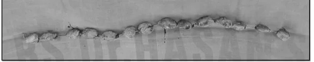
BME-ITB beads, however, release much more fosfomycin sodium than solid bone cement plugs, mainly due to the greatly increased surface area of the many, relatively small beads (see Fig. 1).
Figure 1: A string of hand-made BME-ITB beads.
It consists of multiple various sizes of spherical shape beads which are connecting each other by small diameter of stainless steel wire string.
Mechanism of antibiotic release from bone cements
Bone cements are made up of two primary components: a powder containing copolymers based on the substance polymethylmethacrylate, and liquid monomer, methylmethacrylate. The powder contains a starter, di-benzoil peroxide; while the liquid contains the initiator, N,N-dimethyl-p-toluoidin. Both substances together start the polymerisation process and enable a reaction at room temperature. A special radio-opaque agent (barium sulphate) is added to the powder to provide X-ray contrast. The polymer from which the bone cements are made is capable of taking up very small quantities of dissolution fluid into its outermost layers. This dissolution fluid then slowly transport the antibiotic molecules contained within the polymer out into the surrounding tissue. At the beginning, when the infection is still very active, large amounts of antibiotic are released 9, because it is
particularly easily available at the surface of bone cement. The sustained release of antibiotics from bone cements is largely influenced by the penetration of dissolution fluids into the polymer matrix, which requires a certain porosity of the cement 9,10. A point on which all studies have agreed is that the period of maximum
antibiotic release is limited to the first few hours or days after implantation. The maximum effectiveness of the released antibiotic might thus be expected to occur during this period of time 11. Most, if not all, of the
antibiotic is released from the superficial regions of the cement and fails to be released from the centre 12.
The porosity of the polymer matrix depends on air entrapment during the wetting and stirring of the cement powder during transfer to the cement gum ad on effects of monomer boiling 13 . Baker and Greenham,
concluded that bone cement with greater porosity would be expected to allow more antibiotic release than one with less porosity. Therefore, the methods of cement preparation that are designed to improved mechanical properties by decreasing the porosity could have deleterious effect on elution characteristics. Furthermore, in commercially available antibiotic-loaded bone cement, the antibiotic is evenly dispersed throughout the cement,
Presented at The 7th ASEAN Science and Technology Week (The 7th ASTW), August 5th – 7th, 2005
The State Ministry for Research and Technology (RISTEK), Jakarta,
creating a homogenous mixture. This uniform dispersion of antibiotics is not possible to achieve when adding the antibiotic by hand to the bone cement. Non-uniform dispersion may prevent the antibiotic from eluting properly.
CONCLUSIONS
The concept of BME-ITB beads by using bone cement as a depot for antibiotic make sense, as it allows delivery of antibiotics directly to the site of infection.
The beads having 10 mm in diameter revealed to more homogeneous pores.
REFERENCES
1. Josefsson G, Kolmert L. Prophylaxis with systematic antibiotics versus gentamycin bone cement in total hip arthroplasty. A ten-year survey of 1,688 hips. Clin Orthop 1993; 292: 210-214
2. Sculco TP. The economic impact of infected joint arthroplasty. Orthopaedics 1995; 18: 871-873 3. Buccholz HW, Engelbrecht H. Uber die Depotwirkung einiger Antibiotics bei Vermischung mit dem
Kunstharz Palacos. Chirurg 1970; 40: 511-515
4. Espehaug B, Engesaeter LB, Vollset SE, Havelin LI, Langeland N. Antibiotic prophylaxis in total hip arthroplasty. Review of 10,905 primary cemented total hip replacements reported to the Norwegian
arthroplasty register, 1987 to 1995. J Bone Joint Surg Br 1997; 79: 590-595
5. Wahlig H, Dingeldein E. Antibiotics and bone cements. Experimental and clinical long term observations. Acta Orthop Scand 1980; 51: 495
6. Hendlin D, Stapley EO, Jackson M, Wallick H. Phosphonomycin, a new antibiotic produced by strains of Streptomyces. Science 1969, 166: 122-123
7. Bergeron M, Robert J, and Beauchamp D. Pharmacodynamics of antibiotics in fibrin clot. J Antimicrob. Chemother 1993; 31: 113-136
8. Wahlig H, Dingeldein E, Bergman R, Reuss K. The release of gentamycin from polymethylmethacrylate beads. An experimental and pharmacokinetic study. J Bone and joint Surg Br 1978; 60:270-275
9. Khoury AE, Lam K, Ellis B, Costerton JW. Prevention and control of bacterial infections associated with medical devices. ASAIO J 1992; 383; M174-178
10. Baker AS, Greenham LW. release of gentamicin from acrylic bone cement. J Bone Joint Surg Am 1988; 70: 1551-1557)
11. Tripple SB. Antibiotic-impregnated cement in total joint arthroplasty. J Bone Joint Surg Am 1986; 68: 1297-1302
12. Schurman DJ, Trindale C, Hirschman HP, Moser K, Kajiyama G, Stevens P. Antibiotic-loaded acrylic bone cement composites. J Bone Joint Surg Am 1978; 60: 978-8=984
13. Wixson RL, Lautenschlager EP, Novak MA. Vacuum mixing of acrylic bone cement. J Arthroplasty 1987; 2: 141-149
Presented at The 7th ASEAN Science and Technology Week (The 7th ASTW), August 5th – 7th, 2005
The State Ministry for Research and Technology (RISTEK), Jakarta,