Vol. 43. No. 4 December 2010
Alveolar ridge rehabilitation to increase full denture retention
and stability
Mefina Kuntjoro, Rostiny, and Wahjuni Widajati Department of Prosthodontics
Faculty of Dentistry, Airlangga University Surabaya - Indonesia
abstRact
background:Atrophic mandibular alveolar ridge generally complicates prostetic restoration expecially full denture. Low residual alveolar ridge and basal seat can cause unstable denture, permanent ulcer, pain, neuralgia, and mastication difficulty. Pre-proshetic surgery is needed to improve denture retention and stability. Augmentation is a major surgery to increase vertical height of the atrophic mandible while vestibuloplasty is aimed to increase the denture bearing area. Purpose: The augmentation and vestibuloplasty was aimed to provide stability and retentive denture atrophic mandibular alveolar ridge. case: A 65 years old woman patient complained about uncomfortable denture. Clinical evaluate showed flat ridge in the anterior mandible, flabby tissue and candidiasis, while residual ridge height was classified into class IV. case management:Augmentation using autograph was conducted as the mandible vertical height is less than 15 mm. Autograph was used to achieve better bone quantity and quality. Separated alveolar ridge was conducted from left to right canine region and was elevated 0.5 mm from the previous position to get new ridge in the anterior region. The separated alveolar ridge was fixated by using T-plate and ligature wire. Three months after augmentation fixation appliances was removed vestibuloplasty was performed to increase denture bearing area that can make a stable and retentive denture. conclusion:
Augmentation and vestibuloplasty can improve flat ridge to become prominent.
Key words: Augmentation, vestibuloplasty, athropic ridge, mandible
abstRaK
Latar belakang: Ridge mandibula yang atrofi pada umumnya mempersulit pembuatan restorasi prostetik terutama gigi tiruan lengkap (GTL). Residual alveolar ridge dan basal seat yang rendah menyebabkan gigi tiruan menjadi tidak stabil, menimbulkan ulser permanen, nyeri, neuralgia, dan kesulitan mengunyah. tujuan: Augmentasi dan vestibuloplasti pada ridge mandibula yang atrofi dilakukan untuk menciptakan gigi tiruan yang stabil dan retentive. Kasus: Pasien wanita usia 65 tahun datang dengan keluhan gigi tiruan yang tidak nyaman. Pemeriksaan klinis menunjukkan ridge flat pada anterior mandibula, jaringan flabby dan kandidiasis, sedangkan residual ridge digolongkan menjadi kelas IV. tatalaksana kasus: Augmentasi dilakukan karena ketinggian vertikal mandibula kurang dari 15 mm. Autograf digunakan untuk mendapatkan kuantitas dan kualitas tulang yang lebih baik. Alveolar ridge diambil dari sisi kiri dan kanan region kaninus dan digunakan 0,5 mm dari posisi awalnya untuk mendapatkan ridge baru pada region anterior. Alveolar ridge telah diseparasi difiksasi menggunakan T-plate dan ligature kawat. Tiga bulan setelah fiksasi dilepas, dilakukan vestibuloplasti untuk meningkatkan denture bearing area sehingga gigi tiruan lebih stabil dan retentive. Kesimpulan: Augmentasi dan vestibuloplasti dapat memperbaiki ridge atrofi sehingga menjadi tinggi kembali.
Kata kunci: Augmentasi, vestibuloplasti, ridge atrofi, mandibula
Correspondence: Mefina Kuntjoro, c/o: Departemen Prostodonsia, Fakultas Kedokteran Gigi Universitas Airlangga. Jl. Mayjend. Prof. Dr. Moestopo No. 47 Surabaya 60132, Indonesia. E-mail: [email protected].
intRoduction
Full denture is a denture used for replacing all missing teeth on the maxilla and mandible, supported by mucosa, connective tissue and bone. Full denture can only be used well if it is stable and retentive.1 Full denture could be
stable if during its function the denture only moves slightly above the bone. Meanwhile, the denture could be retentive if the power coming on the denture can not release the adhesion between denture and underneath mucosa. Thus, retention can be defines as the resistance of the denture to its disposition in the mouth.2–5
After tooth extraction, the alveolar bone in the jaw would be atrophic which is called as residual ridge resorbtion. This condition can cause reduction of supporting tissue and bone to make full denture stable and retentive, however this problems will become worse if it occurs in the mandible.6
In order to estimate alveolar ridge resorbtion, radiographic examination could be conducted through cephalometry and panoramic. Cephalometry can be used to examine dimensions of the jaw vertically and horizontally, but the lack of this radiography is the superimposed alveolar ridge. Panoramic is more often used to conduct the evaluation.7
The alveolar ridge height can be calculated from top of the ridge to the anatomical landmark, such as the maxillary sinus or mandibular canal in the posterior region. In the maxillary anterior region, the base of the nose is used, while in the mandible, the inferior border of the mandible is used.8
The initial phase of the residual ridge resorbtion is actually started immediately after the tooth is extracted that can cause lost of periodontal membrane that has the ability to regenerate bone. The lost of the alveolar bone occurs in the labiolingual region and vertical height causing the ridge become narrower, and in some cases also causing it to shape like sharp knife (knife edge). Next, processus alveolaris becomes low, round or flat. As the resorbtion continues, alveolar bone and basal bone can become small, and ridge become shortened. If it occurs in the mandible, it will cause problems for prosthodontics to make full denture.9
The alveolar ridge height can be classified into four classes; class I, the alveolar ridge height is adequate, but less wide and usually accompanied by the deficiency of the lateral or undercut regions; class II, the height and width of the ridge is less, there is sharp ridge like a knife; class III, alveolar ridge resorbtion until basiliar bone that can cause sharp configuration on the ridge; class IV, resorbtion of the basiliar bone occurs, so the mandible become as thin as pencil with ridge in the maxilla become flat.10
There are several techniques that can be conducted to rehabilitate atrophic ridge in the mandible before prostodontics treatment is conducted. The technique is divided into three categories; augmentation with bone or with bone graft continued with vestibuloplasty, using skin or mucosal graft to add surface area; and implant (subperiosteal, transosteal and endosteal implants).11
Augmentation is a major surgery to increased the vertical height of the atrophic ridge, continued with vestibuloplasty to increase denture bearing area. Augmentation is usually performed on the mandibular ridge crest region or on the inferior border. If the vertical height of the mandible less than 15 mm, augmentation is absolutely needed to raised alveolar ridge and make enough retention to make full denture in the mandible. If the height is more that 15 mm, augmentation will not be required, but only vestibuloplasty is needed.12–14
Vestibuloplasty is an additional procedure to repair ridge for achieving additional vertical ridge height. The aim is to improve the height of the atrophic ridge, so the wide basal seat will be enough to make full denture stable.15,16
case
The patient a 65 years old woman, came to Prosthodontics clinic, Faculty of Dentistry, Airlangga University to make a new denture. She had been using full denture since five years ago. Since two years ago her lower denture has become loose and caused pain so she complained difficulty to chew food. She also complained her gums in the front lower denture were often bloody and got bumps. As a result, she want to make a new denture that can make her comfortable and chew food well.
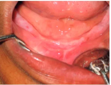
On extra oral examination, the temporomandibular joint, eyes, nose and lips were normal with oval face. Gangrene radix on 12, 14, 17, 22, flabby tissue and candidiasis in the anterior mandibular region were found on intra oral examination. Vestibulum of the maxilla were all quite deep except on the left posterior. Maxillary ridge was ovoid while mandibular ridge was flat. The anterior ridge of mandible was very flat. Torus palatine and mandibula were flat. Palatum was ovoid with large tuber maxilla. No exostosis on the maxilla and mandible. Retromylohyoid on the left and right sides were shallow (Figure 1).
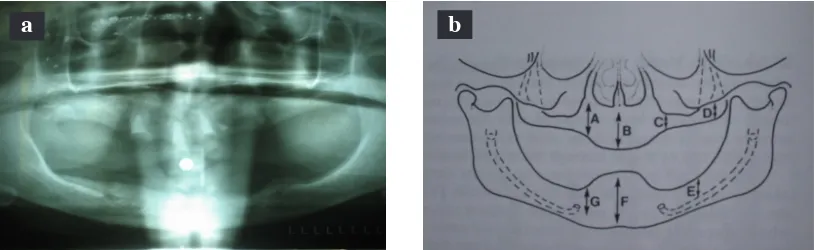
In the radiological examination, gangrene radix on 12, 14, 17, 22 and resorbed processus alveolaris until 1/3 length of the root were found. Residual ridge height in the mandible was measured from the edentulous region to the opposite anatomical landmarks, in this case ridge height was less than 15 mm in all regions of the mandible (Figure 2).
case ManageMent
In this case, gangrene radix on 12, 14, 17, 22 were extracted. Residual ridge height was classified into class IV, since it was below 15 mm, so it was essential to conduct augmentation in the anterior region and followed by vestibuloplasty. After this was done, full denture was made using acrylic base and acrylic artificial teeth.
a
b
Figure 1. The intra-oral condition of the patient: a) Maxilla and mandible; b) Flabby tissue and mandibular candidiasis in the anterior mandible.
(Figure 1a, 3, and 4 published under the courtesy of Prof. Coen P., Department of Oral Surgeon, Faculty of Dentistry, Airlangga University)
b
a
Figure 2. a) Panoramic photo of the patient at 12, 14, 17, 22; b) The height of the available alveolar ridge measured from the edentulous region to the opposite anatomical landmarks. A) Maxillary canine, B) Nose base, C) Maxillary sinus, D) Tuberosity, E) Inferior Mandibular canal, F) Anterior mandible, G) Mandibular canine.
incision was conducted in the anterior mandibular region until the alveolar ridge was exposed by using scalpel.
The alveolar ridge from left canine region to the right was cut by using low speed handpiece and irrigated with saline solution. The cutting process was done until mandibular base. The alveolar ridge was separated from mandibular bone by using chisel.
The separated alveolar ridge was heightened 0.5 mm from the previous position to get a new ridge in the anterior mandibular region. This was done to get retention and stability for the new full denture. Then the separated alveolar ridge was fixated by using T-plate and screwed in four locations, 2 in mandibular regions and 2 in the separated alveolar ridge, then tied by using ligature wire for making it stable and unmovable. Suturing procedure was done by using polyglycolide thread until there was no opened tissues so that food could not be trapped and disturb healing process (Figure 3).
Control I was done 2 weeks after augmentation, the wound was recovering. The surrounding tissue was under normal condition. There was even no visible swelling. Then the suture was opened and evaluated 3 months later to see the augmentation result.
Control II was done 3 months later, the wound was very well, asymptomatic and the newly formed ridge was seen in the anterior mandible. Fixation appliances T plate, screws, and ligature wire was removed by making incision
on the newly formed ridge, then ligature wire, retrieving four screws, and finally removed T plate. As a result of bone reposition, the formation of the new bone joint has occurred in the area below the ridge (Figure 4).
Vestibuloplasty according to Gordon was conducted by making split thickness flap and secondary epithelialization technique in the anterior mandible. Vestibuloplasty was done by moving muscle attachment more inferior in order to obtain the required depth of vestibule. Then suturing procedure was conducted in mucosa and gengigel containing hyaluronic acid was given to accelerate epithelialization. The patient was instructed to rinse her mouth with antiseptics to maintain oral hygiene. One week after vestibuloplasty, primary impression was done to make temporary full denture (transitional denture) to prevent relapse after vestibuloplasty.
Control was done four weeks after vestibuloplasty. The mandibular ridge was recovering very well, so impression was done by using alginate and impression tray to make anatomical cast. The anatomical cast on the maxilla was given a layer of wax used as a spacer to place impression material, meanwhile on the mandible a spacer was not given since the ridge was flat. Then by using this model, individual tray for maxilla and mandible was done.
to improve retention for the full denture. Then impression was done by using polyvinyl siloxane (Figure 6). After recovery time about 30 minutes the impression was filled with dental stone type III to make master cast.
Using master cast, bite rim was done to gain vertical and horizontal jaw relation, then the master cast was put on articulator. The artificial teeth were set in the bite rim and located in the neutral zone to improve stability. The heat cured acrylic was packed and cured. Selective grinding I was done to make sure that the vertical and horizontal jaw relation was correct. Intermaxillary record was done and continued with selective grinding II. Insertion was done, with consideration on the retention and stability of the full denture (Figure 5).
Control I was done one day after insertion. Patient complained pain on the left posterior mandible. On intra oral examination, left retromylohyoid region looked reddish, then grinding on that area was done. Control II was done three days after control I, patient had no complaint and feel comfortable with the new full denture. Control III was done one week after control II. Patient had no complaint and was able to chew food well. Afterwards, the next controls would be periodically every six months.
Six months after insertionl, the anterior ridge in the mandible was looked quite high with no abnormality. The patient was very satisfied with the present condition, the denture was comfortable and was able to chew food properly.
discussion
In this case, there was a problem with the anterior mandibular ridge which was atrophic, as a result, there was not enough basal seat as a support for retention and stability for full denture. This condition could even lead to many problems in the setting of full denture. Patient with flat alveolar ridge could usually have uncomfortable denture because of less retentiton and stability, permanent ulcer on the soft tissues, neuralgia and minimum chewing ability.14
As a consequence, preprosthetics surgery was needed to establish higher anterior mandibular ridge in order to make full denture more stable and retentive. In this case, augmentation was conducted because the height of the mandibular ridge is less than 15 mm in the premolar region and then continued with vestibuloplasty.11
Bone graft is also required in augmentation, its selection must be selective to obtain maximum result. The disadvantage of bone graft is that graft can be resorbed after two to five years. To prevent excessive resorbtion in using bone graft, hydroxyapatite could be used or miced with autogenous bone to reconstruct higher ridge in the flat ridge mandible.12
The use of autograft is more usefull than allograft in augmentation. The reason is the result of using autograft is predictable because better bone quantity and quality Figure 5. The insertion of complete denture in maxilla and
mandible.
Figure 6. The condition of the mandibular ridge 6 months after insertion of the complete denture.
Figure 3. The seperated alveolar ridge was fixated with T-plate screwed and tied with a ligature wire.
could be obtained. Allograft is still usefull because it does not need invasive surgery, but the result is not as better as autograft because of poor bone quality and quantity. Autograft is used in situations of losing many bones, while as allograft is used for small fenestration, labial dehiscence or socket after extraction.17,18
The augmentation in this case was conducted using autograft. The autograft was obtained by separating alveolar ridge and then placing it higher based on the required height. And was fixated using T-plate, screw and ligature wire in order to make the separated ridge unmovable. By using autograft, resorbtion of the ridge was expected to be minimal since the graft was from the patient and in the sama area. After the new bone was formed, vestibuloplasty was conducted.
Vestibuloplasty was done to remove the muscle lower and to provide connective tissue for the remained ridge. To conduct succesfull vestibuloplasty, mucosal flap should be free from any tension to avoid relapse, this can be minimized by making an incision at the base of the sulcus. In this case one week after vestibuloplasty, impression was done to make transitional denture to prevent relaps. It is very important that the full denture should not irritate granulation tissue, the length of the denture should be deep enough to form new sulcus. Then four weeks after vestibuloplasty, final impression was done to make new full denture.20
To make the full denture it is important to make sure high accuracy in each procedure, especially in the case of flat ridge in the mandible. The cause of lack stability and retentive is several factors such as less accurate impression, improper horizontal and vertical jaw relation, setting tooth acrylic not in the neutral zone.21
In the case of extremely flat ridge, the final impression must be conducted properly to get accurate master cast that could support retention and stability of the full denture. The accuracy of the impression could also be obtained by using individual tray and border moulding to get peripheral seal. Then final impression was done by using mucocompresive material to make pressure to the mucosa and the materials could flow to fill complicated part.22
Additional retentiton could be obtained at retromylohyoid region by digging cast so individual tray at that area could be longer, otherwise compound stick could also be used at the time of border moulding. Impression result for retromylohyoid region should have “S” form from lateral part and butterfly wing when seen from above.22 Horizontal
and vertical jaw relation was done and continued with adjust cast with bite rim in the articulator. All of those three phases must be done properly in order to make the new full denture stable, mastication muscle should not get tired easily, create good esthetics, support patient face not
to look older, phonetics not disturbed and prevent Costen syndrome.22
It can be concluded that augmentation and vestibuloplasty could give good result in forming new prominent ridge on atrophic mandibular ridge. As a consequence, it could provide a better prognosis in making full denture.
ReFeRences
1. Henderson D, Mc. Givey Glen, Castleberry D, Mc. Cracken’s. Removable partial prosthodontics. St. Louis, Toronto, Princeton: CV Mosby Company; 1985. p. 1–7.
2. Watt David, Mac Gregor AR. Designing complete denture. Philadelphia: WB Saunders Company; 1986. p. 1–6.
3. Findlay A. Introduction to physical chemistry. 3rd ed. London, Longmans: Green & Company; 1960. p. 534.
4. John S. Complete denture prosthodontics. New York, Toronto, Sydney, London: Mc.Graw-Hill Book Company Inc; 1974. P. 147–52.
5. Baat CA, Albert, Mulder. Prosthetics condition and patient judgement of complete denture. J Prosthet Dent 1997; 78: 472–8.
6. Tina MB, Brian LM. Histological analysis of healing after tooth extraction with ridge preservation using mineralized human bone allograft. J Periodontology 2010; 81(12): 1765–72.
7. Toshiho H. Diagnostic imaging by panoramic radiograph in edentulous patient. J Japan Prosthodontics Society 1999; 43(1): 13–9. 8. Nishimura I, Hosokawa R, Atwood DA. The knife edge tendency
in mandibular residual ridges in women. J Prosthet Dent 1992; 67: 820–6.
9. Misch CE. Contemporary implant dentistry. 2nd ed. St. Louis: Mosby-Year Book, Inc; 1993. p. 123–8.
10. Stafne EC. Oral roentgenographic diagnosis. 3rd ed. Philadelphia: WB Saunders; 1969. p. 2.
11. Pramono CD. Mandible vertical height correction using lingual bone-split pedicle onlay graft technique. Dent J 2006; 39(3): 93–7. 12. Robert EM, Thomas S, James W. Severely resorbed mandible:
predictable reconstruction with soft tissue matric expansion (tent pole) grafts. J Oral Maxillofac Surg 2002; 60: 878–88.
13. Findlay A. Introduction to physical chemistry. 3rd ed. London, Longmans: Green & Company; 1960. p. 534.
14. Joseph E, Michael T. Review of surgical ridge augmentation procedures for the atrophied mandible. J Prosthet Dent 1984; 51(1): 5–10.
15. Hisham FN, Aichelmann-Reidy, Yukna R. Bone and bone subtitutes. Periodontology 2000; 19(2): 74–83.
16. Gordon WP. Buku ajar praktis medah mulut. Cetakan I. Philadelphia:Cetakan I. Philadelphia: WB Saunders; 1996. p. 132–7.
17. Abdel SE, Pipco DJ. Autogenous and allogenous bone grafting techniques to maximize esthetic: A clinical report. J Prosthet Dent 2000; 83: 153–7.
18. Zhdanov E. The innovative approach to the treatment of total edentulism and advance alveolar atrophy. Smile Dental Journal 2009; 4 (Issued 3): 36–9.
19. Ewers VR, Haerle F. Langezeittresultate nach Visieristeotomi. Dtsch Zahnaerl A 1980; 35: 1046–7.
20. Starshak. Preprosthetic oral surgery. United States of America:Starshak. Preprosthetic oral surgery. United States of America: CV Mosby; 1971. p. 157–8.
21. Heartwell CM, Rahn AO. Syllabus of complete denture. 4th ed. Philadelphia: Lea & Febiger; 2004. p. 200–10, 277–305, 327–37. 22. Itjiningsih WH. Geligi tiruan lengkap lepas. Cetakan ke-3. Jakarta: