46 Menara Perkebunan, 200, 69 (2), 46-57.
Morphological variations during the development of somatic
embryos of tea (
Camellia sinensis
L.)
in vitro
Keragaman morfologi selama perkembangan embrio somatik teh (Camellia sinensis L.) in vitro
SUMARYONO, Imron RIYADI & J.S. TAHARDI
Biotechnology Research Unit for Estate Crops, Bogor 16151, Indonesia
Ringkasan
Biak embrio somatik tanaman teh (Camellia sinensis L.) pada medium padat terdiri dari embrio dalam berbagai ukuran, warna dan stadia perkembangan. Satu gram embrio somatik yang sebagian besar dalam stadia globuler telah dibiakkan pada medium padat proliferasi (medium WP dengan IAA 57,1 µM dan BAP 4,4 µM) untuk mengamati keragaman morfologi embrio dalam hal ukuran, warna dan stadia perkembangan dalam satu periode kultur 6 minggu. Berat basah embrio somatik meningkat perlahan pada 4 minggu pertama kemudian meningkat dengan tajam. Pada minggu keempat, jumlah embrio melonjak walaupun beratnya tidak meningkat, hal ini menunjukkan adanya pembentukan embrio sekunder. Ukuran rata-rata embrio somatik tidak berubah secara nyata selama periode kultur, tetapi ukuran embrio sudah sangat beragam sejak awal kultur dan terus meningkat sejalan dengan berkembangnya embrio. Sekitar setengah dari embrio berwarna kuning dan sisanya terdiri dari embrio berwarna hijau dan merah. Pada awal kultur, 60% embrio berada pada stadia globuler, 30% stadia hati dan 10% stadia bentuk-torpedo. Pada umumnya embrio globuler berkembang ke stadia lebih lanjut sejalan dengan waktu, tetapi pada medium proliferasi ini hampir 80% embrio masih dalam stadia globuler dan bentuk-hati pada minggu keenam. Apabila embrio somatik globuler tunggal dengan warna tertentu dibiakkan pada medium padat regenerasi (WP dengan kinetin 0,47 µM, ABA 0,69 µM dan GA3 0,29 µM, sebagian embrio terutama embrio
kuning akan mengalami perubahan warna. Sebagian besar embrio globuler tunggal ini
berkembang secara bertahap kestadia per-kembangan lebih lanjut. Warna awal embrio berpengaruh terhadap kecepatan perubahan stadia perkembangan embrio, dengan embrio globuler awal warna kuning cenderung lebih cepat berkembang kestadia kotiledon dan kecambah dibandingkan dengan embrio hijau dan merah.
Summary
47
Sumaryono et al.
almost 80% of the embryos remained at the globular and heart-shaped stages even after the sixth week. If single globular somatic embryos with a particular color were cultured on a solid regeneration medium of WP with 0.47 µM kinetin, 0.69 µM ABA and 0.29 µM GA3, some of them especially the yellowish embryos underwent color change. Most of these single globular embryos developed gradually into the later stages. While the initial colors of embryos affected the rate of developmental stage changes, yellowish globular embryos tended to develop more rapidly into cotyledonary or germinant stages than the greenish and reddish embryos.
[Keywords: Camellia sinensis, embryo color, embryo development, somatic embryogenesis, tea]
Introduction
In vitro propagation of tea (Camellia sinensis [L.] O. Kuntze) has been achieved through proliferation of axillary buds (Nakamura, 1991; Agarwal et al., 1992; Tahardi, 1994) and more recently through somatic embryogenesis (Wachira & Ogada, 1995; Ponsamuel et al., 1996; Akula & Dodd, 1998; Akula et al., 2000; Tahardi et al., 2000). As a micropropagation system, somatic embryogenesis is considered superior due to its rapid multiplication rate, possibility for large-scale production in bioreactors, and generating embryos with well-developed taproots that are important for plant vigor and tolerance to drought in the field.
Somatic embryogenesis in tea has been induced from nodal or cotyledon explants generally without an intervening callus phase. Somatic embryos cultured on a solid medium
are usually present in all stages of development (globular, heart, torpedo and cotyledonary) at any one time because of repetitive (secondary) embryogenesis (Akula & Dodd, 1998; Akula et al., 2000). Direct production of new embryos on the surface of previously differentiated embryos is very common in Camellia spp. (Vieitez & Barciela, 1990). In most of tea embryogenic systems, somatic embryo development did not require two phase culture procedures, the development and maturation of embryos occurred in the induction medium (Vieitez, 1995).
The morphology of somatic embryos of Camellia japonica was influenced by the concentration of cytokinin in the media (Vieitez & Barciela, 1990). In C. sinensis, Jha et al. (1992) identified three different types of somatic embryos: white ‘seed-like’ embryos, green ‘cup-shape’ embryos and globular embryos. However, the formation of these different types of embryos was not related to plant growth regulator in the media (Jha et al., 1992). The morphological variations of soma-tic embryos limit the scaling up of in vitro mass propagation (Tautorus & Dunstan, 1995) and the development of synthetic seed technology (Attree & Fowke, 1993; Onishi et al., 1994).
48 .
Morphological variations during the development of ….
Materials and Methods
Plant materials and culture conditions
Somatic embryos of tea clone TRI 2025 were produced from nodal cultures (Akula & Dodd, 1998) and subcultured regularly every 5 – 6 weeks on proliferation media. Embryos mostly at the globular stage with different colors were collected and used as experimental materials. All cultures were incubated in the culture room at 25 ± 2°C under cool-white fluorescent lamps providing approximately 30 µmol photon/m2/s over a 14-h photoperiod.
Embryo composition
Mostly globular embryos weighing about 1 g in total were cultured on a 35 mL of proliferation medium in a 300 mL glass jar. The proliferation medium was a Woody Plant (WP) medium (Lloyd & McCown, 1981) with 30 g/L sucrose, 2 g/L Gelrite, 57.1 µM IAA and 4.4 µM BAP. Culture media were adjusted to pH 5.7 and autoclaved at 121°C and 1.0 kg/cm2 for 20 min. A destructive method was used to determine somatic embryo development during 6 weeks of culture by harvesting 5 jars selected randomly every week. Growth and development were measured by embryo fresh weight and number. The length of each embryo was measured using a vernier caliper and its color and developmental stage were determined. Embryo heterogeneity was determined by embryo size and its coefficient of variance (CV). CV is defined as the sample standard deviation divided by the mean and expressed as percent (Steel & Torrie, 1980). In addition, morphological variations of somatic embryos
were also represented by different embryo colors and developmental stages at a specified time of culture.
Embryo development
Single globular embryos were collected and separated according to their colors (yellowish, greenish and reddish) and cultured on solid regeneration media. The regeneration medium was a WP medium with 30g/L sucrose, 2 g/L Gelrite, 0.47 µM kinetin, 0.69 µM ABA and 0.29 µM GA3. As much as 35 mL of the medium was placed in a 10 cm-diameter Petri dish. Twenty single embryos with the same color were placed on the medium and replicated 8 times. The number of embryos was counted weekly for 8 weeks on the basis of color and developmental stages.
Results and Discussion
Embryo composition
Embryo growth
49 Sumaryono et al.
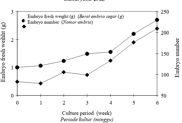
Figure 1. Fresh weight (●) and total number (µ) of tea somatic embryos per jar on a solid medium over a 6-week culture period.
Gambar 1. Berat segar (●) dan jumlah total (µ) somatik embrio teh per jar pada medium padat setelah 6 minggu dikulturkan
very small size. At week-6 there were more than 200 embryos covering almost entire medium surface in a jar from about 80embryos at the beginning (Figure 1). This multiplication rate however, is lower than the results by Tahardi et al. (2000) who obtained a 2.6-fold increase within 4 weeks on a semi-solid half-strength MS medium, and by Akula & Dodd (1998) who obtained 4 – 8-fold within 6 weeks after culturing primary globular-stage embryos on MS basal medium with half-strength macro-salts and devoid of growth regulators. This difference could be attributed to differences in culture and environmental conditions.
Embryo size
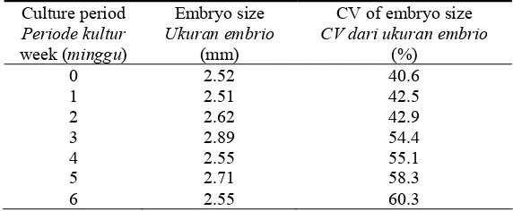
The average size of tea somatic embryos at the initial culture was 2.52 mm (Table 1). The embryo size had increased slowly up to week 3 (2.89 mm) but dropped at week 4 (2.55 mm). This decrease may be due to secondary embryogenesis as indicated by the formation of many small globular embryos on primary embryos. Although the total fresh weight of somatic embryos at week 6 was almost triple of the initial weight (Figure 1), the average size of somatic embryos over one culture passage did not change significantly (Table 1).
0 1 2 3
0 1 2 3 4 5 6
Culture period (week)
Periode kultur (minggu)
Embryo fresh weight (g) (Berat embrio segar (g)
Embryo number (Nomor embrio)
50 100 150 200 250
Emb
ry
o
fre
sh
weih
ht
(g
)
Embr
yo
nu
50 Morphological variations during the development of ….
Table 1. Tea somatic embryo size and its coefficient of variance (CV) over one passage of culture.
Tabel 1. Ukuran dan koefisien varian (CV) somatik embrio teh setelah satu kali disubkultur
Culture period Periode kultur week (minggu)
Embryo size Ukuran embrio
(mm)
CV of embryo size CV dari ukuran embrio
(%)
0 2.52 40.6
1 2.51 42.5
2 2.62 42.9
3 2.89 54.4
4 2.55 55.1
5 2.71 58.3
6 2.55 60.3
The variation in embryo size as indicated by its coefficient of variation (CV) (Table 1) reveals that the size of somatic embryos was already highly varied at the start of the culture. A CV value higher than 30% indicates high variability. CVs of embryo size have increased steadily from 40 to 60% as the culture progressed to the sixth week (Table 1). At the end of the culture passage, many embryos have reached cotyledonary or germinant stages with the size longer than 5 mm, while many more new embryos at globular or heart stages with the size less than 2 mm were also present. Similar results obtained by Akula & Dodd (1998) revealed that the older embryos which increased in size up to 3-5 mm diameter grew into complete plantlets, while most of the other embryos demonstrated the tendency of repetitive embryogenesis by budding off new globular embryos. Therefore, all stages of developing embryos with different sizes were present at any one time over one passage of tea somatic embryo culture (Figure 2). On the other hand,
Wachira & Ogada (1995) did not find secondary embryogenesis in their tea somatic embryo cultures.
Embryo color
51 Sumaryono et al..
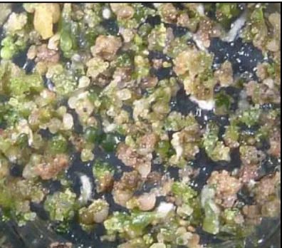
Figure 2. Somatic embryo culture of tea on a solid medium where different sizes, colors and developmental stages of embryos were found at the same time during the culture.
Gambar 2. Kultur somatik embrio teh pada medium padat dengan berbagai ukuran, warna dan fase perkembangan embrio yang ditemukan pada waktu yang sama selama dikulturkan.
Color differences among tea somatic embryos have not been mentioned by other research workers (Wachira & Ogada, 1995; Ponsamuel et al., 1996; Akula & Dodd, 1998; Akula et al., 2000), although Akula & Dodd (1998) reported that brownish trans-lucent cultures produced yellowish globular clumps of primary somatic embryos. Somatic embryo color has been used for quality evaluation of synthetic seeds of carrot (Sakamoto et al., 1992) and for assessing somatic embryo conversion potential of sweet potato (Padmanabhan et al., 1998).
Embryo developmental stage
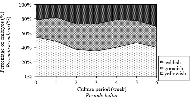
At the initial culture, 60% of the embryos were at globular stage, 30% at heart stage and
52 Morphological variations during the development of ….
Figure 3. Color changes of tea somatic embryo composition over one passage of culture.
Gambar 3. Perubahan warna dan komposisi pasangan somatik embrio teh yang dikulturkan.
Germinant-stage embryos were already found at the third week; however by the end of the experiment at week six there were no plantlets with complete leaves and roots (embryo conversion). Akula & Dodd 1998) reported that 20% of the globular embryos converted into plantlets whereas 60% of the embryos formed secondary embryos. The formation of new globular embryos during the cultures and the development of certain stage of em-bryos into more advanced developmental stages result in the presence of developing embryos of all stages at any time over the culture period (Figure 2). This (highly asyn-chronous culture of tea somatic embryos was derived either from nodal culture (Akula & Dodd, 1998) or seed culture (Akula et al., 2000). The heterogeneous characteristics of
tea culture on a solid medium can be reduced significantly by the use of a suspension culture (Tahardi et al., 1997) or a temporary liquid immersion system (Tahardi et al., 2000).
Embryo development
Single globular embryos with different colors were cultured separately on a solid regeneration medium to observe the changes in term of their colors and developmental stages over one passage of 8-week culture.
Color changes
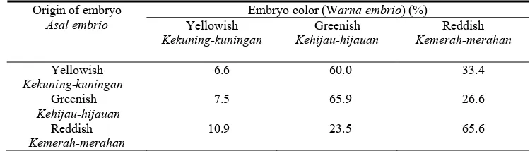
More than half of the originally yellowish embryos turned to greenish and one-third to reddish color after they had been cultured on a solid regeneration medium for 0%
20% 40% 60% 80% 100%
0 1 2 3 4 5 6
Culture period (week)
Periode kultur
( i )
reddish greenish yellowish
Percentage of
embryos (%)
Persentase embrio
53 Sumaryono et al.
Figure 4. Number of different stages of tea somatic embryos over one passage of culture.
Gambar 4. Jumlah dari berbagai fase somatik embrio teh setelah satu kali disubkultur.
eight weeks (Table 2). In contrast, two-thirds of the originally greenish and reddish embryos remained unchanged in color. About one-quarter of the greenish embryos turned reddish and vice versa. By the end of the experiment, most of the embryos were either greenish or reddish and only less than 10% were yellowish. These results show that the color of somatic embryos may change during the culture on a solid medium. Most of the new globular embryos were yellowish as mention-ed by Akula & Dodd (1998) but then turnmention-ed greenish or reddish under light. conditions. Changes in embryo color might be different if the cultures were placed in the dark.
Developmental stage changes
Single embryos with different colors were inoculated at the globular stage. These globular embryos developed gradually into more advanced developmental stages as the cultures progressed. One example of the pattern of developmental stage changes is taken from originally reddish embryos (Figure 5). The figure shows that the proportion of globular embryos reduced steadily while other stages started to appear. At the second week, the globular, heart and torpedo stages of embryos were already found. Cotyledonary stage of originally reddish embryos emerged at 0
10 20 30 40 50 60 70 80 90 100
0 1 2 3 4 5 6
Culture period (week)
Periode kultur
Number of
embr
yos
Nomor embrio
54 Morphological variations during the development of ….
Table 2. The distribution of tea somatic embryos with respect to color after 8 weeks of culture on a solid WP medium.
Tabel 2. Distribusi warna pada somatik embrio teh setelah 8 minggu dikulturkan pada medium padat WP
Embryo color (Warna embrio) (%) Origin of embryo
Asal embrio Yellowish
Kekuning-kuningan
Greenish
Kehijau-hijauan
Reddish
Kemerah-merahan
Yellowish
Kekuning-kuningan
6.6 60.0 33.4
Greenish
Kehijau-hijauan
7.5 65.9 26.6
Reddish
Kemerah-merahan
10.9 23.5 65.6
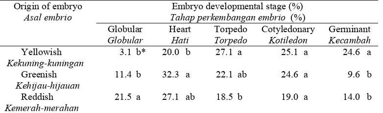
the third week, whereas germinant stage appeared at the fourth week. These results indicate that single somatic embryos are changed into later developmental stages gradually, not at the same time for all embryos. For example only about 10% of the embryos were at cotyledonary stage at the fourth week and increased to 20% by the end of experiment. Asynchronous development of these somatic embryos may be due to differences in embryo sources and culture conditions. Single globular somatic embryos used might not exactly the same in terms of size and developmental stage. In addition, only a part of embryo surface was connected to the surface of solid medium. However, even in suspension cultures where all surface of cells or embryos are exposed to the liquid medium, asynchronous development of somatic embryos of woody species is still very common (Tautorus & Dunstan, 1995). At the end of the experiment, almost three-quarters of the originally yellowish globular embryos were at the torpedo, cotyledonary or germinant stages (Table 3). Only 3% of the embryos
55 Sumaryono et al.
Figure 5. Developmental stage changes of originally reddish globular embryos over 8 weeks of culture on a regeneration solid medium.
Gambar 5. Perubahan fase perkembangan embrio yang berasal dari globular setelah 8 minggu dikulturkan dalam medium padat untuk regenerasi.
Table 3. The distribution of tea somatic embryo with respect to developmental stage after 8 weeks of culture on a solid WP medium.
Tabel 3. Distribusi somatik embrio tehe berdasarkan tingkat perkembangan setelah 8 minngu dikulturkan pada medium WP yang padat.
Embryo developmental stage (%) Tahap perkembangan embrio (%) Origin of embryo
Asal embrio
Globular Globular
Heart Hati
Torpedo Torpedo
Cotyledonary Kotiledon
Germinant Kecambah Yellowish
Kekuning-kuningan
3.1 b* 20.0 b 27.1 a 25.1 a 24.6 a
Greenish Kehijau-hijauan
11.4 b 32.3 a 22.1 ab 24.6 a 9.6 b
Reddish Kemerah-merahan
21.5 a 27.1 ab 18.5 b 19.0 a 14.0 b
*Means in the same column followed by the same letters are not significantly different at P=0.05 according to Duncan’s multiple range test.
* Angka pada kolom yang sama diikuti dengan huruf yang sama tidak berbeda nyata P=0,05 menurut uji jarak berganda Duncan.
0 20 40 60 80 100
0 1 2 3 4 5 6 7 8
Culture period (week)
Periode kultur
( )
56
Morphological variations during the development of ….
In developing synthetic seed production, the availability of somatic embryos at the developmentally mature stages (cotyledonary and germinant) is important as material sources. The experimental results show that higher numbers of originally yellowish embryos had been converted into later developmental stages than those of greenish and reddish embryos. It means that yellowish embryos are better sources of materials forsynthetic seeds. In addition, the germination rate of somatic embryo is also essential to obtain high conversion frequency of synthetic seeds to plants (Redenbaugh et al., 1987). The high variability of tea somatic embryos will restraint the development of mass clonal production and synthetic seed technology of tea. Therefore, attempts to synchronize somatic embryo development of tea have been conducted by using a suspension culture (Tahardi et al., 1997; 2000).
Conclusions
Tea somatic embryos on a solid medium was usually composed of different sizes, colors, and developmental stages at any time during the culture. The average size of tea somatic embryos did not change considerably over one passage of culture, however their size variability increased as the culture progressed. At the initial culture, 50% of the embryos were yellowish, 25% greenish and 25% reddish whereas at the sixth week of culture the proportion of yellowish embryos decreased slightly. The developmental stage of the embryos was changed over the course of culture. Globular embryos of originally yellowish color developed more rapidly into
cotyledonary or germinant stages than the greenish and reddish embryos, therefore more suitable as material sources for synthetic seed production
Acknowledgment
This research was supported by a grant from the Australian Centre for International Agricultural Research (ACIAR), Canberra, Australia.
References
Agarwal, B., U. Singh & M. Banerjee (1992). In vitro clonal propagation of tea (Camellia sinensis (L.) O. Kuntze). Plant Cell Tiss. & Org. Cult., 30, 1-6.
Akula, A., C. Akula & M. Bateson (2000). Betaine a novel candidate for rapid induction of somatic embryogenesis in tea (Camellia sinensis (L.) O. Kuntze). Plant Growth Reg., 30, 241-246.
Akula, A. & W.A. Dodd (1998). Direct somatic embryogenesis in a selected tea clone, ‘TRI-2025’ (Camellia sinensis (L.) O. Kuntze) from nodal explants. Plant Cell Rep. 17, 804-809.
Attree, S.M. & L.C. Fowke (1993). Embryogeny of gymnosperms: advances in synthetic seed technology of conifers. Plant Cell Tiss. & Organ Cult., 35, 1-35.
Cailloux, F., J. Julien-Guerrier, L. Linossier & A. Coudret (1996). Long-term somatic embryogenesis and maturation of somatic embryos in Hevea brasiliensis. Plant Sci.
57 Sumaryono et al.
Jha, T.B., Jha S. & S. Sen (1992). Somatic embryogenesis from immature cotyledons of an elite Darjeeling tea clone. Plant Sci., 84, 209-213.
Lloyd, G. & B. McCown (1981). Commercially feasible micropropagation of mountain laurel,
Kalmia latifolia, by use of shoot-tip culture.
Comb. Proc. Intl. Plant Prop. Soc., 30, 421-427.
Nakamura, Y. (1991). In vitro propagation techniques of tea plants. Japan Agric. Res. Quart., 25(3), 185-194.
Onishi, N., Y. Sakamoto & T. Hirosawa (1994) Synthetic seeds as an application of mass production of somatic embryos. Plant Cell Tiss. & Org. Cult., 39, 137-145.
Padmanabhan, K., D.J. Cantliffe, R.C. Harrell & J. Harrison (1998). Computer vision analysis of somatic embryos of sweet potato [Ipomoea batatas (L.) Lam.] for assessing their ability to convert to plants. Plant Cell Rep., 17, 681-684.
Ponsamuel, J., N.P. Samson, P.S. Ganeshan, V. Sathyaprakash & G.C. Abraham (1996). Somatic embryogenesis and regeneration from the immature cotyledonary tissues of cultivated tea (Camellia sinensis (L.) O. Kuntze). Plant Cell Rep.,16, 210-214.
Redenbaugh, K., D. Slade, P. Viss & J.A. Fujii (1987). Encapsulation of somatic embryos in synthetic seed coats. HortSci., 22(5), 803-809.
Sakamoto, Y., T. Mashiko, A. Suzuki & H. Kawata (1992). Development of encapsulation
technology for synthetic seeds. Acta Hort.,
319, 71-76.
Steel, R.G.D. & J.H. Torrie (1980). Principles and Procedures of Statistics: A Biometrical Approach, 2nd ed. New York, McGraw-Hill Inc. 633p.
Tahardi, J.S. (1994). Micropropagation of tea through shoot proliferation from excised axillary buds. Menara Perkebunan, 62(2), 20-24
Tahardi, J.S., Sumaryono & N. Mardiana (1997). Initiation and maintenance of embryogenic suspension culture of tea (Camellia sinensis
L.). Menara Perkebunan,65(1), 1-8.
Tahardi, J.S., T. Raisawati, I. Riyadi & W.A. Dodd (2000). Direct somatic embryogenesis and plant regeneration in tea by temporary liquid immersion. Menara Perkebunan,68(1), 1-9.
Tautorus, T.E. & D.I. Dunstan (1995). Scale-up of embryogenic plant suspension cultures in bioreactors. In S. Jain, P. Gupta & R. Newton (eds.) Somatic
Embryogenesis in Woody Plants, Vol. 1, p. 265-292.
Vieitez, A.M. (1995). Somatic embryogenesis in
Camellia spp. In S. Jain, P. Gupta & R. Newton (eds.) Somatic Embryogenesis in Woody Plants, Vol. 2, p. 235-276.
Vieitez, A.M. & J. Barciela (1990). Somatic embryogenesis and plant regeneration from embryonic tissue of Camellia japonica L.
Plant Cell Tiss. & Org. Cult., 21, 267-274.
Wachira, F. & J. Ogada (1995). In vitro
regeneration of Camellia sinensis (L.) O. Kuntze by somatic embryogenesis. Plant Cell Rep. ,14, 463-466.