Indo. J. Chem., 2009, 9 (3), 466 - 469 466
ANTI DIABETIC FLAVANONE COMPOUND FROM THE LEAVES OF
Artocarpus communis
Puspa D.N. Lotulung*, Sofa Fajriah, Andini Sundowo, and Euis Filaila
Research Center for Chemistry, Indonesian Institute of Sciences, Kawasan PUSPIPTEK, Serpong, Tangerang, Banten
Received June 10, 2009; Accepted November 1, 2009
ABSTRACT
The Flavanone compound with anti diabetic activity was isolated from ethyl acetate extract of Artocarpus communis leaves using column chromatography techniques. The structure of the flavanone compound was elucidated on the basic of spectroscopic evidence and comparison to published values. This compound, 8-geranyl-4,5,7-trihydroxyflavone, showed strong anti diabetic activity on α-glucosidase inhibition assay with IC50
18.120 µg mL-1.
Keywords: Artocarpus communis, 8-geranyl-4,5,7-trihydroxyflavone, anti diabetic activity
INTRODUCTION
Diabetes mellitus is the most serious chronic metabolic disorder and is characterized by high blood glucose levels [1-2]. At the present time, it is estimated that 150 million people worldwide have diabetes and that this will increase to 220 million by 2010 and 300 million by 2025. Globally, the percentage of type 2 diabetes (non insulin-dependent diabetes mellitus) is greater than 90% [3].
α-Glucosidase inhibitor has been used to treat type 2 diabetes mellitus. This drug does not increase insulin secretion. The antihyperlipidemic activity of α -glucosidase inhibitor comes from reversible inhibition on hydrolase, α-amilase pancreatic enzymes and intestine digestive enzymes, such as isomaltase, sucrase and maltase. These enzymes hydrolyze food carbohydrates
to glucose and other monosaccharide. The α
-glucosidase inhibitor inhibits the glucose absorption in the intestine acting as antihyperglicemia after carbohydrate intake [4-5].
As a number of anti-hyperglycemic agents have been found in plants, research into understanding the scientific basis for plant-based traditional medicine from various cultures has increased as scientists search for clues to discovering new therapeutic drugs for type 2 diabetes mellitus [6-8]. Traditional Indian medicines have long used plant and herbal extracts as anti-diabetic agent [9]. These plants are typically rich in phenolic compounds, which are known to interact with proteins and can inhibit enzymatic activity [10-11]. A number of medicinal plant and herbal extracts have been found to inhibit the enzymatic activity of α-glucosidase and α -amylase, and therein may have potential as dietary anti-diabetic agents to improve the control of postprandial hyperglycemia [12-15].
* Corresponding author.
Email address : [email protected]
The genus Artocarpus (Moraceae), an
exceptionally rich source of prenylated flavonoids,
consists of approximately 50 species that are indigenous to the region of South East Asia, including Indonesia. Different compounds isolated from some species of Artocarpus have been shown to exhibit interesting biological properties [16-17]. Some of these compounds show interesting biological activities, such as cytotoxic [17], antimalarial activity [18], inhibition of tyrosinase and melanin biosynthesis [19-21]. Thus, in a continuation of our studies on the chemistry of Indonesian plants, the chemical constituents of A. communis have been investigated. In this paper, we report the isolation, structure elucidation and biological evaluation of prenylated flavonoid from ethyl acetate extract of the leaves of this species. The structure of this compound was elucidated on the basis of spectroscopic data including 2-D NMR. The isolated compound exhibited α-glucosidase activity using in vitro assay.
EXPERIMENTAL SECTION
Material
Sample of the leaves of Artocarpus communis
was collected in March 2006, from plantation trees growing in Parung, Bogor, Indonesia. The plant was identified by staff at Biology Laboratory, Institute of Technology Bandung, West Java, Indonesia, and a voucher specimen has been deposited at Biology Laboratory.
Instruments
1
H- and 13C-NMR spectra were recorded with JEOL JNM ECA-500 spectrometer, operating at 500 MHz (1H-) and 125.76 MHz (13C-), using TMS (Tetra Methyl Silane) as an internal standard. MS were obtained with Mariner Biospectrometry spectrometer
Indo. J. Chem., 2009, 9 (3), 466 - 469 467
Puspa D.N. Lotulung et al.
Figure 1. The HMBC correlation of compound AC-3-3
using ESI System (Electrospray Ionization) and positive ion mode. Column chromatography was carried out using Merck Silica gel 60 (70-230 mesh ASTM), and TLC (Thin Layer Chromatography) analysis on precoated Silica gel plates (Merck Kieselgel 60 F 254, 0.25 mm).
Procedure
Extraction, Isolation and Identification
The dried leaves (4.95 kg) of A. communis were extracted exhaustively using macerator with ethanol 70%. The ethanol extracts (250 g) were concentrated using vacuum rotary evaporator and then partitioned with hexane-water (1:4). Water extracts added with dichloromethane and then the residue was added with ethyl acetate. Ethyl acetate extract was fractionated by column chromatography on silica gel using gradient elution (hexane-ethyl acetate (8:2)), were resulted 70 fractions. Fractions 16-18, compound AC-3-3, were re-crystallized to give white crystal.
Compound AC-3-3 was identified using LC-MS and
NMR (1H-NMR, 13C-NMR, DEPT 135, HMQC and
HMBC) Spectrometer, to give flavanone compound, 8-geranyl-4,5,7-trihydroxyflavone.
Inhibition assay for α-glucosidase activity
α-Glucosidase inhibitory activity evaluation of the extracts was performed using established procedure [22]. α-Glucosidase enzyme solution was dissolved in phosphate-buffer solution (pH 7) containing 200 mg albumin serum. Before its application, 1 mL of the enzyme solution was diluted 25 times with the buffer solution. The reaction mixture consisting of 250 µL of
20 mM p-nitrophenyl α-D-glucopyranose as the
substrate, 490 µL of 100 mM phosphate buffer (pH 7)
and 10 µL of the extract dissolved in DMSO was prepared. The reaction mixture then was water-bath incubated at 37 °C for 5 min. The enzyme solution (250 µL) was added, and keeps the solution incubated for 15 min. The enzyme reaction was stopped by addition of 1000 µL, 200 mM sodium carbonate solution. The resulted p-nitrophenol from the reaction was measured at λ 400 nm. As positive control, the reaction of 1% of quercetin solution was measured. The commercial of α -glucosidase anti-diabetic drug, glucobay was available in the laboratory only in a form of sustain release tablet, therefore, quercetin is selected for positive control for in vitro evaluation. Samples concentrations for activity evaluation were 3.125, 6.25, 12.5, and 25 µg mL-1, and 6.25, 12.5, and 25 µg mL-1 for quercetin.
RESULT AND DISCUSSION
The ethyl acetate extract from the leaves of A.
communis was fractionated by column
chromatography. A fraction containing the major components was purified by re crystallization to give 8-geranyl-4,5,7-trihydroxyflavone (AC-3-3).
AC-3-3, a colorless crystal, has a molecular formula of C25H28O5 from LC-MS spectrum with [M]+ ion at 409.0832.
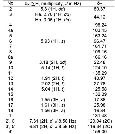
The analysis of its NMR data, including HMQC and HMBC spectra, allowed for an unambiguous assignment of all proton and carbon signals. The 1 H-NMR data indicated the presence of three methyl groups at δ 1.55 (3H, s), 1.56 (3H, s), 1.61 (3H, s), three pairs of methylene protons at δ 1.91 (2H, t), 2.02 (2H, t), 3.18 (2H, dd), and two vinyl protons at δ 5.04 (1H, t) and 5.14 (1H, t) attributed to a geranyl group. In addition, proton signals suggested the existence of two aromatic rings, ring A and B. Ring A have proton aromatics symmetrically, at 6.81 (d, 8.56 Hz, 2 H) and 7.31 (d, 8.56 Hz, 2 H). At ring B, it has a sharp singlet at δ 5.93 indicated a chelated hydroxyl group. The 13 C-NMR spectrum indicated 25 carbons, including three methyl groups, a carbonyl group (δ 198.24), 8 sp2 methine carbons, 4 methylene carbons, 3 methyl groups, and 9 quartenery carbons. These signals from 1
H- and 13C-NMR suggested that this compound
contained a geranyl substituent and flavanone group. The presences of the functional groups above were suggested by the long range coupling HMBC experiment in Fig. 1. The multiplicity of carbons were assigned by the DEPT-135 experiment and correlation of the chemical H and C shift for all protonated carbons was determined based on the HMQC spectrum as summarized in Table 1.
Indo. J. Chem., 2009, 9 (3), 466 - 469
Puspa D.N. Lotulung et al.
468
Table 1. 1H- and 13C-NMR spectral data of compound
AC-3-3*
Figure 2. The molecular structure of compound AC-3-3
Table 2. The α-glucosidase inhibitory activity of
compound AC-3-3 from ethyl acetate extract of A. communis leaves compared with quercetin
* Spectra recorded at 500 MHz for 1H spectrum and 125 MHz for 13C
spectrum in CD3OD. The values are in ppm and J values (Hz) in
parentheses. Abbreviations for NMR signal are as follows: s= singlet, d= doublet, and t= triplet. Correlation of chemical shift H and C were assigned, based on the HMQC spectra.
* % Inhibition: [(Concentration-absorb value)/Concentration] x 100 ** IC50: Concentration of inhibitor to inhibit 50% of its activity.
8-geranyl-4,5,7-trihydroxyflavone ((E)-8-(3,7-
dimethylocta-2,6-dienyl)-5,7-dihydroxy-2-(4-hydroxyphenyl)chroman-4-one) (Fig. 2).
The antidiabetic of this compound was evaluated according to the method previously described [23]. This compound exhibited significant activity in the α -glucosidase inhibitor with IC50 18.120 µg mL-1. This value has higher activity than quercetin as positive control (Table 2).
CONCLUSION
Based on LC-MS, 1H-NMR and 13C-NMR (1D & 2D) spectra, and compared with previous spectral data, compound AC-3-3 from ethyl acetate extract of A. communis leaves was identified as flavanone group, 8-geranyl-4,5,7-trihydroxyflavone ((E)-8-(3,7-dimethylocta- 2,6-dienyl)-5,7-dihydroxy-2-(4-hydroxyphenyl)chroman-4-one). This compound showed significant activity in the
α-glucosidase inhibitor with IC50 18.120 µg mL-1.
ACKNOWLEDGEMENT
This work has been supported by the Indonesian Government budget for the Research Center for
Chemistry, Indonesian Institute of Sciences (LIPI). We also thanks to Dr. Lenny Sutedja for suggesting this research; the staff of Biology Laboratory, Institute of Technology Bandung for identification of the plant specimen.
REFERENCES
1. Corry, D.B and Tuck M.L., 2000, Curr. Hypertens. Rep., 2,154-9.
2. Sratton, I.M., Adler, A.I., Neil, H.A., Matthews, D.R., Manley, S.E., Cull, C.A., 2000, Br. Med. J., 321,
5. Bayer., 2004, Precose (Acarbose Tablets). http://www.drugs.com/PDR/Precose/Tablets.html 6. Jarvill-Taylor, K.J., Anderson, R.A., and Graves,
Indo. J. Chem., 2009, 9 (3), 466 - 469 469
Puspa D.N. Lotulung et al.
2002, J. Ethnopharmacol., 81, 317-320.
8. Elder C., 2003, Altern. Ther. Health Med., 10, 1, 44-50.
9. Grover, J.K., Yadav, S., and Vats, V., 2002, J. Ethnopharm., 81,81-100.
10. Dawra, R.K., Makkar, H.P., and Singh, B., 1988,
Anal. Biochem., 170, 1, 50-53.
11. Suryanarayana, P., Kumar, P.A., Saraswat, M., Petrash, J.M., and Reddy, G.B., 2004, Mol. Vision,
10, 148-154.
12. Hanefeld, M. and Temelkova-Kurktschiev, T., 2002,
Nutr. Metab. Cardiovasc. Dis.,12, 98-107.
13. Vats V., Grover J.K., and Rathi S.S., 2002, J. Ethnopharm., 79, 95-100.
14. McCue, P. and Shetty, K., 2004, J. Clin. Nutr., 13, 1, 101-106
15. McCue, P., Vatten, D., and Shetty K., 2004, J. Clin. Nutr., 13, 4,401-408.
16. Lemmens, R.H.M.J., Soerianegara, I., and Wong, W.C., editors, 1995, Plant resources of South-East Asia No. 5 (2), timber trees: minor commercial
timbers. Indonesia: Prosea Bogor. ISBN Number: 979-8316-18-5
17. Nomura, T., Hano, Y., and Aida, M., 1998,
Heterocycles., 47, 1179-1205.
18. Boonlaksiri, C., Oonanant, W., Kongsaeree, P., Kittakoop, P., Tanticharoen, M., and Thebtaranonth, Y., 2000, Phytochem., 54, 415-417.
19. Likhitwitayawuid, K. and Sritularak, B., 2001, J. Nat. Prod., 64, 11, 1457-1459.
20. Shimizu, K., Fukuda, M., Kondo, R., and Sakai, K., 2000, Planta Med., 66, 1, 16-19.
21. Shimizu, K., Kondo, R., Sakai, K., Lee, S.H., and Sato, H., 1998, Planta Med., 64, 408-412.
22. Lee, D.S. and S.H. Lee., 2001, FEBS Lett., 501, 84-86.
23. Koshihara, Y., Fujimoto, Y., and Inoue, H., 1988,
Biochem. Pharmacol., 37, 2161-2165.
24. McLean, S., Reynolds, W.F., Tinto, W.F., Chan,
W.R., and Shepherd, V., 1996, Magn. Reson.