File Comparison Report
Produced by WCopyfind.4.1.4 with These Settings:
Shortest Phrase to Match: 3 Fewest Matches to Report: 2 Ignore Punctuation: No Ignore Outer Punctuation: No Ignore Numbers: No
Ignore Letter Case: No Skip Non-Words: No Skip Long Words: No
Most Imperfections to Allow: 4 Minimum % of Matching Words: 85
Perfect
Match OverallMatch
View Both
Files File L File R
69 (3% L,
3% R) 76 (3%) L;73 (3%) R Side-by-Side 02.Differences_in_Mean.docx 01.Analyses_of_Nutrients.docx 107 (6%
L, 5% R) 109 (6%) L;112 (5%) R Side-by-Side 03.Differences_in_Malondial.docx 01.Analyses_of_Nutrients.docx 89 (5% L,
4% R) 94 (5%) L;95 (4%) R Side-by-Side 03.Differences_in_Malondial.docx 02.Differences_in_Mean.docx
64 (4% L,
3% R) 68 (4%) L;64 (3%) R Side-by-Side 04.Differences_in_brain.docx 01.Analyses_of_Nutrients.docx
48 (3% L,
2% R) 51 (3%) L;53 (2%) R Side-by-Side 04.Differences_in_brain.docx 02.Differences_in_Mean.docx 77 (5% L,
4% R) 84 (5%) L;78 (4%) R Side-by-Side 04.Differences_in_brain.docx 03.Differences_in_Malondial.docx
Research Article
Differences in Mean Levels of Maternal Resistin Serum between Early Onset Preeclampsia (EOPE) and Late Onset Preeclampsia (LOPE)
Research Article
Differences in Mean Levels of Maternal Resistin Serum between Early Onset Preeclampsia (EOPE) and Late Onset Preeclampsia (LOPE)
INTRODUCTION
Preeclampsia is one of the major causes of morbidity and mortality of the mother and fetus. World Health
Organization (WHO) reported the number of deaths caused by preeclampsia by 16% in developing countries
. Preeclampsia
resulting 3-25 fold increased risk obstetric complications and is the cause of 30-40% of perinatal deaths in Indonesia 1
.
The incidence of preeclampsia ranges between 5-10% of all pregnancies
. Incidence of preeclampsia in the United
States, Canada and Western Europe ranges between 2-5% of all pregnancies and higher, 4-18% in some developing countries in Africa
1
. The incidence of preeclampsia in Indonesia ranged between 3-10% 3
.
In Dr.M. Djamil General Hospital in Padang during the 2
year 2011, the incidence of preeclampsia was 8.31%, on 2012 was 11.47% and on 2013 was 12.02%.
Preeclampsia is divided into early onset preeclampsia (EOPE) (<34 weeks) and late onset preeclampsia (LOPE) (>34 weeks) of pregnancy based on the onset of clinical manifestations of preeclampsia. The EOPE and LOPE has a different pathogenesis. The PEAD is often associated with impaired uteroplacental perfusion caused by disruption of trophoblast invasion, while LOPE is often associated with the presence of extrinsic and maternal factors
.
Preeclampsia is a protean syndrome, in which multiple 4,5
organ systems can be affected compared to the others. Preeclampsia is mainly characterized by hypertension and proteinuria or may be associated with abnormalities in laboratory test results that renal function, hepatic or hemostasis after 20 weeks of pregnancy
.
Analysis of risk factor for preeclampsia is needed to 1,6
maternal risk factor that associated with LOPE .
Resistin is an adipose tissue-specific secretory factor 7
(ADSF), a hormone secreted by adipose tissue that induces insulin resistance in muscle and liver. Resistin stimulated by inflammatory conditions that produce proinflammatory cytokines
.
Preeclampsia is associated with inflammation and insulin 7,8
resistance which is affected by resistin. Resistin is associated with late onset preeclampsia because it was a maternal factors, so there was an increasing of maternal resistin serum levels in late onset preeclampsia
.
Previous study reported an elevated of maternal resistin 7,8
serum levels associated with a systemic inflammatory response and insulin resistance which is both of them are 2
2
increased in preeclampsia compared to normal pregnancy .
The other study showed the mean of Homeostasis Model
Assessment-Insulin Resistance (HOMA-IR) and high sensitivity C-reactive protein (hs-CRP) was higher in late onset
preeclampsia than early onset preeclampsia group. From
various studies on the above it could be estimated that the inflammatory factor and insulin resistance are associated with higher levels of resistin more dominant in late onset preeclampsia group compared to early onset preeclampsia group.
The HOMA-IR could be used as an indicator of insulin resistance, but not for inflammatory states, contrary hs-CRP could be used as an indicator of inflammation, but not for insulin resistance. Resistin is expected to describe both of them, because it was associated with insulin resistance and inflammation
7 .
MATERIALS AND METHODS
This study is an analytical cross sectional study with
2 0 w o me n o f e a r l y o n s e t p r e e c l a mp s i a ( E OP E ) a n d 2 0 w o me n of late onset preeclampsia (LOPE) who met the inclusion
criteria and there were no exclusion criteria. The samples were recruited in Dr.M. Djamil General Hospital, Padang from July-October, 2015.
sugar as >200 mg dLG
), had suffered coronary heart disease,
chronic kidney disease, chronic liver disease and obesity (BMI before pregnancy >25 kg mG
1 ).
Maternal resistin serum was examined by enzyme-linked 2
immunosorbent assay (ELISA) method in Biomedic Laboratory in Medical Faculty of Andalas University. Statistical analysis was conducted by using SPSS program 20th version. The data distribution was normal with p>0.05. The differences mean levels of maternal resistin serum was analyzed by using independent t-test.
RESULTS
Characteristics of research subjects: Forty patients of research subjects consisted 20 patients of early onset preeclampsia (EOPE) and 20 patients of late onset
preeclampsia (LOPE). Mean levels of maternal age in EOPE group is 34.4±5.144 years, whereas in LOPE group is
32.9±6.324 years with p = 0.416. According to maternal age 9-12
Table 1: Characteristics of research subjects between early onset preeclampsia and late onset preeclampsia Characteristics EOPE (n = 20) LOPE (n = 20) Total (%) p
Maternal age group
<20 years 0 (0%) 0 (0%) 0
20-35 years 8 (40%) 10 (50%) 45 0.537 >35 years 12 (60%) 10 (50%) 55
Maternal age (0±SD) years 34.4±5.144 31.9±6.314 0.416 Gravidity group
Primigravid 6 (30%) 7 (35%) 32.5 0.744 Miltigravid 14 (70%) 13 (65%) 67.5
Gravidity (0±SD) 2.4±1.314 2.65±1.461 0.573 BMI group
Underweight 0 (0%) 0 (0%) 0
Normoweight 14 (70%) 12 (60%) 65 0.520 Overweight 6 (30%) 8 (40%) 35
BMI (0±SD) 22.55±1.595 22.59±1.812 0.946
Table 2: Mean levels of maternal resistin serum in early onset preeclampsia and late onset preeclampsia
EOPE LOPE
--- ---Variable Mean SD Mean SD
Maternal resistin serum (ng mLG 1
) 2.526 1.603 8.891 6.219
Table 3: Mean levels difference of maternal resistin serum in early onset preeclampsia and late onset preeclampsia
Variable Mean difference CI (95%) p Maternal resistin serum (ng mLG 1
) 6.365 3.3835-9.3475 0.000
20-35 years old, 12 patients (60%) in group of maternal age >35 years old, none in group of maternal age <20 years old. Research subjects of late onset preeclampsia consists of 1 0 patients (50%) in group of maternal age 20-35 years old, 10 patients (50%) in group of maternal age >35 years old, none in group of maternal age <20 years old.
Mean levels of gravidity in early onset preeclampsia (EOPE) group is 2.4±1.314, whereas in late onset
preeclampsia (LOPE) group is 2.65±1.461 with p = 0.573. According to gravidity groups, study of subjects in early onset preeclampsia consists 6 patients (30%) in group of primigravida, 14 patients (70%) in group of multigravida. Research subjects of l ate onset preecl ampsi a c onsi sts of 7 patients (35%) in group of primigravida, 13 patients (65%). Mean levels of Body Mass Index (BMI) in early onset
preeclampsia (EOPE) group is 22.551±1.595, whereas in late onset preeclampsia (LOPE) group is 22.588±1.4812 kg mG with p = 0.946. According to BMI groups, study of subjects in EOPE consists 14 patients (70%) in group of normal
weight, 6 patients (30%) in group of overweight, none in group BMI underweight. Research subjects of late onset preeclampsia consists 12 patients (60%) in group of normal weight, 8 patients (40%) in group of overweight, none in group BMI underweight.
2 3
There were no statistically significant differences
regarding maternal age, group of maternal age, gravidity, group of gravidity, BMI and group of BMI characteristic between those two groups with p>0.05 showed in Table 1. Mean levels of maternal resistin serum in early onset preeclampsia and late onset preeclampsia: Mean levels of maternal resistin serum was higher in late onset preeclampsia vs
2.526±1.603 ng dLG
than early onset preeclampsia (8.891±6.219 ng mLG 1
), showed in Table 2.
Differences in mean levels of maternal resistin serum between late onset preeclampsia and early onset preeclampsia: The data distribution was normal with Kolmogorov Smirnov test. Analysis was performed with independent t-test. Table 3 showed the differences in mean levels of maternal resistin serum between early onset preeclampsia and late onset preeclampsia as 6.365 ng mLG with Confidence Interval (CI) 95% 3.384-9.347. There is a high significant differences with p<0.001.
DISCUSSION
The mean levels of maternal serum resistin in early
onset preeclampsia (EOPE) group was 2.526±1.603 ng mLG whereas in late onset preeclampsia (LOPE) group was 8.891±6.219 ng mLG
1 1
, 95% CI 3.384-9.347. Statistical test showed
p<0.001, that the mean levels of maternal serum resistin significantly was higher in LOPE than EOPE (Table 3). Resistin is a hormone secreted from adipose tissue that induces insulin resistance in muscle and liver. Resistin stimulated by inflammatory conditions that produce proinflammatory cytokines. Resistin contribute to the inflammatory disorders such as coronary heart disease, 1
1 1
chronic kidney disease and liver cirrhosis as well as insulin resistance in diabetes mellitus through the activation of proinflammatory cytokines varied which depending on the organs affected
.
Insulin resistance and inflammation were the condition 7,8
associated to preeclampsia. This factor related to the role of resistin through the release of proinflammatory cytokines. The relationship between resistin and preeclampsia is connected by those which is a maternal factor in
preeclampsia. Therefore, resistin is associated to late onset preeclampsia, so the levels of maternal resistin serum was increased in late onset preeclampsia
.
Inflammation and insulin resistance have been 7,8
investigated, that the comparasion between HOMA-IR and hs-CRP in EOPE and LOPE. This study reported the results that mean levels of HOMA-IR and hs-CRP were higher in LOPE compared to EOPE (HOMA IR: 4.86±5.50 vs 3.99±5.97 and hsCRP: 123.08±38.67 vs 26.54±34.7 mg LG
). This study
suggested that the inflammatory factor and insulin resistance were more dominant in LOPE compared to EOPE.
1
A s i mi l ar r es ul t s w as r epor t ed i n whi c h t he mean l evel s of maternal resistin serum was higher in preeclampsia
compared to normal pregnancy related to an increase in insulin resistance and the response systemic inflammation that happened in preeclampsia particularly late onset preeclampsia
.
A cross sectional study in which two groups of pregnant
9-12
women with preeclampsia (n = 15) and normal pregnancy (n = 23) demonstrated th mean levels of maternal resistin serum was higher in preeclampsia (5.68±0.41 ng mLG )
p = 0.028. The mean levels of maternal resistin serum related to the mean of HOMA-IR and proinflammatory cytokines that were elevated in preeclampsia compared to normal
pregnancy. T he mean of H OMA-I R ( 2. 5±0. 8 vs 1. 4±0. 1), IL-6 (6.34±1.02 vs 2.80±0.31) and TNF-" (1.89±0.18 vs 1.23±0.10)
.
Another cross-sectional study on two groups of 9
pregnant women, which were preeclampsia (n = 29) and normal pregnancy (n = 30), found that mean levels of maternal resistin serum was higher in preeclampsia 61.98±32.26 ng dLG
, compared to normal pregnancy 38.06±31.26 ng dLG
1 1
, p = 0.013. Insulin resistance is thought
related the increasing the levels of maternal resistin serum, because the mean of HOMA-IR was higher in preeclampsia compared to normal pregnancy (4:44±4:02 vs 3.99±2.82) .
A cross sectional study was conducted on two groups of pregnant women, which are preeclampsia (n = 50) and normal pregnancy (n = 50). The median levels of maternal resistin 1
1 10 4
serum were higher in preeclampsia 61 ng mLG compared to
normal pregnancy 25.5 ng mLG 1
1
, p = 0.033. Insulin resistance
is thought related the increasing the levels of maternal
resistin serum, because the median of HOMA-IR was higher in preeclampsia compared to normal pregnancy (4.7 vs 3.6) .
A cross sectional study was conducted on two groups of pregnant women, which are preeclampsia (n = 16) and normal pregnancy (n = 22). The mean levels of maternal resistin serum was higher in preeclampsia (12.06±0.973 ng mLG )
compared t o n ormal p regnancy ( 7. 35±1. 195 ng mLG ),
p = 0.041. Inflammatory factors is thought related the increasing the levels of maternal resistin serum in preeclampsia. There was an increasing mean levels of TNF" in preeclampsia compared to normal pregnancy (15.23±0.674 vs 12.84±0.348 ng mLG
1
that description, there was a correlation between previous studies and this study
9-12 12 .
The mean levels of HOMA-IR and hs-CRP were higher in late onset preeclampsia than early onset preeclampsia, so according to those studies above, the insulin resistance and inflammation that were higher in late onset preeclampsia than early onset preeclampsia related to the levels of maternal resistin serum that was higher in late onset preeclampsia than early onset preeclampsia. It was appropriate with the results of this research that the mean levels of maternal resistin serum in late onset preeclampsia was significantly higher than early onset preeclampsia with p<0.001.
CONCLUSION
The mean levels of maternal resistin serum was higher significantly in late onset preeclampsia (LOPE) compared to early onset preeclampsia (EOPE).
Analyses of Nutrients and Body Mass Index as Risk Factor for Preeclampsia Introduction
Preeclampsia is an important problem in obstetrics because it is still a major cause of maternal mortality compared to bleeding and infection. Preeclampsia leads to maternal and perinatal morbidity. Preeclampsia is
also associated with high rates of preterm delivery, small for gestational ages, and perinatal death [1]. Little is known about the patho- genesis of preeclampsia. Many factors are identified as risk factors for preeclampsia including parity, multiple preg- nancies, age, family history of preeclampsia, obesity, his- tory of systemic disease, and nutrition.
Since preeclampsia is characterized by reduced perfu- sion of the placenta, oxidative stress, and endothelial dys-function, nutrition has long been hypothesized to have a role in the etiology of preeclampsia [2]. Oxidative stresses are proposed as the linkage between the two stages of preeclampsia. Nutrients can affect oxidative stress by increasing or decreasing free radicals or antioxidants or by providing substrate for the formation of free radicals. Several nutrients, particularly omega-3 (n - 3) fatty acids, antioxidants, and folic acid, have an important roles in modulating endothelial function. It has also been suggested that nutrients such as trace elements, fatty acids, and folic acid can contribute to insulin resistance, a risk factor for preeclampsia. In many studies, decrease in serum magne- sium levels has been considered as the cause of patho- genesis of
preeclampsia. Minerals have an important
influence on the health of pregnant women and growing fetus. Among them, serum or placental zinc (Zn) concen- trations have been reported to be low in PE women. Fur- thermore, decreased levels of zinc, selenium, and copper have been observed in patients with preeclampsia [3]. In another side, the nutrients with antioxidants among high risk women showed a protective effect [4]. Folic acid has been hypothesized as a protective agent of preeclampsia.
Maternal obesity and insulin resistance are also believed to be important risk factors for the development of pla-cental endothelial dysfunction and preeclampsia. Preven- tion of preeclampsia has remained elusive, owing largely to their complex nature. Currently, maternal obesity in prepregnancy is one of the strongest modifiable risk fac- tors. Recent studies have shown a relation between obesity in prepregnancy and the risk of
preeclampsia. The reason for obesity being associated with an increased risk of preeclampsia was explained by increased levels of serum triglycerides and very low-density lipoprotein particles in obese women. This lipid alterations have been suggested to promote oxidative stress caused by ischemia–reperfusion mechanism or activated neutrophils, which leads to endothelial cell dysfunction [1].
The hypothesis about nutritional status and body mass index (BMI) prepregnancy associated with preeclampsia has intrigued us to study the risk factor for preeclampsia in Dr. M. Djamil Hospital, Padang, Indonesia.
Methods
This was a case–control study at the Department of Obstetric and Gynecology in Dr. M. Djamil Hospital, Padang, Indonesia, between January and December 2013. Pregnant women after 20 weeks gestations were included. A total of 140 patients were enrolled in this study with 70 cases and 70 controls. Cases were those diagnosed with preeclampsia; meanwhile, controls were normotensive pregnant women without any other comorbidity.
After providing written informed consent, all subjects completed an interview for their nutritional status and prepregnancy BMI after delivery. The nutritional status was assessed by Food Frequency Questionnaire (FFQ) and then analyzed by Nutrisurvey Program. Calories, protein, fat, carbohydrates, calcium, phosphorus, zinc, sodium, potassium, magnesium, vitamin A, folic acid, vitamin B1, vitamin B2, niacin, vitamin B6, vitamin B12, vitamin C, and vitamin E were assessed. Maternal BMI was catego- rized into two groups: normal BMI and abnormal BMI. Normal BMI was defined as 18.5–24.9 kg/m2. The inde- pendent samples t test was used for nutritional status, and
Chi-square test was used for BMI. Odds ratio (OR) with 95% CI was calculated. A p value \0.05 was considered
statistically significant. For the nutrition variable, if the p value \0.25 then continued by logistic regression back-ward to assess the risk factor.
Results
Most of the subjects had normal weight, both in case and control groups. Table 1 shows that prevalence of abnormal BMI was more common in the preeclampsia group com- pared with those without preeclampsia 19 (27.1%) versus
As shown in Table 2, the mean level for most of the
variables, except calories, fat, and vitamin B1, was lower in subjects with preeclampsia than those without preeclamp- sia. The difference for most of the subjects was also sta- tistically significant with p \ 0.05, except for vitamin B1 and vitamin B2. Table 3 shows the risk factors for preeclampsia obtained by logistic regression analyses. The nutrients that were significantly associated with increased risk of preeclampsia were deficiency of vitamin E, zinc, fat, calcium, and vitamin C. Excess of calories and carbohy- drate also significantly associated with increased risk of preeclampsia. Meanwhile, vitamin A and vitamin B1 were protective factors.
Discussion
The reason for obesity being associated with an increased risk of preeclampsia was explained by increased levels of serum triglycerides, very low-density lipoproteins, and formation of small low-density lipoprotein particles in obese women. This lipid profile was also found in women with preeclampsia. These lipid alterations have been suggested to promote oxidative stress, caused by ische- mia–reperfusion mechanism or activated neutrophils, which leads to endothelial cell dysfunction [1]. Moreover, dyslipidemia also can cause atherothrombosis and induce the aggregation of the thrombocytes than can lead to coagulopathy which is a characteristic of
preeclampsia.
Obesity is accompanied by oxidative stress. The origin of oxidative stress is proposed to be secondary to increased free fatty acids and inflammation. It is also suggested that diet can contribute to oxidative stress. Obese individuals have lower blood concentrations of antioxidants. This could be due to reduced dietary intake of antioxidants, but increased consumption by reactive oxygen species is also possible [5].
In this study, we found no relationship between BMI with preeclampsia. As mentioned before, dyslipidemia is the important factor that can lead into preeclampsia. But our study did not assess profile lipid of the subjects. Fur- thermore, central obesity has a higher risk of preeclampsia. Central obesity is characterized by visceral fat. Visceral fat produces C-reactive protein (CRP), PAI-1, and leptin that contributes to oxidative stress. People with central obesity have a higher risk to get preeclampsia. In this study, we did not assess the central obesity of the subjects. Measures of body composition, including percent body fat, may very likely identify the obese woman at risk of preeclampsia more accurately.
Our study found that carbohydrate intake in preeclampsia group was significantly higher than the non-preeclampsia group. The subjects that have higher carbo- hydrate will have lower protein level. Meanwhile, protein is needed in the process of trophoblast invasion so the protein-energy malnutrition increases the risk of preeclampsia. We also found that intake of proteins in preeclampsia was significantly lower in the preeclampsia group.
Folic acid and vitamin B12 are also a protector factors against preeclampsia. Those micronutrients play an important role in suppressing the metabolism of homo- cysteine, whereas the excess of homocysteine was a cau-sative factor of endothelial damage and became one of the causes of preeclampsia. In addition, folic acid-supple- ments preconception enhance the placentation process and can prevent preeclampsia. Three earlier cohort studies assessed the effect of folic acid containing multivitamins (including folic acid) and gestational hypertension (in- cluding preeclampsia), and all showed a protective effect of folic acid supplementation on preeclampsia [2, 6, 7].
Table 1 Asssociation between body mass index and the risk of preeclampsia BMI
Preeclampsia %
Non-preeclampsia %
p* OR CI 95% Min. Max.
Abnormal BMI 19
0.222 1.801 0.797 4.067
Normal BMI 51
72.9 58 82.9 70 100 70 100
BMI body mass index, OR odds ratio, CI confidence interval * p value were obtained by Chi-square
Table 2 Differences of mean nutritions level in preeclampsia and non-preeclampsia group Preeclampsia
Mean (95% CI) Non-preeclampsia Mean (95% CI) p*
Calorie (Kcal)
1269.22 (745.2–2214) 1171.15 (649.1–2447.6) 0.048
Protein (g)
42.37 (23.03–95.42) 54.9 (24.2–200.6) 0.000
Fat (g)
26.8 (4.65–58.9) 32.4 (10.79–93.12) 0.009
KH (g)* 219.5 ± 56.8 163.49 ± 46.5 0.000
Calcium (mg) 234.7 (63.7–758.3) 9362.9 (63.7–922.4) 0.000
Phosphor (mg) 589.4 (296.5–1444.5) 714.6 (324.6–2353.4) 0.006
Fe (mg)
9.4 (2.84–23.9) 11.06 (2.84–35.6) 0.005
Zinc (mg) 4.8 (2.39–12.87) 5.5 (2.4–16.5) 0.005
299.3 (76.9–970.24) 533.3 (105.9–1847.60) 0.000
Kalium (mg)* 595.4 ± 237.2 732.15 ± 367.8 0.010
Magnesium (mg) 181.1 (97.1–370.14) 210.0 (97.1–554.5) 0.010
Vitamin A (lg) 544.9 (174.9–1567.4) 763.9 (174.9–3354.5) 0.000
Folic acid (lg) 108.6 (56.8–421.6) 163.5 (56.83–497.9) 0.000
Vitamin B1 (mg) 0.7 (0.27–2.15) 0.65 (0.27–2.01) 0.085
Vitamin B2 (mg) 0.94 (0.25–2.45) 1.1 (0.25–2.7) 0.046
Niacin (mg) 4.3 (0.6–11.9) 5.8 (2.1–23.7) 0.000
Vitamin B6 (mg) 1.1 (0.66–2.14) 1.22 (0.67–3.8) 0.009
Vitamin B12 (mg) 1.1 (0.15–9.15) 2.8 (0.27–21.1) 0.000
Vitamin C (mg) 43.4 (8.4–136.8) 68.2 (19.2–262.7) 0.000
Vitamin E (mg TE) 1.9 (0.2–4.4) 3.6 (1.8–8.2) 0.000
CI confidence interval
* p value was obtained by independent samples t test
A recent large cohort study from Denmark also showed that regular use of folic acid in pregnancy was related to a reduced risk of preeclampsia among normal-weight women [8]. But in this study, we did not found the folic acid and vitamin B12 as a protective agent of preeclampsia. The different result maybe caused by
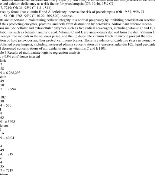
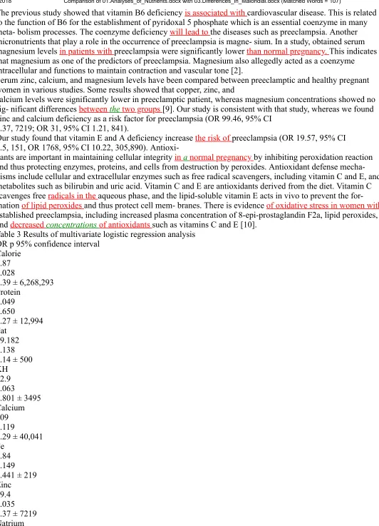
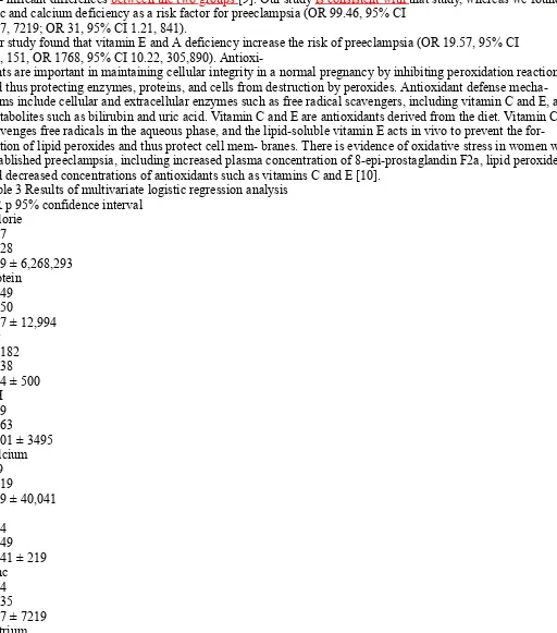
The previous study showed that vitamin B6 deficiency is associated with cardiovascular disease. This is related to the function of B6 for the establishment of pyridoxal 5 phosphate which is an essential coenzyme in many meta- bolism processes. The coenzyme deficiency will lead to the diseases such as preeclampsia. Another micronutrients that play a role in the occurrence of preeclampsia is magne- sium. In a study, obtained serum
magnesium levels in patients with preeclampsia were significantly lower than normal pregnancy. This indicates that magnesium as one of the predictors of preeclampsia. Magnesium also allegedly acted as a coenzyme intracellular and functions to maintain contraction and vascular tone [2].
Serum zinc, calcium, and magnesium levels have been compared between preeclamptic and healthy pregnant women in various studies. Some results showed that copper, zinc, and
calcium levels were significantly lower in preeclamptic patient, whereas magnesium concentrations showed no sig- nificant differences between the two groups [9]. Our study is consistent with that study, whereas we found zinc and calcium deficiency as a risk factor for preeclampsia (OR 99.46, 95% CI
1.37, 7219; OR 31, 95% CI 1.21, 841).
Our study found that vitamin E and A deficiency increase the risk of preeclampsia (OR 19.57, 95% CI 2.5, 151, OR 1768, 95% CI 10.22, 305,890).
Antioxi-dants are important in maintaining cellular integrity in a normal pregnancy by inhibiting peroxidation reaction and thus protecting enzymes, proteins, and cells from destruction by peroxides. Antioxidant defense mecha-nisms include cellular and extracellular enzymes such as free radical scavengers, including vitamin C and E, and metabolites such as bilirubin and uric acid. Vitamin C and E are antioxidants derived from the diet. Vitamin C scavenges free radicals in the aqueous phase, and the lipid-soluble vitamin E acts in vivo to prevent the for-mation of lipid peroxides and thus protect cell mem- branes. There is evidence of oxidative stress in women with established preeclampsia, including increased plasma concentration of 8-epi-prostaglandin F2a, lipid peroxides, and decreased concentrations of antioxidants such as vitamins C and E [10].
Table 3 Results of multivariate logistic regression analysis OR p 95% confidence interval
Calorie 3.87 0.028
2.39 ± 6,268,293 Protein
0.049 0.650
0.27 ± 12,994 Fat
59.182 0.138 3.14 ± 500 KH
52.9 0.063
0.801 ± 3495 Calcium 109 0.119
0.29 ± 40,041 Fe
9.84 0.149 0.441 ± 219 Zinc
0.62 0.731 0.041 ± 9.37 Kalium 0.005 0.054 0.000 ± 1.08 Magnesium 0.015 0.528
0.000 ± 7321 Vitamin A 0.002 0.055 0.000 ± 1.14 Folic acid 42.9 0.193
0.15 ± 12,383 Vitamin B1 0.000 0.009
0.000 ± 0.014 Vitamin B2 3.75
0.550 0.049 ± 285 Niacin 2.39 0.068
0.571 ± 1E ? 007 Vitamin B6 0.299 0.591 0.004 ± 24.3 Vitamin B12 4.511
0.243 0.36 ± 56.4 Vitamin C 19.5 0.004 2.52 ± 151 Vitamin E 1.76 0.004 10.2 ± 30.5 OR odds ratio Conclusions
Differences in Malondialdehyde and Catalase Activity Levels Between Abortion and Normal Pregnancy
INTRODUCTION
Abortion is defined as a termination of a pregnancy before 20 weeks of gestation or when the fetal weight is less than
500 g. Clinically, the most frequent abortion encountered in the hospital is incomplete abortion. Patients usually come with bleeding and severe abdominal pain. The second one is threatened abortion. Threatened abortion is the most common complication in the first half of pregnancy and have incidence about 20-25%. Less than 30% of the women who experience threatened abortion will end in spontaneous abortion1.
There are many factors that associated with abortion, so it is hard to determine the exact mechanism. In spite of many possibilities, there is now a clear evidence that abortion is associated with placental oxidative stress. The abnormal placentation will lead to placental oxidative stress with resultant destructive effects on the
syncytiotrophoblast. The theory has been proposed as a mechanism involved in the etiopathogenesis of abortion2.
The body has many antioxidant systems to defend the excessive Reactive Oxygen Species (ROS) production3. In a healthy body, ROS and antioxidant remain in balance. When the body has an excessive amount of ROS, Oxidative Stress (OS) then occurs. An increase in the expression of oxidative stress marker in the trophoblast was detected in abortion and this was speculated to be a cause of early pregnancy loss4.
Antioxidant system divided into enzymatic and nonenzymatic group. Catalase (CAT) is one of enzymatic antioxidant that can remove hydrogen peroxide (H2O2), prevents lipid peroxidation in the cell membrane and works as free radicals binding5. The CAT role in pregnancy is very important, in early pregnancy CAT plays a role for implantation by protecting the blastocysts from superoxide radicals in the endometrium. The CAT is also important for embryonic development and the maintenance of early pregnancy. The CAT level was found
increased during pregnancy and low CAT activity in plasma or placenta found in cases of spontaneous abortion6. Another study also suggests that low antioxidant level increases the risk of spontaneous abortion7.
Free radicals are thus highly unstable molecules that have electrons available to react with a various organic substrate such as lipids, proteins and DNA8. Accumulation of lipid peroxides may cause not only tissue damage but also some biological events to accelerate the termination of pregnancy. It has been reported that ROS or lipid peroxide stimulate synthesis of PGF2α that causes uterine contraction6. Oxidative stress has been variably determined by many ways. The most reliable assessment of free radicals synthesis is using assays directly to detect the superoxide or other free radical. However, this technique is difficult and require specialized document. In another side, measurement of the products of oxidative modification product provides the most direct and possible assessment of oxidative stress. Malondialdehyde
(MDA) is a metabolite of lipid peroxides that detectable in plasma and was used as an indicator of lipid peroxidation9. This study was carried out to estimate the mean difference of CAT and MDA in normal pregnancy, threatened abortion and incomplete abortion.
MATERIALS AND METHODS
This was an observational study with cross-sectional design on February-December, 2014. The population was all pregnant women before 20 weeks gestations were enrolled in the maternity ward of Dr. M. Djamil Hospital and Dr. Reksodiwiryo Hospital, Padang, Indonesia. Subjects were taken by consecutive sampling method. Cases were those diagnosed with threatened and incomplete abortion by a specialist doctor. Meanwhile, controls were normal pregnant women without any other comorbid.
Exclusion criteria were a mother with hydatidiform mole, uterus disorder, myoma, history of provocating and recurrent abortion. After providing informed, written consent, subject’s blood was taken from antecubital vein for analyzed the CAT activity and MDA concentration with spectrophotometry method in Biomedical
Laboratory of Medical Faculty of Andalas University.
The significant difference among the three groups was analyzed with ANOVA test. If p<0.05 was found, then the analysis continued with post hoc multiple comparison test to determined the differences between two group. Statistical analysis was conducted using a computer program.
RESULTS
During the study period, 42 blood samples were collected, consisting of 14 subjects with threatened abortion, 14 subjects with incomplete abortion and 14 subjects with normal pregnancy before 20 weeks gestations. Table 1 shows the activity of CAT enzyme in incomplete abortion group was
66.46±9.11 IU mgG1, whereas normal pregnancy group was
78.26±8.88 IU mgG1. The analysis with statistical tests ANOVA found that p<0.001. To saw the specific difference between two subjects, the analysis continued by post hoc multiple comparisons test. As presented in Table 2, there were significant differences of CAT activity between incomplete abortion group and threatened abortion (p<0.001), incomplete abortion and normal pregnancy (p<0.001), threatened abortion and normal pregnancy (p = 0.01).
Table 1: Difference of catalase activity in incomplete abortion, threatened abortion and normal pregnancy Catalase activity
Parameters Mean±SD (IU mgG1) 95% CI p-value* Incomplete abortion 51.10±12.20 44.06-58.15 <0.001 Threatened abortion 66.46±9.11 61.20-71.72
Normal pregnancy 78.26±8.88 73.13-83.38
*p-values are obtained by ANOVA test, SD: Standard deviation, CI: Confidence interval Table 2: Post hoc test for the difference of the mean level of catalase activity
in incomplete abortion, threatened abortion and normal pregnancy Incomplete Threatened Normal Parameters abortion abortion pregnancy
Incomplete abortion <0.001* <0.001* Threatened abortion <0.001* 0.01* Normal pregnancy <0.001* 0.01* -*p-value obtained by Tukey’s test to determine the difference between two groups
Table 3: Difference of malondialdehyde level in incomplete abortion, threatened abortion and normal pregnancy Malondialdehyde level Parameters Mean±SD (nmol mLG1) 95% CI p-value*
Incomplete abortion 4.02±0.36 3.81-4.23 <0.001 Threatened abortion 3.46±0.39 3.24-3.68
Normal pregnancy 2.83±0.21 2.71-2.96
*p-values are obtained by ANOVA test, SD: Standard deviation, CI: Confidence interval
Table 4: Post hoc Test for the difference of the mean level of malondialdehyde in incomplete abortion, threatened abortion and
normal pregnancy Incomplete Threatened Normal Parameters abortion abortion pregnancy
Incomplete abortion - <0.001* <0.001* Threatened abortion <0.001* - <0.001* Normal pregnancy <0.001* <0.001*
-*p-value obtained by Tukey’s test to determine the difference between two groups
As presented in Table 3, the mean level of MDA in incomplete abortion group was 4.02±0.36 nmol mLG1, the threatened abortion group was 3.46±0.39 nmol mLG1, whereas in normal pregnancy group was 2.83±0.21 nmol mLG1 with p = 0.00 by ANOVA test. To saw the difference between two group analysis continued with post hoc test. According to the Table 4, it was found that there were significant differences of MDA level between
incomplete abortion with threatened abortion (p<0.001), incomplete abortion and normal pregnancy group (p<0.001), threatened abortion and normal pregnancy (p<0.001).
DISCUSSION
In this study, it was found that the mean of MDA level of incomplete abortion was higher than threatened abortion and normal pregnancy (p<0.05). Meanwhile, the mean level of CAT activity was lower in incomplete abortion than threatened abortion and normal pregnancy (p<0.05).
This study was accordance with a study conducted by Abdul-Barry et al.8 that found the serum MDA level was significantly higher in patients with a history of recurrent spontaneous abortion than women with a healthy pregnancy. Ozkaya et al.10 also reported that the spontaneous abortion before 8 weeks gestation was associated with higher serum of MDA than healthy pregnancy (66.4±13.7 vs. 40.3±16.1, p<0.01). Because MDA is a side product of lipid peroxidation, the increase in MDA level may reflect an overproduction of lipid peroxides and alter antioxidant defend mechanism.
Jauniaux et al.11 suggested the theory of how the free radical is higher in abortion than in normal pregnancy. They found that the intervillous O2 flow appeared much earlier in the abortion group so it can lead to excessive free radicals production that can cause abortion11.
the lipid peroxidation injury in the embryo. Second, the increasing free radicals can change oxygen partial pressure in embryonic cells. And the last, increased of free radicals can lead to ischemia-reperfusion injury in the fetus12.
Actually, increased of antioxidants activity also found in early healthy pregnancy. These indicate that the oxidative stress actually also occurs in normal pregnancy but the sufficient antioxidants are defended the free radicals and prevented the damage6,13,14. In this study, the mean activity of CAT enzyme of incomplete abortion was lower than normal pregnancy and threatened abortion (p<0.05). It means that there were
differences between the CAT enzyme activity in incomplete abortion, threatened abortion and normal pregnancy. It was suggested that the excess of free radicals and the decreased of antioxidants in this study were associated with the abortion.
Sugino et al.6 found that CAT total activity was decrease
and prostaglandin F2α synthesis was increase in decidua in cases of spontaneous abortion with vaginal bleeding. They proposed that the termination of pregnancy was caused by decreasing activity of CAT which stimulates the synthesis of prostaglandins6. This study was accordance with the study by Yigenoglu et al.15 that found the total antioxidant capacity is lower in pregnant women with history of spontaneous abortion than healthy pregnant women. It also accordance with a study conducted by Biri et al.16 that found the CAT activity was higher in abortion group than in control group (87.62±8.19 vs
44.47±3.79 IU mgG1). CONCLUSION
Abortion group had lower antioxidant concentration and higher free radicals than normal pregnancy. There was an imbalance of free radical and antioxidant concentration in abortion.
SIGNIFICANT STATEMENT
The imbalance of free radical and antioxidant has been proposed to one of many theories that can cause abortion. This study tried to determine the comparison of antioxidant and free radical level by analyzed the catalase
activity and malondialdehyde concentration in normal pregnancy and
abortion. Malondialdehyde (MDA) is a metabolite of lipid peroxides that used as an indicator of lipid
Analyses of Nutrients and Body Mass Index as Risk Factor for Preeclampsia Introduction
Preeclampsia is an important problem in obstetrics because it is still a major cause of maternal mortality compared to bleeding and infection. Preeclampsia leads to maternal and perinatal morbidity. Preeclampsia is also associated with high rates of preterm delivery, small for gestational ages, and perinatal death [1]. Little is known about the patho- genesis of preeclampsia. Many factors are identified as risk factors for preeclampsia including parity, multiple preg- nancies, age, family history of preeclampsia, obesity, his- tory of systemic disease, and nutrition.
Since preeclampsia is characterized by reduced perfu- sion of the placenta, oxidative stress, and endothelial dys-function, nutrition has long been hypothesized to have a role in the etiology of preeclampsia [2]. Oxidative stresses are proposed as the linkage between the two stages of preeclampsia. Nutrients can affect oxidative stress by increasing or decreasing free radicals or antioxidants or by providing substrate for the formation of free radicals. Several nutrients, particularly omega-3 (n - 3) fatty acids, antioxidants, and folic acid, have an important roles in modulating endothelial function. It has also been suggested that nutrients such as trace elements, fatty acids, and folic acid can contribute to insulin resistance, a risk factor for preeclampsia. In many studies, decrease in serum magne- sium levels has been considered as the cause of patho- genesis of
preeclampsia. Minerals have an important
influence on the health of pregnant women and growing fetus. Among them, serum or placental zinc (Zn) concen- trations have been reported to be low in PE women. Fur- thermore, decreased levels of zinc, selenium, and copper have been observed in patients with preeclampsia [3]. In another side, the nutrients with antioxidants among high risk women showed a protective effect [4]. Folic acid has been hypothesized as a protective agent of preeclampsia.
Maternal obesity and insulin resistance are also believed to be important risk factors for the development of pla-cental endothelial dysfunction and preeclampsia. Preven- tion of preeclampsia has remained elusive, owing largely to their complex nature. Currently, maternal obesity in prepregnancy is one of the strongest modifiable risk fac- tors. Recent studies have shown a relation between obesity in prepregnancy and the risk of
preeclampsia. The reason for obesity being associated with an increased risk of preeclampsia was explained by increased levels of serum triglycerides and very low-density lipoprotein particles in obese women. This lipid alterations have been suggested to promote oxidative stress caused by ischemia–reperfusion mechanism or activated neutrophils, which leads to endothelial cell dysfunction [1].
The hypothesis about nutritional status and body mass index (BMI) prepregnancy associated with preeclampsia has intrigued us to study the risk factor for preeclampsia in Dr. M. Djamil Hospital, Padang, Indonesia.
Methods
This was a case–control study at the Department of Obstetric and Gynecology in Dr. M. Djamil Hospital, Padang, Indonesia, between January and December 2013. Pregnant women after 20 weeks gestations were included. A total of 140 patients were enrolled in this study with 70 cases and 70 controls. Cases were those diagnosed with preeclampsia; meanwhile, controls were normotensive pregnant women without any other comorbidity.
After providing written informed consent, all subjects completed an interview for their nutritional status and prepregnancy BMI after delivery. The nutritional status was assessed by Food Frequency Questionnaire (FFQ) and then analyzed by Nutrisurvey Program. Calories, protein, fat, carbohydrates, calcium, phosphorus, zinc, sodium, potassium, magnesium, vitamin A, folic acid, vitamin B1, vitamin B2, niacin, vitamin B6, vitamin B12, vitamin C, and vitamin E were assessed. Maternal BMI was catego- rized into two groups: normal BMI and abnormal BMI. Normal BMI was defined as 18.5–24.9 kg/m2. The inde- pendent samples t test was used for nutritional status, and
Chi-square test was used for BMI. Odds ratio (OR) with 95% CI was calculated. A p value \0.05 was considered
statistically significant. For the nutrition variable, if the p value \0.25 then continued by logistic regression back-ward to assess the risk factor.
Results
Most of the subjects had normal weight, both in case and control groups. Table 1 shows that prevalence of abnormal BMI was more common in the preeclampsia group com- pared with those without preeclampsia 19 (27.1%) versus
As shown in Table 2, the mean level for most of the
variables, except calories, fat, and vitamin B1, was lower in subjects with preeclampsia than those without preeclamp- sia. The difference for most of the subjects was also sta- tistically significant with p \ 0.05, except for vitamin B1 and vitamin B2. Table 3 shows the risk factors for preeclampsia obtained by logistic regression analyses. The nutrients that were significantly associated with increased risk of preeclampsia were deficiency of vitamin E, zinc, fat, calcium, and vitamin C. Excess of calories and carbohy- drate also significantly associated with increased risk of preeclampsia. Meanwhile, vitamin A and vitamin B1 were protective factors.
Discussion
The reason for obesity being associated with an increased risk of preeclampsia was explained by increased levels of serum triglycerides, very low-density lipoproteins, and formation of small low-density lipoprotein particles in obese women. This lipid profile was also found in women with preeclampsia. These lipid alterations have been suggested to promote oxidative stress, caused by ische- mia–reperfusion mechanism or activated neutrophils, which leads to endothelial cell dysfunction [1]. Moreover, dyslipidemia also can cause atherothrombosis and induce the aggregation of the thrombocytes than can lead to coagulopathy which is a characteristic of
preeclampsia.
Obesity is accompanied by oxidative stress. The origin of oxidative stress is proposed to be secondary to increased free fatty acids and inflammation. It is also suggested that diet can contribute to oxidative stress. Obese individuals have lower blood concentrations of antioxidants. This could be due to reduced dietary intake of antioxidants, but increased consumption by reactive oxygen species is also possible [5].
In this study, we found no relationship between BMI with preeclampsia. As mentioned before, dyslipidemia is the important factor that can lead into preeclampsia. But our study did not assess profile lipid of the subjects. Fur- thermore, central obesity has a higher risk of preeclampsia. Central obesity is characterized by visceral fat. Visceral fat produces C-reactive protein (CRP), PAI-1, and leptin that contributes to oxidative stress. People with central obesity have a higher risk to get preeclampsia. In this study, we did not assess the central obesity of the subjects. Measures of body composition, including percent body fat, may very likely identify the obese woman at risk of preeclampsia more accurately.
Our study found that carbohydrate intake in preeclampsia group was significantly higher than the non-preeclampsia group. The subjects that have higher carbo- hydrate will have lower protein level. Meanwhile, protein is needed in the process of trophoblast invasion so the protein-energy malnutrition increases the risk of preeclampsia. We also found that intake of proteins in preeclampsia was significantly lower in the preeclampsia group.
Folic acid and vitamin B12 are also a protector factors against preeclampsia. Those micronutrients play an important role in suppressing the metabolism of homo- cysteine, whereas the excess of homocysteine was a cau-sative factor of endothelial damage and became one of the causes of preeclampsia. In addition, folic acid-supple- ments preconception enhance the placentation process and can prevent preeclampsia. Three earlier cohort studies assessed the effect of folic acid containing multivitamins (including folic acid) and gestational hypertension (in- cluding preeclampsia), and all showed a protective effect of folic acid supplementation on preeclampsia [2, 6, 7].
Table 1 Asssociation between body mass index and the risk of preeclampsia BMI
Preeclampsia %
Non-preeclampsia %
p* OR CI 95% Min. Max.
Abnormal BMI 19
0.222 1.801 0.797 4.067
Normal BMI 51
72.9 58 82.9 70 100 70 100
BMI body mass index, OR odds ratio, CI confidence interval * p value were obtained by Chi-square
Table 2 Differences of mean nutritions level in preeclampsia and non-preeclampsia group Preeclampsia
Mean (95% CI) Non-preeclampsia Mean (95% CI) p*
Calorie (Kcal)
1269.22 (745.2–2214) 1171.15 (649.1–2447.6) 0.048
Protein (g)
42.37 (23.03–95.42) 54.9 (24.2–200.6) 0.000
Fat (g)
26.8 (4.65–58.9) 32.4 (10.79–93.12) 0.009
KH (g)* 219.5 ± 56.8 163.49 ± 46.5 0.000
Calcium (mg) 234.7 (63.7–758.3) 9362.9 (63.7–922.4) 0.000
Phosphor (mg) 589.4 (296.5–1444.5) 714.6 (324.6–2353.4) 0.006
Fe (mg)
9.4 (2.84–23.9) 11.06 (2.84–35.6) 0.005
Zinc (mg) 4.8 (2.39–12.87) 5.5 (2.4–16.5) 0.005
299.3 (76.9–970.24) 533.3 (105.9–1847.60) 0.000
Kalium (mg)* 595.4 ± 237.2 732.15 ± 367.8 0.010
Magnesium (mg) 181.1 (97.1–370.14) 210.0 (97.1–554.5) 0.010
Vitamin A (lg) 544.9 (174.9–1567.4) 763.9 (174.9–3354.5) 0.000
Folic acid (lg) 108.6 (56.8–421.6) 163.5 (56.83–497.9) 0.000
Vitamin B1 (mg) 0.7 (0.27–2.15) 0.65 (0.27–2.01) 0.085
Vitamin B2 (mg) 0.94 (0.25–2.45) 1.1 (0.25–2.7) 0.046
Niacin (mg) 4.3 (0.6–11.9) 5.8 (2.1–23.7) 0.000
Vitamin B6 (mg) 1.1 (0.66–2.14) 1.22 (0.67–3.8) 0.009
Vitamin B12 (mg) 1.1 (0.15–9.15) 2.8 (0.27–21.1) 0.000
Vitamin C (mg) 43.4 (8.4–136.8) 68.2 (19.2–262.7) 0.000
Vitamin E (mg TE) 1.9 (0.2–4.4) 3.6 (1.8–8.2) 0.000
CI confidence interval
* p value was obtained by independent samples t test
A recent large cohort study from Denmark also showed that regular use of folic acid in pregnancy was related to a reduced risk of preeclampsia among normal-weight women [8]. But in this study, we did not found the folic acid and vitamin B12 as a protective agent of preeclampsia. The different result maybe caused by
The previous study showed that vitamin B6 deficiency is associated with cardiovascular disease. This is related to the function of B6 for the establishment of pyridoxal 5 phosphate which is an essential coenzyme in many meta- bolism processes. The coenzyme deficiency will lead to the diseases such as preeclampsia. Another micronutrients that play a role in the occurrence of preeclampsia is magne- sium. In a study, obtained serum magnesium levels in patients with preeclampsia were significantly lower than normal pregnancy. This indicates that magnesium as one of the predictors of preeclampsia. Magnesium also allegedly acted as a coenzyme intracellular and functions to maintain contraction and vascular tone [2].
Serum zinc, calcium, and magnesium levels have been compared between preeclamptic and healthy pregnant women in various studies. Some results showed that copper, zinc, and
calcium levels were significantly lower in preeclamptic patient, whereas magnesium concentrations showed no sig- nificant differences between the two groups [9]. Our study is consistent with that study, whereas we found zinc and calcium deficiency as a risk factor for preeclampsia (OR 99.46, 95% CI
1.37, 7219; OR 31, 95% CI 1.21, 841).
Our study found that vitamin E and A deficiency increase the risk of preeclampsia (OR 19.57, 95% CI 2.5, 151, OR 1768, 95% CI 10.22, 305,890).
Antioxi-dants are important in maintaining cellular integrity in a normal pregnancy by inhibiting peroxidation reaction and thus protecting enzymes, proteins, and cells from destruction by peroxides. Antioxidant defense mecha-nisms include cellular and extracellular enzymes such as free radical scavengers, including vitamin C and E, and metabolites such as bilirubin and uric acid. Vitamin C and E are antioxidants derived from the diet. Vitamin C scavenges free radicals in the aqueous phase, and the lipid-soluble vitamin E acts in vivo to prevent the for-mation of lipid peroxides and thus protect cell mem- branes. There is evidence of oxidative stress in women with established preeclampsia, including increased plasma concentration of 8-epi-prostaglandin F2a, lipid peroxides, and decreased concentrations of antioxidants such as vitamins C and E [10].
Table 3 Results of multivariate logistic regression analysis OR p 95% confidence interval
Calorie 3.87 0.028
2.39 ± 6,268,293 Protein
0.049 0.650
0.27 ± 12,994 Fat
59.182 0.138 3.14 ± 500 KH
52.9 0.063
0.801 ± 3495 Calcium 109 0.119
0.29 ± 40,041 Fe
9.84 0.149 0.441 ± 219 Zinc
0.62 0.731 0.041 ± 9.37 Kalium 0.005 0.054 0.000 ± 1.08 Magnesium 0.015 0.528
0.000 ± 7321 Vitamin A 0.002 0.055 0.000 ± 1.14 Folic acid 42.9 0.193
0.15 ± 12,383 Vitamin B1 0.000 0.009
0.000 ± 0.014 Vitamin B2 3.75
0.550 0.049 ± 285 Niacin 2.39 0.068
0.571 ± 1E ? 007 Vitamin B6 0.299 0.591 0.004 ± 24.3 Vitamin B12 4.511
0.243 0.36 ± 56.4 Vitamin C 19.5 0.004 2.52 ± 151 Vitamin E 1.76 0.004 10.2 ± 30.5 OR odds ratio Conclusions
Differences in Malondialdehyde and Catalase Activity Levels Between Abortion and Normal Pregnancy
INTRODUCTION
Abortion is defined as a termination of a pregnancy before 20 weeks of gestation or when the fetal weight is less than
500 g. Clinically, the most frequent abortion encountered in the hospital is incomplete abortion. Patients usually come with bleeding and severe abdominal pain. The second one is threatened abortion. Threatened abortion is the most common complication in the first half of pregnancy and have incidence about 20-25%. Less than 30% of the women who experience threatened abortion will end in spontaneous abortion1.
There are many factors that associated with abortion, so it is hard to determine the exact mechanism. In spite of many possibilities, there is now a clear evidence that abortion is associated with placental oxidative stress. The abnormal placentation will lead to placental oxidative stress with resultant destructive effects on the
syncytiotrophoblast. The theory has been proposed as a mechanism involved in the etiopathogenesis of abortion2.
The body has many antioxidant systems to defend the excessive Reactive Oxygen Species (ROS) production3. In a healthy body, ROS and antioxidant remain in balance. When the body has an excessive amount of ROS, Oxidative Stress (OS) then occurs. An increase in the expression of oxidative stress marker in the trophoblast was detected in abortion and this was speculated to be a cause of early pregnancy loss4.
Antioxidant system divided into enzymatic and nonenzymatic group. Catalase (CAT) is one of enzymatic antioxidant that can remove hydrogen peroxide (H2O2), prevents lipid peroxidation in the cell membrane and works as free radicals binding5. The CAT role in pregnancy is very important, in early pregnancy CAT plays a role for implantation by protecting the blastocysts from superoxide radicals in the endometrium. The CAT is also important for embryonic development and the maintenance of early pregnancy. The CAT level was found
increased during pregnancy and low CAT activity in plasma or placenta found in cases of spontaneous abortion6. Another study also suggests that low antioxidant level increases the risk of spontaneous abortion7.
Free radicals are thus highly unstable molecules that have electrons available to react with a various organic substrate such as lipids, proteins and DNA8. Accumulation of lipid peroxides may cause not only tissue damage but also some biological events to accelerate the termination of pregnancy. It has been reported that ROS or lipid peroxide stimulate synthesis of PGF2α that causes uterine contraction6. Oxidative stress has been variably determined by many ways. The most reliable assessment of free radicals synthesis is using assays directly to detect the superoxide or other free radical. However, this technique is difficult and require specialized document. In another side, measurement of the products of oxidative modification product provides the most direct and possible assessment of oxidative stress. Malondialdehyde
(MDA) is a metabolite of lipid peroxides that detectable in plasma and was used as an indicator of lipid peroxidation9. This study was carried out to estimate the mean difference of CAT and MDA in normal pregnancy, threatened abortion and incomplete abortion.
MATERIALS AND METHODS
This was an observational study with cross-sectional design on February-December, 2014. The population was all pregnant women before 20 weeks gestations were enrolled in the maternity ward of Dr. M. Djamil Hospital and Dr. Reksodiwiryo Hospital, Padang, Indonesia. Subjects were taken by consecutive sampling method. Cases were those diagnosed with threatened and incomplete abortion by a specialist doctor. Meanwhile, controls were normal pregnant women without any other comorbid.
Exclusion criteria were a mother with hydatidiform mole, uterus disorder, myoma, history of provocating and recurrent abortion. After providing informed, written consent, subject’s blood was taken from antecubital vein for analyzed the CAT activity and MDA concentration with spectrophotometry method in Biomedical
Laboratory of Medical Faculty of Andalas University.
The significant difference among the three groups was analyzed with ANOVA test. If p<0.05 was found, then the analysis continued with post hoc multiple comparison test to determined the differences between two group. Statistical analysis was conducted using a computer program.
RESULTS
During the study period, 42 blood samples were collected, consisting of 14 subjects with threatened abortion, 14 subjects with incomplete abortion and 14 subjects with normal pregnancy before 20 weeks gestations. Table 1 shows the activity of CAT enzyme in incomplete abortion group was
66.46±9.11 IU mgG1, whereas normal pregnancy group was
78.26±8.88 IU mgG1. The analysis with statistical tests ANOVA found that p<0.001. To saw the specific difference between two subjects, the analysis continued by post hoc multiple comparisons test. As presented in Table 2, there were significant differences of CAT activity between incomplete abortion group and threatened abortion (p<0.001), incomplete abortion and normal pregnancy (p<0.001), threatened abortion and normal pregnancy (p = 0.01).
Table 1: Difference of catalase activity in incomplete abortion, threatened abortion and normal pregnancy Catalase activity
Parameters Mean±SD (IU mgG1) 95% CI p-value* Incomplete abortion 51.10±12.20 44.06-58.15 <0.001 Threatened abortion 66.46±9.11 61.20-71.72
Normal pregnancy 78.26±8.88 73.13-83.38
*p-values are obtained by ANOVA test, SD: Standard deviation, CI: Confidence interval Table 2: Post hoc test for the difference of the mean level of catalase activity
in incomplete abortion, threatened abortion and normal pregnancy Incomplete Threatened Normal Parameters abortion abortion pregnancy
Incomplete abortion <0.001* <0.001* Threatened abortion <0.001* 0.01* Normal pregnancy <0.001* 0.01* -*p-value obtained by Tukey’s test to determine the difference between two groups
Table 3: Difference of malondialdehyde level in incomplete abortion, threatened abortion and normal pregnancy Malondialdehyde level Parameters Mean±SD (nmol mLG1) 95% CI p-value*
Incomplete abortion 4.02±0.36 3.81-4.23 <0.001 Threatened abortion 3.46±0.39 3.24-3.68
Normal pregnancy 2.83±0.21 2.71-2.96
*p-values are obtained by ANOVA test, SD: Standard deviation, CI: Confidence interval
Table 4: Post hoc Test for the difference of the mean level of malondialdehyde in incomplete abortion, threatened abortion and
normal pregnancy Incomplete Threatened Normal Parameters abortion abortion pregnancy
Incomplete abortion - <0.001* <0.001* Threatened abortion <0.001* - <0.001* Normal pregnancy <0.001* <0.001*
-*p-value obtained by Tukey’s test to determine the difference between two groups
As presented in Table 3, the mean level of MDA in incomplete abortion group was 4.02±0.36 nmol mLG1, the threatened abortion group was 3.46±0.39 nmol mLG1, whereas in normal pregnancy group was 2.83±0.21 nmol mLG1 with p = 0.00 by ANOVA test. To saw the difference between two group analysis continued with post hoc test. According to the Table 4, it was found that there were significant differences of MDA level between
incomplete abortion with threatened abortion (p<0.001), incomplete abortion and normal pregnancy group (p<0.001), threatened abortion and normal pregnancy (p<0.001).
DISCUSSION
In this study, it was found that the mean of MDA level of incomplete abortion was higher than threatened abortion and normal pregnancy (p<0.05). Meanwhile, the mean level of CAT activity was lower in incomplete abortion than threatened abortion and normal pregnancy (p<0.05).
This study was accordance with a study conducted by Abdul-Barry et al.8 that found the serum MDA level was
significantly higher in patients with a history of recurrent spontaneous abortion than women with a healthy pregnancy. Ozkaya et al.10 also reported that the spontaneous abortion before 8 weeks gestation was associated with higher serum of MDA than healthy pregnancy (66.4±13.7 vs. 40.3±16.1, p<0.01). Because MDA is a side product of lipid peroxidation, the increase in MDA level may reflect an overproduction of lipid peroxides and alter antioxidant defend mechanism.
Jauniaux et al.11 suggested the theory of how the free radical is higher in abortion than in normal pregnancy. They found that the intervillous O2 flow appeared much earlier in the abortion group so it can lead to excessive free radicals production that can cause abortion11.
the lipid peroxidation injury in the embryo. Second, the increasing free radicals can change oxygen partial pressure in embryonic cells. And the last, increased of free radicals can lead to ischemia-reperfusion injury in the fetus12.
Actually, increased of antioxidants activity also found in early healthy pregnancy. These indicate that the oxidative stress actually also occurs in normal pregnancy but the sufficient antioxidants are defended the free radicals and prevented the damage6,13,14. In this study, the mean activity of CAT enzyme of incomplete abortion was lower than normal pregnancy and threatened abortion (p<0.05). It means that there were
differences between the CAT enzyme activity in incomplete abortion, threatened abortion and normal pregnancy. It was suggested that the excess of free radicals and the decreased of antioxidants in this study were associated with the abortion.
Sugino et al.6 found that CAT total activity was decrease
and prostaglandin F2α synthesis was increase in decidua in cases of spontaneous abortion with vaginal bleeding. They proposed that the termination of pregnancy was caused by decreasing activity of CAT which stimulates the synthesis of prostaglandins6. This study was accordance with the study by Yigenoglu et al.15 that found the total antioxidant capacity is lower in pregnant women with history of spontaneous abortion than healthy pregnant women. It also accordance with a study conducted by Biri et al.16 that found the CAT activity was higher in abortion group than in control group (87.62±8.19 vs
44.47±3.79 IU mgG1). CONCLUSION
Abortion group had lower antioxidant concentration and higher free radicals than normal pregnancy. There was an imbalance of free radical and antioxidant concentration in abortion.
SIGNIFICANT STATEMENT
The imbalance of free radical and antioxidant has been proposed to one of many theories that can cause abortion. This study tried to determine the comparison of antioxidant and free radical level by analyzed the catalase
activity and malondialdehyde concentration in normal pregnancy and
abortion. Malondialdehyde (MDA) is a metabolite of lipid peroxides that used as an indicator of lipid
Research Article
Differences in Mean Levels of Maternal Resistin Serum between Early Onset Preeclampsia (EOPE) and Late Onset Preeclampsia (LOPE)
Research Article
Differences in Mean Levels of Maternal Resistin Serum between Early Onset Preeclampsia (EOPE) and Late Onset Preeclampsia (LOPE)
INTRODUCTION
Preeclampsia is one of the major causes of morbidity and mortality of the mother and fetus. World Health
Organization (WHO) reported the number of deaths caused by preeclampsia by 16% in developing countries
. Preeclampsia
resulting 3-25 fold increased risk obstetric complications and is the cause of 30-40% of perinatal deaths in Indonesia 1
.
The incidence of preeclampsia ranges between 5-10% of all pregnancies
. Incidence of preeclampsia in the United
States, Canada and Western Europe ranges between 2-5% of all pregnancies and higher, 4-18% in some developing countries in Africa
1
. The incidence of preeclampsia in Indonesia ranged between 3-10% 3
.
In Dr.M. Djamil General Hospital in Padang during the 2
year 2011, the incidence of preeclampsia was 8.31%, on 2012 was 11.47% and on 2013 was 12.02%.
Preeclampsia is divided into early onset preeclampsia (EOPE) (<34 weeks) and late onset preeclampsia (LOPE) (>34 weeks) of pregnancy based on the onset of clinical manifestations of preeclampsia. The EOPE and LOPE has a different pathogenesis. The PEAD is often associated with impaired uteroplacental perfusion caused by disruption of trophoblast invasion, while LOPE is often associated with the presence of extrinsic and maternal factors
.
Preeclampsia is a protean syndrome, in which multiple 4,5
organ systems can be affected compared to the others. Preeclampsia is mainly characterized by hypertension and proteinuria or may be associated with abnormalities in laboratory test results that renal function, hepatic or hemostasis after 20 weeks of pregnancy
.
Analysis of risk factor for preeclampsia is needed to 1,6
maternal risk factor that associated with LOPE .
Resistin is an adipose tissue-specific secretory factor 7
(ADSF), a hormone secreted by adipose tissue that induces insulin resistance in muscle and liver. Resistin stimulated by inflammatory conditions that produce proinflammatory cytokines
.
Preeclampsia is associated with inflammation and insulin 7,8
resistance which is affected by resistin. Resistin is associated with late onset preeclampsia because it was a maternal factors, so there was an increasing of maternal resistin serum levels in late onset preeclampsia
.
Previous study reported an elevated of maternal resistin 7,8
serum levels associated with a systemic inflammatory response and insulin resistance which is both of them are 2
2
increased in preeclampsia compared to normal pregnancy .
The other study showed the mean of Homeostasis Model
Assessment-Insulin Resistance (HOMA-IR) and high sensitivity C-reactive protein (hs-CRP) was higher in late onset
preeclampsia than early onset preeclampsia group. From
various studies on the above it could be estimated that the inflammatory factor and insulin resistance are associated with higher levels of resistin more dominant in late onset preeclampsia group compared to early onset preeclampsia group.
The HOMA-IR could be used as an indicator of insulin resistance, but not for inflammatory states, contrary hs-CRP could be used as an indicator of inflammation, but not for insulin resistance. Resistin is expected to describe both of them, because it was associated with insulin resistance and inflammation
7 .
MATERIALS AND METHODS
This study is an analytical cross sectional study with
2 0 w o me n o f e a r l y o n s e t p r e e c l a mp s i a ( E OP E ) a n d 2 0 w o me n of late onset preeclampsia (LOPE) who met the inclusion
criteria and there were no exclusion criteria. The samples were recruited in Dr.M. Djamil General Hospital, Padang from July-October, 2015.
sugar as >200 mg dLG
), had suffered coronary heart disease,
chronic kidney disease, chronic liver disease and obesity (BMI before pregnancy >25 kg mG
1 ).
Maternal resistin serum was examined by enzyme-linked 2
immunosorbent assay (ELISA) method in Biomedic Laboratory in Medical Faculty of Andalas University. Statistical analysis was conducted by using SPSS program 20th version. The data distribution was normal with p>0.05. The differences mean levels of maternal resistin serum was analyzed by using independent t-test.
RESULTS
Characteristics of research subjects: Forty patients of research subjects consisted 20 patients of early onset preeclampsia (EOPE) and 20 patients of late onset
preeclampsia (LOPE). Mean levels of maternal age in EOPE group is 34.4±5.144 years, whereas in LOPE group is
32.9±6.324 years with p = 0.416. According to maternal age 9-12
Table 1: Characteristics of research subjects between early onset preeclampsia and late onset preeclampsia Characteristics EOPE (n = 20) LOPE (n = 20) Total (%) p
Maternal age group
<20 years 0 (0%) 0 (0%) 0
20-35 years 8 (40%) 10 (50%) 45 0.537 >35 years 12 (60%) 10 (50%) 55
Maternal age (0±SD) years 34.4±5.144 31.9±6.314 0.416 Gravidity group
Primigravid 6 (30%) 7 (35%) 32.5 0.744 Miltigravid 14 (70%) 13 (65%) 67.5
Gravidity (0±SD) 2.4±1.314 2.65±1.461 0.573 BMI group
Underweight 0 (0%) 0 (0%) 0
Normoweight 14 (70%) 12 (60%) 65 0.520 Overweight 6 (30%) 8 (40%) 35
BMI (0±SD) 22.55±1.595 22.59±1.812 0.946
Table 2: Mean levels of maternal resistin serum in early onset preeclampsia and late onset preeclampsia
EOPE LOPE
--- ---Variable Mean SD Mean SD
Maternal resistin serum (ng mLG 1
) 2.526 1.603 8.891 6.219
Table 3: Mean levels difference of maternal resistin serum in early onset preeclampsia and late onset preeclampsia
Variable Mean difference CI (95%) p Maternal resistin serum (ng mLG 1
) 6.365 3.3835-9.3475 0.000
20-35 years old, 12 patients (60%) in group of maternal age >35 years old, none in group of maternal age <20 years old. Research subjects of late onset preeclampsia consists of 1 0 patients (50%) in group of maternal age 20-35 years old, 10 patients (50%) in group of maternal age >35 years old, none in group of maternal age <20 years old.
Mean levels of gravidity in early onset preeclampsia (EOPE) group is 2.4±1.314, whereas in late onset
preeclampsia (LOPE) group is 2.65±1.461 with p = 0.573. According to gravidity groups, study of subjects in early onset preeclampsia consists 6 patients (30%) in group of primigravida, 14 patients (70%) in group of multigravida. Research subjects of l ate onset preecl ampsi a c onsi sts of 7 patients (35%) in group of primigravida, 13 patients (65%). Mean levels of Body Mass Index (BMI) in early onset
preeclampsia (EOPE) group is 22.551±1.595, whereas in late onset preeclampsia (LOPE) group is 22.588±1.4812 kg mG with p = 0.946. According to BMI groups, study of subjects in EOPE consists 14 patients (70%) in group of normal
weight, 6 patients (30%) in group of overweight, none in group BMI underweight. Research subjects of late onset preeclampsia consists 12 patients (60%) in group of normal weight, 8 patients (40%) in group of overweight, none in group BMI underweight.
2 3
There were no statistically significant differences
regarding maternal age, group of maternal age, gravidity, group of gravidity, BMI and group of BMI characteristic between those two groups with p>0.05 showed in Table 1. Mean levels of maternal resistin serum in early onset preeclampsia and late onset preeclampsia: Mean levels of maternal resistin serum was higher in late onset preeclampsia vs
2.526±1.603 ng dLG
than early onset preeclampsia (8.891±6.219 ng mLG 1
), showed in Table 2.
Differences in mean levels of maternal resistin serum between late onset preeclampsia and early onset preeclampsia: The data distribution was normal with Kolmogorov Smirnov test. Analysis was performed with independent t-test. Table 3 showed the differences in mean levels of maternal resistin serum between early onset preeclampsia and late onset preeclampsia as 6.365 ng mLG with Confidence Interval (CI) 95% 3.384-9.347. There is a high significant differences with p<0.001.
DISCUSSION
The mean levels of maternal serum resistin in early
onset preeclampsia (EOPE) group was 2.526±1.603 ng mLG whereas in late onset preeclampsia (LOPE) group was 8.891±6.219 ng mLG