Purification and Characterization of Amyloglucosidase Enzyme
from the Thermophilic
Endomycopsis fibuligera
Using Sago Starch as a Substrate
Ahyar Ahmad
1,2,*and Harningsih Karim
31Department of Chemistry, Faculty of Natural Sciences, Hasanuddin University, Makassar 90245, Indonesia
2Laboratory of Research Centre and Developing of Sciences, Faculty of Natural Sciences
Hasanuddin University, Makassar 90245, Indonesia
3Department of Pharmacy, School of Pharmacy YAMASI, Jl. Mapala 2 Blok D5 No.10, Makassar 90222, Indonesia
Received December 9, 2015; Accepted May 24, 2016
ABSTRACT
An investigation on purification and characterization of amyloglucosidase enzyme from Endomycopsis fibuligeraby fermentation of sago starch has been carried out. This enzyme is inductive and can be produced by fermenting sago starch in a medium containing E. fibuligera. Crude enzyme was obtained by centrifuging the medium cultures containing E. fibuligera at 6,000 rpm for 20 min and then adding with 0.15 M acetate buffer (pH 5.0). Enzyme activity was determined using Somogyi-Nelson method by quantifying the released glucose from amyloglucosidase catalysis of starch (0.2%) as substrate. Prepurification process was conducted by ammonium sulphate fractionation and it showed that the ammonium sulphate fractionation with the degree of saturation of 40-60% produced a maximum activity of enzyme. Purification by DEAE-Cellulose and Sephadex G-75 column chromatography produced three and one fractions with purifity 17.4 and 22.5 times, respectively, compared to the crude extract enzyme. Characterization of this enzyme showed the optimum condition at pH 5.0 and 55 °C with 0.2% starch as substrate. The amyloglucosidase activities was strongly increased by addition of Co2+ and Mn2+ions, whereas the activities were weakly decreased by addition of K+, Mg2+, and Fe3+ions.
Keywords:enzyme; purification; amyloglucosidase; chromatography
ABSTRAK
Telah dilakukan penelitian tentang pemurnian dan karakterisasi enzim amiloglukosidase dari Endomycopsis fibuligerasecara fermentasi dari tepung sagu sebagai substrat. Enzim amiloglukosidase bersifat induktif yang dapat diproduksi pada medium fermentasi yang mengandung khamir E. fibuligera. Ekstrak kasar enzim diperoleh dari kultur media fermentasi khamirE. fibuligera setelah disentrifugasi pada kecepatan 6.000 rpm selama 20 menit lalu supernatannya ditambahkan 0,15 M buffer asetat (pH 5,0). Aktivitas enzim ditentukan dengan metode Somogyi-Nelson dengan mengukur kadar glukosa hasil hidrolisis larutan amilum (0,2%) sebagai substrat. Pra-pemurnian enzim dilakukan dengan fraksionasi ammonium sulfat, menunjukkan aktivitas enzim yang paling tinggi ditemukan pada fraksi 40-60% kejenuhan. Pemurnian dengan kromatografi kolom DEAE-sellulosa dan Sephadex G-75 menghasilkan tiga dan satu fraksi dengan tingkat kemurnian masing-masing sebesar 17,4 dan 22,5 kali dibandingkan dengan ekstrak enzim kasar. Karakterisasi enzim menunjukkan kondisi optimum pada pH 5.0 dan suhu 55 °C menggunakan larutan amilum 0,2% sebagai substrat. Penambahan ion Co2+ dan Mn2+ paling kuat meningkatkan aktivitas enzim sedangkan penambahan ion K+, Mg2+, dan Fe3+ dapat menurunkan aktivitas enzim amiloglukosidase.
Kata Kunci:enzim; pemurnian; amiloglukosidase; kromatografi
INTRODUCTION
Amylase, an important enzyme in food industries, is widely used to hydrolyze starch into dextrin and then further into maltose and glucose, respectively. This enzyme can be produced by fermentation of yeast cells in media containing corn starch or agricultural biomass resources [1-4]. Although plants, fungi, and bacteria
absence of effects brought about by seasonality, and rapid fermentation culture development methods. The above characteristics make microbial enzymes suitable as biocatalysts for various food industrial applications [5]. The production of enzymes from thermophilic microbes can be explored further to produce the enzymes that have stability at high temperatures. Thermophile microbe is often found in an extreme environment of hot springs with temperatures between 50 to 80 °C. Therefore, identification and dissemination of new thermophilic microbial sources, mainly those which are non-toxic to humans, are of high strategic interest. Besides, to guarantee enzyme supply for different industrial processes, the development of new enzymatic systems which cannot be obtained from plants or animals for possible so important progress in the food industry may be achieved.
In Indonesia, abundance of sago trees can be found uncultivated and become a source of carbohydrates other than rice by some people. To optimize the utilization of sago, its starch is converted into glucose by the manufacturers to produce fructose syrup and other chemicals, such as, acetone, propanol, butanol, and ethanol.
The process of converting polysaccharides into monosaccharides (glucose) enzymatically involves a group of the amylase enzymes. In recent years, a great interest in yeast amylases has been taken because of the ability of these enzymes to convert starch completely to glucose. This enzyme use ranges from bakery, textile, beer, liquor, infant feeding cereals, starch liquefaction saccharification and animal feed industries to chemical and pharmaceutical ones. Therefore, the amylase enzymes many isolations, and studies, particularly their nature and characteristics have been carried out. Gasperik and Minaricova, 1991 [1] have isolated and characterized the amylase enzyme produced extra cellular from the liquid culture of E. fibuligera containing sago starch. Further separation by a HPLC method showed that amylase was composed of several
enzymes including α-amylase (EC.3.2.1.1) and
amyloglucosidase (EC.3.2.1.3). In addition, they found
that α-amylase and amyloglucosidase had a different
thermal stability. Amyloglucosidase seems to be more
stable than the α-amylase [1]. Amyloglucosidase
enzyme is a glycoprotein which contains carbohydrate residues that are glycosidically linked through D-mannose to the hydroxyl groups of serine and threonine in the polypeptide chain of the enzyme. Enzyme activity assay of amyloglucosidase was calculated based on the amount of glucose obtained from the hydrolysis of starch; the amount of glucose was measured by a Nelson-Somogyi method. They also found that the
optimum α-amylase and amyloglucosidase activities
were achieved at pH 5.8 and 5.4, respectively.
Research on the isolation and purification of amyloglucosidase enzymes of thermophilic yeast E. fibuligera in sago starch fermentation media as substrate has not been widely reported so far. Mittal et al. [6] produced and purified an extracellular amyloglucosidase from non-thermophilic yeast Aspergillus awamori NA21 by ammonium sulphate fractionation and Sephadex G-100 gel filtration column chromatography, which gave a single band by SDS-PAGE analysis.
In this study, we are describing the purification and characterization of amyloglucosidase enzyme using thermophilic yeast E. fibuligera fermented in media containing sago starch as the substrate.
EXPERIMENTAL SECTION
Materials
E. fibuligera stock culture was obtained from Laboratory of Bioprocess, Department Chemical Engineering, Polytechnic State Ujung Pandang. Yeast extract and chemicals with the degree of purity in accordance with the requirements were obtained from Difco and E. Merck, DEAE-Cellulose and Sephadex G-75 were purchased from Aldrich.
Instrumentation
Instruments that used in this study were Electrophoresis apparatus and Shimadzu UV/Vis spectrometer model UV-2600. Autoclave, clean bench, shaker incubator, Centrifuge, and column chromatography were used for yeast cells preparation and protein purification.
Procedure
Preparation of E. fibuligera culture
E. fibuligerastrain was cultured on a sterile potato Dextrose Agar (PDA) slant and allowed to grow in an incubator at 50 °C for 4 days. After the optimum growth achieved, the culture was stored at 4 °C in refrigerator for further use.
collected than adding with 0.15 M acetate buffer (pH 5.0), and concentrated by ultrafiltration then used as a crude enzyme extract.
Purification of amyloglucosidase enzyme
Prepurification began with 0-20, 20-40, 40-60, and 60-80% ammonium sulphate fractionation [7]. Subsequent enzyme was precipitated by centrifugation at 4 °C at 15,000 rpm for 15 min, dissolved in 0.15 M acetate buffer pH 5.0, and then dialyzed using a cellophane visking bag (Sigma). The ammonium sulphate fraction having the highest activity determined using the method described below was further purified by a column chromatography on DEAE-cellulose and the fractions with amyloglucosidase activity were further subjected to a gel filtration chromatography using Sephadex G-75-column. Before activity assay, enzyme solution was dialyzed using the cellophane visking bag in 0.15 M acetate buffer (pH 5.0) for 48 h.
The Enzyme activity assay
Amyloglucosidase enzyme activity was determined using a Fukui method described by Shimazaki et al. [4]. Unit activity was calculated based on the amount of glucose obtained from the hydrolysis of 0.2% starch. One unit of amyloglucosidase activity was expressed as
a release of 1 μmol of glucose from starch per min under
the assay condition by 1 mL of enzyme solution. Glucose levels were calculated by comparing the absorbance of the solution with a calibration curve standard solution of glucose (0.1–1.0 mg/mL). Specific activity was expressed in terms of amyloglucosidase activity unit/mg of protein. All the experiments were carried out independently in triplicate and results presented are means of the three values.
Determination of protein content
To calculate the specific activity, the protein content of the purified enzyme was determined by a Lowry method [8] using bovine serum albumin (BSA) with concentration of 20–200 µg/mL as the standard solution. To determine the pattern of the proteins in all the fractions from chromatography column separation, the optical densities of the enzyme solutions were
monitored at λmax 280 nm (OD280nm) using a UV-Vis
Spectrophotometer [9].
The Enzyme purity assay
Enzyme solutions obtained from each stage of purification were tested for the purity by a gel electrophoresis using 10% sodium dodecyl sulphate-polyacrylamide gel electrophoresis (SDS-PAGE) [10].
Characterization of enzymes
Characteristics of amyloglucosidase included the determinations of the optimum pH and temperature of the enzyme from the purified fraction obtained by Sephadex G-75 column. The optimum pH was determined by the enzyme activity assay at the pH ranging from 4.0 to 6.0, while the optimum temperature was determined by the enzyme activity assay in the temperature ranging from 45 to 70 °C. Effect of metal ions on enzyme activity assay at the concentrations of 1.0 mM and 2.0 mM, with metal ions used of KCl; MgCl2; CaCl2; MnCl2; CoCl2; ZnCl2, FeCl3, and CuCl2
as activator and/or inhibitor.
RESULT AND DISCUSSION
Production and Isolation of Amyloglucosidase Enzyme
According to the researches that have been reported [1,4 ], generally amylase enzymes group is an extracellular enzyme that is inductive. Besides that amyloglucosidase enzymes can be produced from the fermentation of yeast containing 1% starch and 1% yeast extract. Fermentation is done either by batch systems (batch culture) for 108 h at 50 °C. In this study, the inducer used was starch obtained from sago. The activity assay of the enzyme at every inducer level of sago starch performed from a preliminary experiment step, showed that higher amyloglucosidase enzyme activity occurred from E. fibuligera cells fermented with in the medium containing sago starch of 0.75% (w/v) with a specific activity of 0.96 U/mg protein. Thus the optimum sago starch content for the next amyloglucosidase enzyme production from E. fibuligeracell was 0.75% (w/v).
Purification of the Amyloglucosidase Enzyme
Fig 1. The protein pattern (curve I, λ = 280 nm) and
amyloglucosidase activity (curve II, λ = 620 nm) in
fractions from DEAE-cellulose column with a size of 2.5 x 25 cm using a gradient elution of 0–0. 25 M NaCl (in 0.25 M acetate buffer pH 5.0) with a flow speed of 57 mL/h
Fig 2. The protein pattern (curve I, λ = 280 nm) and
amyloglucosidase activity (curve II, λ = 620 nm) in
fractions from Sephadex G-75 column with the size of 2.5 x 25 cm using a gradient elution of 0–0. 25 M NaCl (in 0.25 M acetate buffer pH 5.0) with a flow speed of 10 mL/h
Table 1.The enzyme activities of each stage of amyloglucosidase purification process
Purification Stage Volume(mL) Total Protein(mg)
Enzyme Activity
Percent Recovery
(%)
Fold Purification
(X) Total (U)
Specific Activity (U/mg prot)
Crude Extract 2870 4034 3840 0.96 100 1.0
Ultrafiltration 440 1165 2325 2.02 61 2.1
40-60% (NH4)2SO4 36 161 1074 6.68 28 7.0
DEAE-Cellulose 13.5 45 724 16.72 19 17.4
Sephadex G-75 14.5 24 519 21.60 14 22.5
Fig 3. Electrophoretograms of proteins from 4 purification stages. Column 1, crude extract enzyme. Column 2, 40-60% ammonium sulphate enzyme fraction; Column 3, enzyme fraction from DEAE-Cellulose column; Column 4, enzyme fraction from Sephadex G-75 column. Column M, molecular weight of marker proteins (from the bottom): 31; 45; 66; 97; 123 kDa)
The results from the chromatogram showed three peaks of protein, but only the peak between fractions numbers of 38-48 had an amyloglucosidase activity, while other fractions showed no activity. Enzyme fractions that had the activity were pooled and then dialyzed. The total volume of the pooled enzyme fraction solution was 13.5 mL with the specific activity of 16.72 U/mg of protein. So it can be concluded that the
process of purification by chromatography column using DEAE-Cellulose could increase the amyloglucosidase purity 17.4 times in comparison to the crude enzyme.
Further purification of the pooled fraction above was conducted using a Sephadex G-75 chromatography column and the obtained protein pattern and amyloglucosidase activity is shown in Fig. 2. The separation by this column only yielded one peak (fraction numbers 4-10). After these fractions were pooled and then dialyzed, the volume of the obtained enzyme solution was 14.5 mL with the specific activity of 21.6 U/mg protein. Thus at this stage of purification, the purity of the enzyme increased by 22.5 times compared to the crude enzyme. The amyloglucosidase purification process of each stage was shown in Table 1.
The purity Assay of Amyloglucosidase Enzyme
Fig 4.Effect of pH on amyloglucosidase enzyme activity on substrate concentration of 0.2% and a temperature of 55 °C
Fig 5. The temperature effect towards amyloglucosidase enzyme activity on substrate concentration of 0.2% and a pH of 5.0
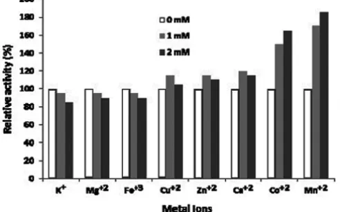
Fig 6.Effect of various metal ions on the relative activity of amyloglucosidase enzyme on substrate concen-tration of 0.2%, pH of 5.0, and temperature of 55 °C
of DEAE-cellulose column chromatography was shown a band with high density and the other band with very low density. While the fractions of chromatography column Sephadex G-75 only seen as single band with a very high density corresponding to molecular weight of 85 kDa (Fig. 3 in column 4), so that it can be concluded that the step of purification by column chromatography on Sephadex G-75 produces an enzyme with the highest purity compared to the previous purification step. The exact molecular weight of this enzyme will be analysis using 2D SDS-PAGE electrophoresis or ESI-MS spectroscopy in the future.
Similarly, the amyloglucosidase was produced from thermophilicPaecilomyces varioti, purified by Sephadex G-100 filtration and followed by ion exchange chromatography. It was found a single band protein with molecular weight of 86.5 kDa [11]. The molecular weight of amyloglucosidase in different organism sources have been reported in ranging from 49 to 112 kDa [12].
Characterization of Amyloglucosidase Enzyme
The result of the effect of pH on the enzyme activity of the purified enzyme from Sephadex G-75 column is showed in Fig. 4. The figure shows that the optimum pH of amyloglucosidase activity tested on starch substrate at 0.2% (w/v) was 5.0.
Aithal et al. [12], reported that amyloglucosidase from Rhizopus, sp had an enzyme activity with the optimum pH of 4.5 on raw starch of wheat, rice, jowar, corn, and soybean.
When the effects of temperature on the enzyme activity was evaluated (Fig. 5), the result showed that the enzyme activity increased when the temperature went up to 55 °C and after passing this temperature, the activity of enzyme decreased most possible due to the protein of enzyme started to get denaturated at higher temperatures. Thus the optimum temperature for the amyloglucosidase enzyme produced by E. fibuligeracells incubated in sago starch substrate of 0.2% (w/v) is 55 °C. Mittal et al. [6] studied amyloglucosidase production by Aspergillus awamori NA21 and characterization of the enzyme using tapioca powder as its substrate. The enzyme produced showed a high specific activity with the activity value of 0.347 U/mg protein under standardized optimum conditions. The optimum activity of this enzyme was achieved at a temperature of 60 °C and at pH 5.0. Another researcher reported that amyloglucosidase from Corticium rolfsii had an optimum temperature of 65 °C when the bacteria was incubated in a raw starch substrate [14].
The pattern of activator or inhibitor effects of metal ions on the amyloglucosidase activity of E. fibuligera cells was studied by using K+, Ca+2, Mg+2,
Cu+2, Zn+2, Fe+3, Co+2, and Mn+2 ions. Results of
research showed that the amyloglucosidase activities was strong activated by addition of 2 mM Co2+ (165%)
and 2 mM Mn2+ (185%) ions, whereas the activities
(89%), and 2 mM Fe3+(90%) ions when compared
without addition this ions as control (Fig. 6). The result was similar to the one given by amyloglucosidase of soybean [15], the enzyme could be activated by added 1 mM Mn2+ and Co2+. Another researcher reported that 1
mM Mn2+ and Cu2+, respectively, could activate of the
amyloglucosidase enzyme [16]. Activator or inhibitor compounds at the certain amount can enhance the rate of enzyme catalyzed enzymatics reaction, but the excess amount of the activator can cause the competition of the free activator or inhibitor and the activator or inhibitor substrate complex to free enzyme.
CONCLUSION
The addition of sago starch as much as 0.75% (w/v) in the fermentation medium of E. fibuligera could produce amyloglucosidase with the highest activity resulting in the enzyme activity of 0.96 U/mg protein. Amyloglucosidase enzyme purification by Sephadex G-75 column provided the highest purity which was 22.5 times greater than the crude enzyme. Characterization of the purified amyloglucosidase to determine the optimum conditions revealed that the pH and temperature, values of 5.0 and 55 °C, respectively, could be considered as its optimum conditions, when the medium contained the sago starch substrate at a level of 0.2% (w/v). The amyloglucosidase activities was strongly increased by addition of Co2+ and Mn2+ ions, whereas
the activities were weakly decreased by addition of K+,
Mg2+, and Fe3+ions.
Based on the obtained results above, to further increase the activity of the enzyme produced it is necessary to determine the optimum conditions for other factors such as concentration of substrate and enzyme and time of incubation. In addition, the comparison between the activity of an amyloglucosidase enzyme naturally produced by the yeast cells with a genetically modified one needs to be evaluated. In the future, it also needs to investigate the optimal conditions of this enzyme preparation using sago starch as its medium substrate to produce glucose that can be used as a basic material for bioethanol production.
ACKNOWLEDGEMENT
The authors thank the Head of Laboratory of Bioprocess, Department Chemical Engineering, and Polytechnic State Ujung Pandang, Indonesia for sample
preparation. The authors also thank to Ika Septiany for technical assistance to conduct this work and Ronny Horaks for editorial reading of the manuscript.
REFERENCES
1. Gašperík, J., Kováč, L., and Mináriková, O., 1991,
Int. J. Biochem., 23 (1), 21–25.
2. Manera, A.P., Kamimura, E.S., Brites, L.M., and Khalil, S.J., 2008,Braz. Arch. Biol. Technol., 51 (5), 1015–1024.
3. Mohamed, L., Zakaria, M., Ali, A., Youssfie, E.K., Mohamed, E., Mohamed, O., El Hassan, B., and Mohamed, J., 2007, Pak. J. Biol. Sci., 10 (19), 3322–3329.
4. Shimazaki, T., Hara, S., and Sato, M., 1984, J. Ferment. Technol., 62, 165–170.
5. Hasan, F., Shah, A.A., and Hameed, A., 2006, Enzyme Microb. Technol., 39 (2), 235–251.
6. Mittal, A., Aggarwal, N.K., Gupta, V., Singh, G., and Yadav, A., 2013, Octa J. Biosci., 1 (2), 122– 131.
7. Scopes, R.K., 1982,Protein Purification: Principles and Practices, Springer Verlag, New York. 39–59. 8. Colowick, S.P., and Kaplan, N.O., 1957, Methods
in Enzymology, vol. I, Academic Press Inc. Publisher, New York.
9. Deutscher, M.P., 1990, Methods in Enzymology: Guide to Protein Purification, vol. 128, Academic Press Inc. Publisher, New York.
10. Sambrook, J., Fritsch, E.F., and Maniatis, T., 1982. Molecular Cloning, Cold Spring Harbor Laboratory, New York, 73–177.
11. Michelin, M., Ruller, R., Ward, R.J., Moraes, L.A.B., Jorge, J.A., Terenzi, H.F., and Polizeli, L.T.M., 2008,J. Ind. Microbiol. Biotechnol., 35 (1), 17–25. 12. Selvakumar, P., Ashakumary, L., Helen, A., and
Pandey, A., 1996, Lett. Appl. Microbiol., 23 (6), 403–406.
13. Aithal, S.C., Sawalikar, A.A., and Manwar, A.V., 2011,Bionano Front., 4 (1), 31–35.
14. Nagasaka, Y., Kurosawa, K., Yokota, A., and Tomita, F., 1998, Appl. Microbiol. Biotechnol., 50, 323–330.
15. Prakash, O., Jaiswal N., and Pandey, K., 2011, Asian J. Biochem., 2 (1), 1–9.