RESPIRATORY PHY SIOLOGY TEACHING:
DETERMINATION OF RESIDUAL
VOLUME BY APPLY ING THE
INDICATOR-DILUTION TECHNIQUE
Hartmut Heller, Klaus Granitza, and Bernd Eixmann
Depa rtm ent of Physiology, University of Bonn, 53115 Bonn, Germ a ny
A
part from the current teaching of spirometric methods in laboratory courses on respiratory physiology, we have included an experiment in which medical students determine their own residual volume by applying the indicator-dilution technique. For hygienic reasons we used a bag-in-the-box system to dilute helium within alveolar space by performing the single-breath method. Although each participant independently underwent only one single-breath maneuver, we gained a reliable relationship between residual volume and subjects’ height and body weight in 68 female (r50.6,P,0.0001) and 99 male (r50.42,P,0.0001) students. From this successful outcome and with the opportunity to discuss the limitations of the single-breath method as well, we inferred that this experiment affords a transparent and instructive approach to interpreting the determination of lung volumes on the basis of the indicator-dilution technique.AM. J. PHYSIOL. 274 (ADV. PHYSIOL. EDUC. 19): S53–S56, 1998.
Key words:single-breath method
Most medical students are familiar with spirometric methods of depicting lung volumes graphically. Al-though those measurements are commonly included as an essential part of teaching physiology, methods of determining residual volume (RV), whether by apply-ing body plethysmography or spirometry, are not easy to demonstrate. We have successfully included the relatively transparent and instruc tive indic ator-dilution technique to determine RV in our physiology laboratory courses as described in this report. For hygienic reasons, we used the single-breath method to dilute helium (He).
MATERIALS AND METHODS
The experiments described here were carried out in the Department of Physiology of the University of
Bonn by groups consisting of 12–16 medical students and 1 medical doctor. The students gave their in-formed consent and were introduced to the experimen-tal procedure but were not specially trained. Each student who participated in the experiments was assisted by a colleague who performed all measure-ments required for calculating RV. In addition, the students underwent common spirometry (Keilbalg-spirometer, Vitalograph, Hamburg, Germany) to deter-mine their respective values for vital capacity, forced expired volume (1 s), and expiratory peak flow.
For RV experiments, we used an inspiratory gas mixture containing 9% He in compressed air. This test gas was put into the bag of a self-made bag-in-the-box system immediately before registration of each
I N N O V A T I O N S A N D I D E A S
1043 - 4046 / 98 – $5.00 – COPYRIGHTr1998 THEAMERICAN PHYSIOLOGICAL SOCIETY
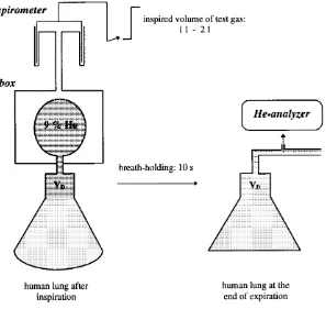
measurement. After expiring their vital capacity in a sitting position, the volunteers were asked to inhale 1–2 liters of test gas (with nose closed), to hold their breath for 10 s, and to finally expire maxi-mally through a stainless steel tube (volume 5 0.5 liter). From this tube, the alveolar gas sample was continuously sucked into a He analyzer (He-Test, E. Jaeger, Wu¨ rzburg, Germany) to measure the frac-tional concentration of He within the end-tidal gas mixture (FAHe). The fractional concentration of in-spired He (FIHe5 9%) and an inspired volume of 1–2 liters turned out to provide an optimal consistency of results, reducing the cross talk contribution of oxygen to the He measurements. A spirometer (Spiro-Junior, E. Jaeger), which was connected to the inside of the box (i.e., the outside of the bag), was used as a volumeter to indicate inspired volume of test gas (VI). A scheme of the experimental procedure is given in Fig. 1.
Before these experiments were established within our laboratory courses, 20 single-breath maneuvers were respectively performed by two scientists of the Depart-ment of Physiology, yielding a coefficient of variation (SD/mean) of ,4% in the determination of RV. In addition, each of the two subjects underwent a further set of 20 measurements of RV by applying the multiple-breath nitrogen washout method without obtaining significantly different results: 1,756645 (mean6SD) vs. 1,733 6 50 ml (He dilution) and 1,620 6 47 vs. 1,597660 ml (He dilution). Nonetheless, this compari-son indicates that the He measurements tend to reveal underestimates of true RV. This may be caused by a more adequate indicator gas mixing within alveolar space by the multiple-breath methods (1).
Using the indicator-dilution technique, we had the opportunity to teach the development of the mass-balance equation, required to calculate RV. Because
FIG. 1.
Scheme of ex perimental pr ocedur e to deter mine r esidual volume by applying He-dilution technique. VD, anatomical dead space.
I N N O V A T I O N S A N D I D E A S
He represents an inert gas of low solubility, the amount of test gas, diluted within RV, should not change significantly during the breath-holding period of 10 s, and therefore
(VI2VD).FIHe5(RV1VI2VD).FAHe (1)
where VDis anatomical dead space. SolvingEq. 1 for RV then reveals
RV5(VI2VD).
FIHe2FAHe
FAHe
(2)
To keep the experimental setup simple, VD was not measured separately, but was assumed to be 150 ml in each subject.
Each participant was asked to compare experimen-tally determined and predicted values of RV, using known equations from the literature (2).
RESULTS
Table 1 contains the volunteers’ respective character-istics, given as means6SD. As shown here, we found the expected significant differences between women and men in height, weight, body surface, body mass index, and, of course, in RV when values were subjected to Student’s pairedt-test.
Figure 2 shows the interrelation between RV and height as well as body weight obtained from the female and male partic ipants. Linear regression
TABLE 1 Subject characteristics
Women Men P
Age, yr 2463.2 2463.7
Height, m 1.7060.06 1.8360.06 0.001 Weight, kg 5966 7468 0.001 Body surface, m2 1.6860.10 1.9660.13 0.001
BMI, kg/m2 20.662.2 22.361.8 0.001
RV, liters 1.4660.27 1.5760.26 0.01 Values are means6SD of 68 female and 99 male medical students. BMI, body mass index; RV, residual volume.
FIG. 2.
Dependence of r esidual volume on height and body weight as deter mined in 68 female (A) and 99 male (B) medical students.
I N N O V A T I O N S A N D I D E A S
analyses revealed
residual volume5 21.7812.63.height
20.02.weight (3)
as well as
residual volume51.8610.73.body surface
20.079.body mass index (4)
(Eqs. 3 and4: n 5 68, r 50.6, P, 0.0001) for the female students and
residual volume5 21.7212.29.height
20.012.weight (5)
as well as
residual volume51.6110.75.body surface
20.068.body mass index (6)
(Eqs. 5 and6: n 599,r50.42, P,0.0001) for the male students.
DISCUSSION
During each seminar the pros and cons of the indicator-dilution technique, as applied here, were discussed.
1) Under the assumption that VDalways amounts to 150 ml and with an inspiratory volume averaging 1.5 liters, even large errors in assuming VD were only weighted by 10%. For our purposes in this case such a degree of uncertainty was quite acceptable.
2) When single-breath maneuvers are performed in cases of obstructive or restrictive ventilation disor-ders, an inhomogeneous distribution of inert gases within alveolar space is very likely (1), leading to a false determination of residual volume. Therefore, those of our students who were suffering from a common cold were not included in the measurements
(our findings thus represent an unbiased sample of healthy, young volunteers).
3) In the case of patients with pulmonary diseases, multiple-breath or plethysmographic tec hniques should be used to exclude inadequate gas mixing (1).
4) With the use of the bag-in-the-box system, the test gas was always inhaled from the bag, which was always filled up from the same gas bottle. Because inspiratory and expiratory pathways were separated from each other, this equipment enabled us to attend to one student group within an hour but without any danger of airborne infection.
5) Only 5% of the participants showed a significant deviation between measured and predicted values (2) of RV.
6) Our students were able to gauge their own total lung capacity without having to employ extensive means.
CONCLUSION
Although each medical student normally underwent only one single-breath maneuver, and despite the fact that this technique particularly depends on complying meticulously with the experimental procedure, we gained reliable results when comparing measured and predicted values of residual volume in each subject. This successful outcome may reflect the eager partici-pation of our volunteers. We therefore believe that the determination of residual volume was and is well accepted by our medical students.
Address for reprint requests: H. Heller, Dept. of Physiology, Univ. of Bonn, Nussallee 11, D-53115 Bonn, Germany.
Received 26 March 1997; accepted in final form 12 March 1998.
Refer ences
1. Anthonisen, N. R.Tests of mechanical function. In:Ha ndbook of Physiology.The Respira tory System.Mecha nics of Brea th-ing. Bethesda, MD: Am. Physiol. Soc., 1986, sect. 3, vol. III, pt. 2, chapt. 44, p. 753–784.
2. Islam, M. S., and W. T. Ulmer. Referenzwerte der ventilato-rischen Lungenfunktion.Pra x. Klin. Pneum ol. 37: 9–14, 1983.
I N N O V A T I O N S A N D I D E A S