i
IDENTIFICATION OF GENETIC DEFECTS
INVOLVED IN X-LINKED MENTAL RETARDATION
THESIS
Submitted to fulfill the assignment and fit-out requisite in passing
Post-graduate Program Majoring Genetic Counselling
Diponegoro University Semarang
Magister of Biomedical Sciences
ADITIA RETNO FITRI
G4A007042
Post Graduate Program
Diponegoro University Semarang
ii Revised Thesis
IDENTIFICATION OF GENETIC DEFECTS IN X-LINKED MENTAL RETARDATION
By Aditia Retno Fitri
G4A007042 Approved by,
The Netherland Indonesia
Principal Supervisor, Principal Supervisor,
Arjan de Brouwer, PhD Prof Sultana MH Faradz, MD, PhD NIP. 130 701 415
Supervisor, Supervisor,
Janneke Schuurs-Hoeijmakers, MD DR. dr. Mexitalia Setiawati EM, SpA (K) NIP. 19670227 1995092001 Supervisor,
Helger Yntema, PhD
Recognition,
Head of Master’s Degree Program in Biomedical Science Post Graduate Program Diponegoro University
iv TABLE OF CONTENTS
TITLE i
APPROVAL SHEET ii
REVISION APPROVAL FORM iii
TABLE OF CONTENTS iv
DECLARATION
ACKNOWLEDGEMENT
LIST OF FIGURES, TABLES AND APPENDIX
vi
CHAPTER I INTRODUCTION 1
I.1 Backgrounds 1
I.2 Research questions 3
I.2.1 General research questions 3
I.2.2 Specific research questions 3
I.3 Research purpose 3
I.4 Research advantages 4
I.5 Research originality 4
CHAPTER II LITERATURE REVIEW 7
II.1 Mental Retardation 7
II.1.1. Definition 7
II.1.2. Classification 7
II.1.3 Prevalence 8
II.1.4. Etiology 8
II.2. X-linked Mental Retardation 11
II.2.1. Definition 11
II.2.2. Prevalence 12
II.2.3. Classification of XLMR 12
II.2.4. Identification of genetic defects involved in XLMR 13
II.2.4.1. Positional Cloning 13
II.2.4.2. Positional Candidate Gene Analysis 17
II.2.4.3. Mutation analysis of known gene 19
II.2.4.4. Array technology 20
II.2.4.5. Next generation sequencing 20
II.3. X-Chromosome Linkage Analysis
II.6. Theoritical Framework 25
CHAPTER III RESEARCH METHOD 26
v
III.1.1. Research Field 26
III.1.2. Research Location 26
III.1.3. Research Period 26
III.1.4. Research Design 26
III.1.5. Variables 26
III.1.6. Operational Definition 27
III.1.7. Research Protocol 28
III.2. Method 29
III.2.1.Population 29
III.2.2. Samples 29
III.2.2.1 Inclusion Criteria 29
III.2.2.2 Exclusion Criteria 29
III.2.2.3. Clinical Examination 29
III.2.2.4. Sample Collection 30
III.2.2.5 Minimum samples required 30
III.3. Work-flow 30
III.3.1 General 30
III.4 Collected Data 31
III.4.1 Primary Data 31
III.4.2. Secondary Data 31
III.5 Data analysis 31
III.6. Ethical Implication 31
CHAPTER IV. RESULTS AND DISCUSSION 33
IV.1 Clinical Findings 33
IV.2 Conventional Cytogenetic analysis 34
IV.3 Fragile-X exclusion test 34
IV.4 Linkage Analysis 34
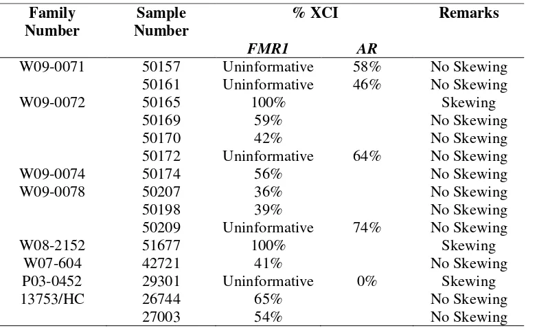
IV.5 X-Chromosome Inactivation Analysis 35
IV.6 Mutation analysis in Candidate Genes 36
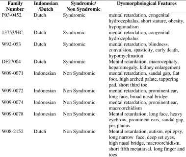
IV.7 Results and discussion for each family 37
Syndromic XLMR Families
Family W92-053 (XLMR and hypomyelination family) 37
Family P03-0452 and 13753/HC (XLMR with Hydrocephalus families) 44
Family W07-604 53
Family DF27004 (MR and Overgrowth Features) 57
Non Syndromic XLMR Families
CHAPTER V CONCLUSION AND SUGGESTION 78
REFERENCES xvii
vi DECLARATION
I hereby declare that this submission is my own work and that to the best of my knowledge and belief, it contains no material previously published or written by another person nor material which to a substantial extent has been accepted for the award of any other degree or diploma of the university or other institute of higher learning, except where due acknowledgement is made in the text.
Semarang, August 2010
vii ACKNOWLEDGEMENTS
The report will not be accomplished without assistance from various
individuals and organizations both in Indonesia and Netherlands. First and
foremost, I would like to express my immeasurable thanks to Arjan PM de
Brouwer, PhD as my supervisor for all his guidance, knowledge sharing, also time
and patience to teach me. His wide knowledge and his logical way of thinking
have been of great value for me. His understanding, encouraging and personal
guidance have provided tremendous basis for the present thesis. I would like to
express my deepest and sincere gratitude to my supervisor, Prof. dr.Sultana MH
Faradz, PhD who gave me this precious opportunity to study and work in this
project. I would like to thank her for all the valuable guidance, attention, time and
continuous support. Her hard effort and willingness to motivate contributed
tremendously to this research. I heartily thankful to my supervisor,
Janneke-Schuurs Hoeijmakers, M.D. whose encouragement, intensive guidance and
support enabled me to develop an understanding of the subject. She gives me a
lifetime unforgettable memory of her caring, patience, encouragement and
untiring help during my difficult moments. I wish to express my warm and sincere
thanks to Dr.dr.Mexitalia Setiawati EM SpA(K) as my supervisor for all her
guidance and also detailed and constructive comments throughout this work. I am
deeply grateful to Helger Yntema, PhD as my supervisor for all her guidance and
knowledge sharing.
My sincere thanks are due to dr. Tri Indah Winarni, MsiMed for her
valuable guidance and friendship help in cytogenetic analysis. I am grateful to
dr.Farmaditya EP Mundhofir, Msi.Med for his kind help in referring samples. I
wish to extend my warmest thanks to all those who have helped me with my work
in the Division of Human Genetics CEBIOR Faculty of Medicine Diponegoro
University in Semarang Indonesia for your kind help and friendship. Particularly I
would like to thank Ardina Apriliani, Rita Indriyati, Wiwik Lestari, Lusi Suwarsi
viii During this work I have undertake a precious opportunity to do internship
in Department of Human Genetics Radboud University Nijmegen Netherlands. I
would like to express my warmest gratitude to Prof Ben Hammel, M.D, PhD for
giving me this precious opportunity to do internship in Department of Human
Genetics RUNMC Netherlands. I would like to express my sincere thank to Prof.
Dr. Ir Hans van Bokhoven for allowing me to join his group. It is my honorable
pleasure to be able to work in Mental Retardation Group Department of Human
Genetics, Radboud University Nijmegen Netherlands. I would like to thank to all
the staff of Department of Human Genetics for their kind friendship and
cooperation during my stay in Nijmegen. My warm thanks are due to Jaap
Oostrik, Martina van Ruiterkamp, Erwin Khuny and Rosangela Artuso for their
guidance and assistance in molecular genetic techniques. I wish to thank dr. Bert
van de Vries, M.D for his kind help in clinical dysmorphology analysis. I wish to
thank to my group member, Linda Peters, Nick Rossen, Frank tel Est, Zafar
Iqbal,Msc, Drs Bregje van Bonn for their kind hospitality and for making my feel
at home even in The Netherlands. I wish my thanks to Will Groenen, Inneke
Zaalmink, and Bregje van Hellemondt for their sympathetic help in arranging my
stay in Nijmegen.
This work would have been impossible without generous help and support
of my colleagues in Indonesia. I would like to express my gratitude to Prof.
Soesilo Wibowo, MD, PhD (The President of Diponegoro University Semarang
Indonesia), Soejoto M.D. (The Dean of Faculty of Medicine Diponegoro
University Semarang Indonesia), Dr.dr Winarto SpMK, SpM(K) (Head of
Biomedical Science Post Graduate Program Faculty of Medicine Diponegoro
University Semarang Indonesia) and Director of Center for Biomedical Research
Prof Sultana MH Faradz, MD, PhD. I wish to thanks dr. Alifiani Hikmah SpA(K),
dr Hendriani Selina SpA(K), MARS, Dr.dr.Andrew Johan, Msi, dr. Suhartono
Mkes for their suggestion and advice to improve my thesis.
In the last two years I met and worked with people that became a second
family for me and gave me a lot of love. My special thanks to all my Genetic
ix SpPD, dr Stefanus SpA, Amal, Costrie, Santoso, Liha, Mahayu, Meira, Safrina,
Dewi and Galuh for the knowledge sharing, friendship, and support during the
difficult moments. Besides, I would like to thank the authority of Indonesian
Ministry of National Education for providing us with Excellence scholarship as
facilities to complete this master study.
Above all, I most thankful my husband who stood beside me and
encouraged me with unconditional love constantly throughout this study. Finally,
an honorable mention goes to my parents for always support and prays for me.
Without their encouragement and understanding it would have been impossible
for me to finish this work.
Finally, I would like to give my special gratitude to all the families who
participated in this study. Thank you for give me a chance to learn.
x LIST OF FIGURES
Figure 1. Etiology of mental retardation
Figure 2. Main genetic causes of mental retardation
Figure 3. Ideogram of human X-chromosome showing genetic heterogeneity of XLMR.
Figure 4. Concept of data fusion
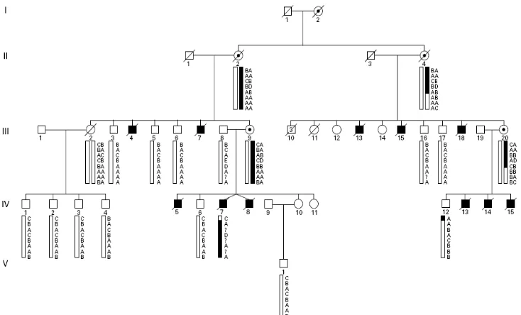
Figure 5. Multiple steps in linkage analysis Figure 6. Haplotypes within family W92-053
Figure 7. Top 25 candidate genes according to candidate gene prioritization with ENDEAVOUR for family W92-053.
Figure 8. Pedigree and haplotypes within family P03-0452. Figure 9. Pedigree and haplotype within family 13753/HC.
Figure 10. Candidate genes in the overlapping interval between family P03- 0452, 13753/HC, and previous reported family by Strain et al. Figure 11. Top 20 candidate genes according to candidate gene prioritization
with ToppGene for hydrocephalus families.
Figure 13. 250K SNP array data on the family W07-604 patient III.9 Figure 14. Family DF27004: Pedigree and haplotypes.
Figure 15. Family W090-0071: Pedigree and haplotype. Figure 16. Clinical pictures of family W09-00071. Figure 17. Family W090-0072: Pedigree and haplotypes. Figure 18. Clinical picture of family W09-072:
Figure 19. Family W090-0074: Pedigree and haplotype. Figure 20. Pasien 50176 (A) and patient 50177 (B). Figure 21. Family W090-0078: Pedigree and haplotypes. Figure 22. Patient 50196 from Family W09-078. Figure 23. Family W08-2152: Pedigree and haplotypes.
xi LIST OF TABLES
Table1. Research originality in matriks form
Table 2. Genes known to be mutated in non syndromic XLMR Table 3. Comparisson between Endeavour and ToppGene Table 4. Summary of Dysmorphological Features
Table 5. CGG repeat sizes in subject screened by FMR1 analysis Table 6. Linkage analysis result in all families
Table 7. XCI status for Females Table 8. Candidate genes sequencing
Table 9. XLMR syndromes associated with neurological features and early death Table 10. Clinical Comparisson between Family W92-053 and previous patients
with mutation in HSD17B10
Table 11. Two-Point LOD Scores for 16 X-Chromosomal Markers of family P03- 0452
Table 12. Two-Point LOD Scores for 16 X-Chromosomal Markers of family 13753/HC
Table 13. Summary of known loci of hydrocephalus in vertebrates
Tabel 14. Comparisson of clinical features of patient IV.9 from family W07-604 with previous reported cases of 9p deletion syndromes.
Table 15. Two-Point LOD Scores for 16 X-Chromosomal Markers of family DF27004
Table 16. Two-Point LOD Scores for X-Chromosomal Markers of family W09-071 Table 17. Two-Point LOD Scores for 16 X-Chromosomal Markers of family W09-
072
Table 18. Two-Point LOD Scores for 16 X-Chromosomal Markers of family W09-074
Table 19. Two-Point LOD Scores for X-Chromosomal Markers of family W09-078 Table 20. Two-Point LOD Scores for X-Chromosomal Markers of family W08-
2152 LIST OF APPENDIX
Appendix I Chromosomal Preparation Appendix II DNA Isolation
Appendix III FMR1 gene amplification
Appendix IV X-Chromosome Linkage Analysis
Appendix V X-Chromosome inactivation (XCI) analysis Appendix VI Candidate Gene Selection
Appendix VII Proposed Workflow for XLMR studies
Appendix VIII Proposed Workflow for X-linked Hydrocephalus and MR Appendix IX Form of clinical examination
Appendix IX Informed consent form for Indonesian Patients
xii ABBREVIATIONS
ACSL4 Acyl-CoA synthetase long chain family member 4 AGTR2 Angiotensin II receptor type 2
AP1S2 Adaptor-related protein complex 1. sigma 2 subunit AR Androgen Receptor gene
ARHGEF6 Rac/Cdc42 guanine nucleotide exchange factor 6 ARX Aristaless-related homeobox
ATRX α-thalassemia-mental retardation, X-linked
BRWD3 Bromodomain and WD repeat domain containing 3 CASK Calcium/calmodulin-dependent serine protein kinase CNV Copy Number Variations
CUL4B Cullin 4B
DLG3 Dics, large homolog 3 DNA Deoxyribo Nucleic Acid
DSM-IV Diagnostic and Statistical Manual of Mental Disorders-IV FGD1 FYVE, RhoGEF and PH-domain-containing 1
FISH Fluorescence In Situ Hybridization FMR1 Fragile X Mental Retardation 1 FMR2 Fragile X Mental Retardation 2 FTSJ1 Ftsj homolog 1
GDI1 GDP dissociation inhibitor 1
GRIA3 Glutamine receptor, ionotrophic, AMPA3 HUWE1 HECT, UBA-and WWE-domain containing 1 IL1RAPL1 Interleukin 1 receptor accessory protein-like 1 IQ Intelligence Quotient
JARID1C Jumonji, AT rich interact domain 1C LOD Logarithm of Odds
MAGT1 Magnesium transporter 1 MECP2 Methyl-CpG binding protein 2
MLPA Multiplex Ligation-Dependent Probe Amplification MR Mental Retardation
MRX Nonsyndromic X-linked mental retardation MRXS Syndromic X-linked mental retardation NLGN3 Neuroligin 3
NLGN4X Neuroligin 4, X-linked
OMIM Online Mendelian Inheritance in Man OPHN1 Oligophrenin 1
PAK3 p21 protein (Cdc42/Rsc)-activated kinase 3 PQBP1 Polyglutamine-binding protein 1
RPL10 Ribosomal protein L10
RPS6KA3 Ribosomal protein S6 kinase, 90kDa, polypeptida 3 RUNMC Radboud University Nijmegen Medical Center SHROOM4 Shroom family member 4
xiii SLC9A6 Solute carrier family 9 (sodium-hydrogen exchanger)
STR Short Tandem Repeats SYP Synaptophysin
TM4SF2 Transmembrane 4 superfamily member 2
UPF3B UPF3 regulator of nonsense transcripts homolog B XCI X-Chromosome Inactivation
XLMR X-linked mental retardation
ZDHHC9 Zinc finger, DHHC-type containing 9 ZNF41 Zinc finger protein 41
ZNF674 Zinc finger family member 674 ZNF711 Zinc finger protein 711
xiv CURRICULUM VITAE
Name : Aditia Retno Fitri Place/Date of Birth : Semarang/30 April 1984
Educational Background:
1990-1995 Karang Kumpul Public Elementary school, Semarang 1995-1998 Junior High School 2(SMP 2) Semarang
1998-2001 Senior High School 3 (SMA 3) Semarang 2001-2005 Bachelor of Medical Science
Faculty of Medicine Diponegoro University Semarang 2005-2008 Medical Doctor
Faculty of Medicine Diponegoro University Semarang 2008-2010 Master of Biomedical Science
Faculty of Medicine Diponegoro University Semarang
Working experience
2003-2004 Student asistant of laboratory, Department of Microbiology Faculty of Medicine Diponegoro University
2006-2006 Co-Assistant Fellowship in Department of Neurology and Neurosurgery University of Hiroshima, Japan
2008-present Lecture assistant, Department of Pharmacology Faculty of Medicine Diponegoro University 2009-2010 Intern student, Master student
Mental Retardation Group, Division of MolecularGenetics Department of Human Genetics
Radboud University Nijmegen Netherlands
Scholarship:
2006 Fellow Scholarship
Department of Neurology and Neurosurgery University of Hiroshima, Japan
2008 Excellence Scholarship, Master Program Indonesian Ministry of National Education
Scientific Article
2005 Fitri AR, Susilaningsih N.Nitric Oxide Production of C3H mice inoculated with adenocarsinoma mammae cell in the administration of Typhonium flagelliforme juice in
stratified doses. Undergraduate thesis. Faculty of Medicine Diponegoro University
2007 Frequency of Hepatitis B vaccination in Neonatals 0-2 months old on Primary Health Care Region of Balapulang 2007-2008. Clerkship thesis. Faculty of Medicine
xv IDENTIFIKASI DEFEK GENETIK PADA
X-LINKED MENTAL RETARDATION
Latar Belakang: X-linked mental retardation (XLMR) berperan pada 40% pria penderita retardasi mental (RM). Defek genetik berperan pada 50% kasus MR. Terdapat 56 loci XLMR non-sindromik (MRX) dan 35 loci XLMR-sindromik (MRXS) yang belum diketahui gen penyebabnya.
Tujuan: Identifikasi defek genetik pada keluarga XLMR.
Metode: Pemeriksaan klinis dan analisis sitogenetik konvensional dilakukan pada 4 keluarga MRXS dan 6 keluarga MRX, dilanjutkan analisis pengulangan CGG pada regio promoter FMR1. Analisis linkage dilakukan dengan STR-markers polimorfik pada kromosom X dilanjutkan perhitungan skor LOD. Dilakukan pemeriksaan status inaktivasi kromosom X wanita pembawa dengan metode FMR1 dilanjutkan metode AR bila tidak informatif untuk FMR1. Dilakukan pemilihan kandidat gen dalam linkage interval dan analisis mutasi.
Hasil: Tidak dijumpai adanya kelainan kromosom numerikal dan Fragile-X pada 10 keluarga ini. Terdapat variasi linkage interval antara 20 Mb hingga 121 Mb. Tidak terdapat mutasi HSD17B10, UBQLN2, SYP, ARGHEF pada keluarga W92-053 (XLMR dan hipomielinasi). Tidak terdapat mutasi SLITRK2 dan SLITRK4 pada keluarga P03-0452 dan 13753/HC (XLMR dan hidrosefalus). Tidak terdapat mutasi GPC3 pada keluarga DF27004 (XLMR dan pertumbuhan berlebih). Keluarga W092-053, PO3-0452, DF27004, dan W08-2152 menunjukkan penyimpangan inaktivasi kromosom-X pada wanita pembawa.
xvi IDENTIFICATION OF GENETIC DEFECTS INVOLVED IN
X-LINKED MENTAL RETARDATION
Backgrounds: X-linked mental retardation (XLMR) has been the focus of MR research because of 40% excess of males with MR. Genetic defects are estimated to account for 50% MR cases. There are still 56 non-syndromic (MRX) and 35 syndromic XLMR (MRXS) loci with unknown causative genes.
Aims: Identification of the genetic defects in XLMR families.
Methods: Four MRXS and 6 MRX families were studied. Clinical dysmorphologic examination and conventional cytogenetic analysis were performed followed by Fragile-X exclusion. Linkage analysis was conducted with highly polymorphic STR-markers on the X-chromosome followed by LOD scores calculation. An FMR1 X-inactivation assay was performed in 15 females from all families, followed by AR method if the result were uninformative for FMR1. Candidate genes were selected in linkage interval and mutation analysis was performed.
Results: Gross numerical chromosomal abnormalities and Fragile-X were excluded in all 10 families. Ten XLMR families showed intervals varying from 20 Mb to 121 Mb. Family W92-053 (mental retardation and hypomyelination) showed no mutation in HSD17B10, UBQLN2, SYP, ARGHEF. Two families with MR and congenital hydrocephalus (P03-0452 and 13753/HC) showed no mutations in SLITRK2 and SLITRK4. Family DF27004 (MR and overgrowth features) showed no mutations in GPC3. Family W092-053, PO3-0452, DF27004, and W08-2152 showed skewed X-inactivation in the obligate carrier female.
Conclusions: Genetic defects identification in ten families showed varying linkage intervals from 20 Mb to 121 Mb with varying LOD scores from 0,17 to 3.3, skewed X-inactivation in 4 families, and no mutation in the candidate genes. STR markers analysis was useful in determining linkage intervals, narrowing down the region of interest for further studies, and genetic counselling.
1 CHAPTER I
INTRODUCTION
I.1 Backgrounds
Mental Retardation (MR) is defined by IQ below 70 and adaptive behavior
limitations, which manifest before 18 years of age (Schalock et al. 2007). The prevalence of mental retardation is estimated to be about 1 to 3% of the general
population (Brosco et al. 2006). Mental retardation is the most common reason for referral to genetic services and one of the important unsolved problems in
healthcare (de Vries et al. 1997; Yeargin-Allsopp et al. 1997). Genetic defects are estimated to account for approximately 50% of cases (Leonard and Wen, 2002).
Genetic defects in MR consist of chromosomal abnormalities (structural and
numerical), single gene disorders, and multifactorial defect (Basel-Vanegeite,
2008).
X-linked mental retardation (XLMR) is characterized as mental retardation
with a distinctive pattern of inheritance, associated with X-chromosome (Ijntema,
2001). XLMR has been the focus of MR research for over three decades because
of the fact that there is 40% excess of MR males (Yeargin-Allsopp et al. 1997; Leonard and Wen, 2002). In this case, linkage to the X-chromosome can be
established in families with only 2 male patients and one obligate female carrier,
such as nephew uncle families. In addition, instead of the whole genome, only
X-chromosome needs to be considered (de Brouwer et al. 2007). XLMR is a heterogeneous disorder for which many of the causative genes are still unknown,
although 69 genes have been identified as causing syndromic XLMR and 33
genes as causing non syndromic XLMR (18 causing both syndromic and
non-syndromic XLMR) (Greenwood Genetic Center, 2010). However, there are still
56 non syndromic and 35 syndromic XLMR loci for which the gene defect is still
unknown (Gecz et al. 2009). It is assumed that most of these loci represent separate, novel XMLR genes, suggesting that there are still more than 80 MR
2 Genetic defect identification in MR is a challenging process. The first step
in elucidating genetic defect in MR is a thorough clinical work-up, which could
screen acquired factor from anamnesis and also largely known syndrome for
example Down Syndrome and Fragile X from the clinical features (Lugtenberg et al. 2006). The pedigree taken also could show the possible mode of inheritance of the disorder. The next step is exclusion other cause of mental retardation.
Considering the large role of chromosomal aberration in MR (11%; Stevenson et al. 2003), in the following step patients are routinely screened for large chromosomal aberration by conventional karyotyping. In addition, considering
that Fragile X is the most common inherited MR syndrome with incidence of
1/3000 male, in the next step all the patients are screened for CGG repeat
expansion in 5’ untranslated region of FMR1 that cause Fragile X syndrome (de Vries et al. 1997). Before researchers could identify and finally sequence the gene responsible for a disease, the gene location first must be mapped in the genome.
Linkage analysis is a method that allow to rule out regions of chromosomes that
are likely to contain a risk gene in the linkage interval, and determine areas where
there is a low chance of finding a risk gene (Massanet, 2009). This approach use
the polymorphism character of microsatellite markers (STRs: short tandem
repeats) -a short blocks (often less than 150 bp) of simple repetitive sequences (1-4 bp) dispersed randomly across all chromosomes- for mapping the linkage interval (Stopps and McDonald, 1998). Once a linkage interval is located in a chromosome, candidate genes within the interval could be selected based on its characteristics,
for example its expression in brain, inactivation status by X chromosome
inactivation (XCI) mechanism, homology to known MR genes, etc (Lugtenberg et al. 2006). This approach is called positional candidate gene analysis approach, which is assumed to be able to largely reduce cost needed compared to screening
the whole genomes (Stratchan and Read, 1999).
National Survey, SUSENAS in 2000 reported 384.818 person with MR in
Indonesia (0,19% (National Survey, 2000)). Underreporting is likely considering
the widespread stigma (Komardjaja, 2005; Gabel, 2004) and discrimination (Kats,
3 in Asian cultures and the lack of research on mental retardation in Indonesia. As a
developing country, there is no health care insurance system for all citizens
making costs for medical care for MR patients unbearable for their families.
Associated mental impairment, high risk of recurrence, and no therapy available
makes genetic counseling essential for families with mental retardation to prevent
recurrence of similarly affected children in the family. The understanding of the
molecular basis of MR will lead to improvement in diagnostic testing, genetic
counselling and also future therapeutics (Basel-Vanagaite, 2008). In contrast to
the extensive research performed on XLMR in European countries, so far there
are only a few XLMR studies performed in the Indonesian population, for
example Fragile X screening by Faradz et al in 1998 and subtelomeric duplication
screening by Mundhofir et al 2008, and no study about other XLMR. Currently,
there are no researchprotocols nor diagnostic workflows for XLMR in general in
Indonesia. Thus, this study aims to identify genetic defects involved in XLMR by
using positional candidate gene analysis approach which can be used for genetic
counselling purposes and to set up a basic workflow for future XLMR studies in
Indonesia.
I.2 Research questions
I.2.1. General research question
What genetic defects found in the XLMR families of this study?
I.2.2. Specific research questions
1. What linkage intervals do we observe in the XLMR families?
2. What is the X-inactivation status of the females carriers within these
families?
3. What are the best candidate genes within the linkage intervals in these
families?
I.3 Research purposes
I.3.1. General research purposes
4 I.3.2. Specific research purposes
1. Identify linkage intervals within XLMR families.
2. Measure X-inactivation status of the females carriers in these families.
3. Select new candidate genes within the linkage intervals in these families.
I.4 Research advantages
1. This study will contribute to the understanding of the molecular basis of
MR in XLMR families
2. Considering the current absence of an XLMR workflow in research and
diagnostics in Indonesia, this study will establish a starting point for
XLMR research and diagnostics by developing the first steps for a
diagnostic workflow for XLMR in Indonesia.
3. Linkage result from this study will help to show potential carrier which
can be used to perform genetic counselling prevent recurrence of similarly
affected children in the family
4. The technical knowledge, gained by this research will help to introduce
new techniques in genetic research in Semarang: e.g linkage analysis by
STR-markers.
5. Regarding the limited XLMR research in Indonesia, this study will
encourage other researchers to perform further studies in mental
retardation in Indonesia.
I.5 Research originality
1. To our knowledge this is the first study in Indonesia to identify the genetic
defects that cause X-linked mental retardation in Indonesian patients by
using linkage analysis.
2. Screening of Fragile-X and conventional cytogenetic abnormalities in
individual with mental retardation in Indonesia have been performed by
Faradz et al in 1998, but no other study known about other XLMR in
5 3. Mundhofir et al.in 2008 performed population screening of chromosomal
abnormalities, CGG repeat expansion in FMR1, subtelomeric deletion and
duplication (STD), and Prader Willy/Angelman Syndrome in mentally
retarded pupil in Semarang. No other study known about other MR in
Indonesia.
Table1. Research originality in matriks form
Title Author Method Result
6 patient showed fully
CGG repeat
7 CHAPTER II
LITERATURE REVIEW
II.1. Mental Retardation II.1.1. Definition
Mental retardation has been classified as disease category three decades
ago. According to DSM-IV in 1994, American Psychiatric Association defines
mental retardation as sub average intellectual functioning and concurrent deficits
or impairments in present adaptive functioning in at least two of the following
skill areas: communication, self-care, home living, social/interpersonal skills, use
of community resources, self-direction, functional academic skills, work, leisure,
health, and safety with onset before 18 years (APA, 1994). In 1996, WHO in the
guideline ICD-10, described mental retardation as a reduced level of intellectual
functioning, which decrease the ability to adapt with the daily needs of normal
social environment (WHO, 1996). Nowadays, we used definition developed by
AAMR on the 2002 AAMR Manual, which describe mental retardation as
significant limitation in both intellectual functioning and adaptive behavior, which
presents before age of 18 (Luckasson et al. 2002). Lately, term of “intellectual disability” is increasingly being used instead of “mental retardation” (Schallock et al. 2007).
II.1.2. Classification
There are several classification system of intellectual disability that are
currently used. The first classification is developed by WHO on 1996, and
summarized in ICD-10. WHO classified intellectual disability in axis I of five
axes of ICD-10 as: mild (IQ 50-69), moderate (IQ 35-49), severe (IQ 20-34), and
profound (IQ under 20) (WHO, 1996). The second classification is developed by
American Psychiatric Association on 2002 in multiaxial system DSM-IV TR,
which divided intellectual disability as mild (IQ 50-55 to 70), moderate (IQ 35-40
8 unspecified (strong presumption of MR but the intellegence untestable by
standard test) (First, 2004).
Based on the clinical features, intellectual disability could also be divided
into syndromic and non syndromic form. Syndromic forms of MR are
characterized by MR accompanied by either malformations, dysmorphic features,
or neurological abnormalities. Non syndromic MR are characterized by MR
without any additional features (Basel-Vanagaite, 2008). Nowadays, the
boundary between syndromic and non syndromic forms of MR is becoming
blurred due to the finding that in several genes, different mutations in the same
gene can result in both syndromic and non syndromic form of MR (Frints, 2002).
II.1.3 Prevalence
In 2002, Leonard et al made a meta-analysis about MR prevalence, and
estimated that MR is affecting 1-3% of general population (Leonard and Wen,
2002). The prevalence of intellectual disability in Asia seemed to be consistent
with Western population, which account about 0, 06-1,3% of total population,
except for China (6,68%) (Jeevanandam, 2009).
II.1.4. Etiology
The etiology factors of mental retardation is heterogeneous. In 2003,
Stevenson et al, based on a study cohort of 10,997 individuals with MR, found
that, a specific cause for the MR could be found in 43.5% of the cohort and that
genetic causes accounted for 28% of all cases and 63% of cases in which the
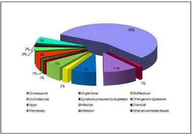
9 Figure 1. Etiology of mental retardation (adopted from Stevenson et al, 2003)
The etiology of intellectual disability are basically categorized as genetic,
acquired, and unknown causes (Moog, 2005). Acquired causes of MR can be
divided based on the timing of defect as: prenatal (for example: fetal alcohol
syndrome, teratogen exposure, toxoplasmosis), perinatal (for example:
intrapartum metabolic acidosis, early onset severe neonatal encepalopathy,
perinatal distress), and post natal (for example: traumatic brain damage, lead
intoxication) (Moog, 2005). Genetic causes of MR include chromosomal
abnormalities, monogenic disorder, and multifactorial causes (Figure 2; Moog,
10 Figure 2. Main genetic causes of mental retardation (Adopted from Basel-Vanegeite, 2008)
Chromosomal abnormalities as a cause of intellectual disability has been
recognized for many years. Trisomy 21 that cause Down syndrome is one of
recurrent chromosomal abnormality that cause mental retardation with the
incidence of 1/600 newborns (Hulten et al. 2008). Chromosomal abnormalities
cause cognitive impairment, which is also frequently with defects of heart
formation and dysmorphic features (Raymond and Tarpey, 2006), which
represent the most frequent cause of syndromic MR (Basel-Vanegeite, 2008).
Chromosome abnormalities with size of 3-5 megabases (Mb) can be detected by
conventional microscopic analysis of chromosomes isolated from peripheral blood
lymphocytes in ∼5% of patients with unexplained MR (Anderson et al. 1996; de Vries et al. 1997). In the early 1990s, with the introduction of fluorescence in situ hybridization (FISH), recurrent small microdeletions of the genome (with
maximum resolutin of 150 kb) not visible by light microscopy were identified
associated with characteristic syndromic MR (Raymond and Tarpey, 2006). By
the development of molecular cytogenetic techniques, such as FISH and multiplex
ligation-dependent probe amplification (MLPA) (Schouten et al. 2002), it is shown that causative submicroscopic rearrangements of the subtelomeric regions
11 al. 2004). In 2005, Van Karnebeek estimated that the frequency of deteced
chromosomal abnormalities is about 10%, ranging from 2% to 50% depending on
the variation in the study design among published report. (Van Karnebeek, 2005).
Nowadays, the focus of MR research has been shifted to identify smaller
chromosome abnormalities associated with disease, especially after introduction
of high-resolution array. During the past three years, numerous copy number
variations (CNVs) have been identified that are associated with MR and
developmental delay (Stankiewicz and Beaudet, 2007). Zahir and Friedman in
2007 estimated that pathogenic CNVs can be found in 10–15% of individuals with
idiopathic mental retardation (Zahir and Friedman, 2007). By the development of
array technology, even it is assumed that up to 25% of all cases of MR may be
explained by copy number-dependent gene dosage variations, although not all of
these variants will be fully penetrant, which create a challenge in clinical
interpretation (Vissers et al. 2009).
Monogenic disorders include autosomal dominant disorders, autosomal
recessive disorders, and X-linked disorders. Single-gene disorders have been
increasingly recognized to cause MR over the past half century. Searching in
McKusick catalogue of genes and phenotypes (Online Mendelian Inheritance in
Man (OMIM); OMIM, 2010) on January 2010 show 1629 entries associated with
“mental retardation”. In cohort study of 10,997 individuals with MR in 2003 by
Stevenson et al, it was found that 8% of MR in the cohort was caused by
single-gene disorders.
II.2. X-Linked Mental Retardation II.2.1. Definition
XLMR is defined as proportion of mental retardation indicating distinctive
pattern of inheritance associated with X-chromosome (Ijntema, 2001). General
characteristic of XLMR recessive inheritance are demonstrating the following
pattern: (Kingston, 2002)
− Only male affected almost exclusively.
12 − No male to male transmission.
− All daughters of affected males will be carriers.
II.2.2. Prevalence
Contribution of X-chromosome mutations to the spectrum of mental
retardation has become subject of interest for many years. It was Penrose in 1938
who reported for the first time that mental retardation is significantly more
common in males than in females, with the ratio of affected males to females
being 1.3:1 (Penrose, 1938). Following studies described large families with
X-linked inheritance pattern arising concept that X-X-linked genetic defects play an
important role in the etiology of MR. It was predicted that XLMR (including
monogenic and multiple gene XLMR) might be contribute to up to 20-25% of
mental retardation (Turner, 1996). In 2005, Roper and Hamel predicted that
monogenic XLMR might be contribute to up to 10-15% of mental retardation
(Ropers and Hamel, 2005). In 1980, Herbst and Miller estimate that the
prevalence of XLMR was about 1.83/1000 males (Herbst and Miller, 1980), with
the fragile-X syndrome being considered as the most prevalent condition (20% of
all XLMR cases) (Fishburn,1983). Later on, the estimation was reduced into 10–
12% of all MR cases in males by the finding of a much smaller contribution of
individual genes other than FMR1, to XLMR (Mandel and Chelly, 2004; Ropper
and Hamel, 2005).
II.2.3. Classification of XLMR
Kerr in 1991 suggested classification of XLMR into syndromic (MRXS)
and non-syndromic (MRX) (Kerr, 1991). Syndromic MRXS refers to condition
associated with distinctive clinical features. Nonsyndromic MRX is associated
with nonprogressive condition that affects cognitive function without any other
distinctive features (Gecz and Mulley, 2000). Trinucleotide repeat expansion on
FMR1 gene that cause Fragile X syndrome is generally regarded as the most
common cause of XLMR with the prevalence of 1/4000-1/8000 (Hagerman,
13 disorders have been described : 149 with specific clinical findings, including 98
syndromes and 51 neuromuscular conditions, and 66 nonspecific (MRX) forms
(Chiurazzi et al. 2008). More than 90 XLMR-associated genes have been identified, which at least 53 were for syndromic, 27 for nonsyndromic, and 11 for
both syndromic and nonsyndromic forms of mental retardation, which show the
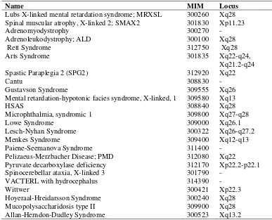
heterogeneity of XLMR (Figure 3; Table 2; Chiurazzi et al., 2008; Tarpey et al., 2009; Greenwood Genetic Center 2010; XLMR Website 2010).
II.2.4. Identification of genetic defects involved in XLMR
The effort to identification genetic defects involved in XLMR has been
developed since many years ago. There are several methods developed to address
this effort, namely positional cloning, candidate gene, mutation analysis of the
known gene, array method, and the newest, next generation sequencing.
II.2.4.1. Positional Cloning
Positional cloning is intended to localize determinants of disease
susceptibility in the DNA sequence prior to determining their function (Maniatis
et al., 2004). This method identifies a disease gene based on no information except its approximate chromosomal location. Linkage mapping is routinely used
to get the position information. In this method, it is important to define the
candidate region as tightly as possible, considering the disadvantage of this
method of being expensive and time- and resource- consuming (Strachan and
Read, 1999; Zhu and Zhao, 2007).
Later on, it was found that chromosomal aberrations can provide a useful
short-cut to locating a disease gene. Translocation could give a chance to clone
the X-chromosome gene which is disrupted by the translocation (Strachan and
Read, 1999). Small-scale deletions (microdeletions) are also valuable for
positional cloning in which the deletion could encompass gene that cause XLMR.
Using positional cloning methods, several MRX genes have been identified, for
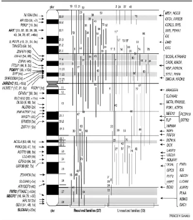
14 Figure 3. Ideogram of human X-chromosome showing genetic heterogeneity of XLMR. Genes in left side are currently known to be mutated in NS-XLMR
(n=38). Genes in the right side are known to be mutated in syndromic XLMR
(n=52). Vertical lines shows linkage interval in MRX families. Asterisks sign near
the gene names show genes which is mutated in both syndromic and
15 Table 2. Genes known to be mutated in non syndromic XLMR
Gene Symbol
Gene Name Protein Function Years Found OPHN1 Oligophrenin 1 Axon guidance,
signal transduction,
2000 Gibbons and Higgs, 2000
SLC6A8 Solute carrier family 6 (creatine), member
ZNF41 Zinc finger protein 41
Transcription regulation
16
Gene Symbol
Gene Name Protein Function Years Found
NLGN3 Neuroligin 3 Cell adhesion molecule, synaptic transmission
2003 Laumonnier et al. 2004
ZNF81 Zinc finger protein 81
Transcription regulation
2004 Kleefstra et al. 2004
DLG3 Dics, large homolog 3
Signal transduction, kinase, NMDA receptor localization
2004 Tarpey et al. 2004
FTSJ1 Ftsj homolog 1 Nucleolar protein, modification of rRNA
2004 Freude et al. 2004
JARID1C Jumonji, AT rich interact domain 1C
ZNF674 Zinc finger family member 674
Transcription regulation
2006 Lugtenberg et al. 2006
AP1S2 Adaptor-related
CUL4B Cullin 4B E3 ubiquitin ligase, proteolysis of DNA
UPF3B UPF3 regulator of nonsense transcripts
SLC9A6 Solute carrier family 9 (sodium-hydrogen exchanger)
Sodium ion transport, pH regulation
17
Gene Symbol
Gene Name Protein Function Years Found
ZNF711 Zinc finger protein 711
DNA replication 2009 Molinari et al. 2008
CASK
Calcium/calmodulin-SYP Synaptophysin Synaptic vesicle maturation and membrane organization
2009 Tarpey et al. 2009
(adopted from Gecz, 2009)
II.2.4.2. Positional Candidate Gene Analysis
A purely positional approach is often inefficient because candidate regions
identified by positional cloning usually contain dozens of genes, which will be
time-consuming and labour-consuming to screen them all. This matter can be
resolved by combining both positional and non positional information in a
positional candidate gene approach. This method uses mutation analysis of the
most promising functional candidate genes encompassed by linkage intervals
(Strachan and Read, 1999). Several genes that have been found by this approach
are: GDI1, PAK3, and RSK2 (Mulley, 2008). This method brings advantages for being effective and economical method for direct gene discovery. However, the
practicability of this approach is limited by its reliance on prior knowledge about
the known or presumed biology of the phenotype under investigation, necessity of
discrete phenotypic differences, and also necessity of highly subjective in the
process of choosing specific candidates from numbers of potential possibilities.
(Zhu and Zhao, 2007).
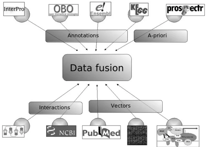
Nowadays, the technology development emerges several bioinformatic
tools that could help in candidate gene prioritization. This tools use concept of
data-fusion (Figure 4) which prioritizing candidate genes based on combined
information from many sources, including converging actual experimental data,
web database-based resources (including literature-based resources and biological
18 molecular interaction principles, e.g., gene structure variation, homologs,
orthologs, SNPs data, protein-DNA interactions, protein-protein interactions
(interactome), molecular module, pathway and gene regulatory network (Aerts et
al. 2006). Several bioinformatic tools that use data-fusion for prioritizing
candidate genes are Endeavour (Aerts et al. 2006) and ToppGene (Chen, 2007).
Figure 4. Concept of data fusion (adopted from KU Leuven, 2010).
Both Endeavour and ToppGene use the training genes, genes already
known to be involved in the process under study, as model. Then, the model is
used to score the candidate genes based on the similarity and rank them according
to their score (Chen, 2007). The basic difference of those software is that
Endeavour use Blast, cis-element and transcriptional motifs in sequence features
and annotation, while ToppGene not. The other difference is that ToppGene use
mouse phenotype in the annotation, and PubMed ID for literature information,
while Endeavour not using mouse phenotype and use keywords in abstract for
literature data (Table 3; Chen, 2007). In 2008, Endeavour extend the framework
19 and C. elegans, and also developing the versions for D. rerio and D. Melanogaster (Tranchevent et al., 2008). However, it is important to remember that prioritizing candidate genes is only worked for syndromic MR.
Table 3. Comparisson between Endeavour and ToppGene
Feature type ENDEAVOUR ToppGene
Sequence Features &
Gene Annotations Gene Ontology Gene Ontology Mouse Phenotype Transcript Features Gene expression
EST expression
Gene expression
Protein Features Protein domains Protein interactions Pathways
Protein domains Protein interactions Pathways
Literature Keywords in abstracts Co-citation (PMIDs) (adopted from Chen et al., 2007)
II.2.4.3. Mutation analysis of known gene
XLMR is a clinically complex and genetically heterogeneous disorder
arising from many mutations along the X chromosome. Lately, two large studies
by de Brouwer et al and Raymond et al showing the contribution of point
families with LOD <2.0. Tarpey et al in 2009 screened the coding regions of 718
genes in probands from 208 families and detected 1,858 different coding sequence
variants (Tarpey et al., 2009). In this study, the proportion of resolved brother
pairs and larger families were quite similar, 21% versus 23%, which indicating
that a considerable proportion of affected brother pairs might result from X-linked
20 II.2.4.4. Array technology
This technology appeared seven years ago, when Veltman et al described
microarray-based copy number analysis of all human telomeres in patients with
mental retardation (Veltman et al., 2002). Later on, microarrays have developed
and target not only the telomeres, but even entire genome at varying resolution
levels (Menten et al., 2006). In the beginning, the array technology used
clone-based genomic microarrays was only available to researchers with dedicated
microarray facilities. Nowadays, these microarrays have been replaced by
commercially available microarrays using oligonucleotide probes with higher
genome coverage that can easily be put into practice in clinical diagnostic
laboratories (Koolen et al., 2009). Increasing resolution of the different array
platforms open up the possibility to detect smaller and smaller genomic copy
number variations (CNVs) (Vissers et al.,2009). There were several different
chromosome X specific DNA microarrays developed and applied for screening of
XLMR families in search for new causative mutations (Bashiardes et al., 2009).
The first chromosome X-specific array CGH study using tiling resolution BAC
array gave causal hit in 3 of 40 patients with nonspecifix-XLMR (Lugtenberg et
al.,2006a), later followed by identification of novel nonspecific XLMR gene by this approach (Lugtenberg et al, 2006b), indicating that this method is useful in XLMR. However, the array practice is still hampered by the high cost needed and
challenging interpretation of the CNV results. Nowadays, 2.7M array is available,
With unbiased, whole-genome coverage and the density of 2.7 million copy
number markers, this array enables detection of the smallest submicroscopic
aberrations, including those that would have been missed with classical array
techniques (Affymetrix).
II.2.4.5. Next generation sequencing
The newest advances in DNA sequencing technologies, called
next-generation sequencing (NGS) technologies, are now enabling the comprehensive
analysis of whole genomes, transcriptomes and interactomes. This method
21 al., 2009), with capability of reading 400 K—4 M sequences compared with the
traditional 96 capillary, and reading length from 25 to 450 basepairs, depending
on the platform (Mardis, 2008). In shotgun sequencing, the genome is cut up into
smaller fragments of DNA which can be massively sequenced in parallel.
Subsequently, the sequenced fragments are assembled into contigs based on the
overlap in the sequence reads or, alternatively, aligned and compared to a
reference genome which will bring to disease-gene identification. This promising
method, however, still limited by its high cost. Clinical and biological
interpretation the variants resulted from this method will require large
international and multidisciplinary collaborative efforts (Visser et al., 2009)
II.3. X-Chromosome Linkage Analysis
Before researchers could elucidate and finally sequence the gene
responsible for a disease, it must be first mapped, located in the Genome. Genetic
linkage analysis plays role in identification regions of the genome containing
genes (locus) that predispose to disease by use of observations of related
individuals (Teare and Barret, 2005). This method works using short tandem
repeat (STR)-markers or microsatellite, a well-characterized regions of DNA that
consist of multiple repeats of a short sequence (typically 2–8 bp) and highly show
genetic variation (polymorphism) in nature (Weber, 1990). Researcher are looking
for a marker that is consistently present in those that are affected, and is not
present in non-affected relatives, assuming that a causative genetic variant is
likely to lie close to that marker (Burton et al. 2005). Linkage analysis work on
the principle of cosegregation of stretches of DNA in families rearranged by
recombination events in meiosis. The probability of recombination between two
loci at meiosis is called recombination fraction ( ), which can be utilized as a stochastic measure for the genetic distance between two genes (Massanet, 2009).
The further apart two loci are from each other on a chromosome, the greater the
probability is that a recombination will occur between them(hypothesis null
assumes no linkage, or =0.5) (Teare and Barret, 2005). Two loci segregate
22
indicates how much higher the likelihood of the data is under linkage than under
the absence of linkage (Massanet, 2009).
( )
( )
Morton (1955) proposed a critical value of LOD score=2 for significant linkage in
X-linked locus (Morton, 1955). Linkage can be excluded from the region if the
LOD score is below -2. This approach is called exclusion mapping (Massanet, 2009). In mental retardation, linkage analysis is often used as first stage to narrow
down region of interest into linkage interval in effort to find evidence of
containing a disease gene (Teare and Barret, 2005). Candidate gene present in the
linkage interval can be used as a target of sequencing to find the disease causing
genes (Lugtenberg et al., 2006).
II.4. X-Chromosome Inactivation
X-chromosome inactivation (XCI) is described as the transcriptional
silencing of one of the two X-chromosomes in female mammalians (Orstavik,
2009). Males have one copy whereas females have two copies of the X
chromosome, and this potential dosage difference from the two X-chromosomes
in females is equalized by inactivating one X in humans and other mammals at 1N
(Agrelo and Wutz, 2009; Nora and Heard, 2009). As the result, females are
mosaics for two cell populations cells with either the paternal or the maternal X in
the active form (Kristiansen et al., 2005). This mechanism occurs in early
embryonic life at the preimplantation stage following early whole-genome
activation, and is stochastic and permanent for all descendants of a cell (Berg et
23 the X-chromosome (Royce-Tolland and Panning, 2008). The silencing
mechanism of the X-chromosome is a complex mechanism involving interplay
between noncoding transcripts such as Xist, chromatin modifiers, and factors
involved in nuclear organization (Chow and Heard, 2009). Most of the
X-chromosome, with exception of pseudoautosomal regions at Xpter and Xqter,
participates in the inactivation (Miller et al., 1995).
Generally, X-chromosome inactivation is a random process, which result
in 50% of cells expressing the paternal and the remaining 50% expressing the
maternal genes (Migeon, 2007). Once this ratio is established, it remains fixed for
all descendants of a particular cell. This random inactivation is altered in the
presence of certain gene mutations and genomic alternations, where the
chromosome bearing the mutated gene or region is preferentially inactivated. If
there is a marked deviation from this 50:50 ratio, then it will be called skewing of
XCI, arbitrarily defined as preferential inactivation of either the maternally or
paternally inherited X-chromosome in 30:70 or more of cells (Plenge et al., 2002).
A ratio of inactivation of >90:10 is defined as marked skewing of
X-inactivation (Stevenson and Schwartz, 2009).
In XLMR, skewed X-chromosome inactivation is often observed in
phenotypically normal females who carry the mutant gene. This phenomena is
presumed to work as selection against cells that express the mutant allele during
early development and the degree of skewing can vary between different tissues
(Muers, 2007). Previous studies of families with XLMR indicated skewed XCI in
all carriers in three of 19 (Raynaud et al., 2000) and four of 20 families (Plenge et
al., 2002). Skewed X-chromosome inactivation is more or less consistently seen in
carriers of genomic duplications and X-linked alpha-thalassemia mental
retardation syndrome (ATRX) mutations. Also, marked skewing of X-inactivation
is less consistently present in carriers of other XLID disorders (Plenge et
al.,2002). So, skewed XCI in the mother of an affected male may indicate the
presence of XLMR. However, random XCI does not exclude the possibility of an
24 II.5. Genetic Counselling
The implication of a genetic diagnosis on an individual will also affect the
entire family. Thus, genetic counselling is crucial in genetic condition. Genetic
counseling is described as “... the process of helping people understand and adapt
to the medical, psychological and familial implications of genetic contributions to
disease,”. (National Society of Genetic Counselors’ Definition Task Force et al.
2006). In this process, genetic counselors play pivotal roles in risk assessment and
patient counseling, consultation and case management, and education for patients
and providers (O’Daniel, 2009). Risk assessment is important for prospective
parents, especially couples who already have a child with mental retardation.
Parents are keen to know the risk of their next child being affected. This
information may help them make informed decisions about having the next child.
(WHO, 2010).
Despite the importance and advantages of genetic counselling, many
children who should be receiving geneticcounseling and testing often do not
receive all of the servicesthey require (Wang and Watts, 2007). Data from
American Academy of Paediatric have indicated that families of children with
mental retardation perceive significantly higher need for genetic counselling
compared to other children with special need. Data from 2005–2006 National
Survey of Children With Special Health Care Needs also showed that access to
genetic counselling services is affected by several barriers: the lack of a medical
home, the lack of insurance, low family income and low education attainment
(McGrath et al., 2009). There are also several factors influencing transmission of
genetic counselling information inside family members. First-degree family
members are more frequently informed compared to second- or third-degree
family member (Claes et al., 2003). Gender is also play role in this process, as
women are more likely to communicate (d’Agincourt-Canning, 2001). Intrafamily
mode of communication and emotional bond, mode of inheritance of the genetic
condition, positive family history and the perception ofthe ability to act on the
genetic information are also affecting the transmission of information (Forrest et
25 II.6. Theoritical Framework
Mental Retardation Acquired
Prenatal
Perinatal
Postnatal
Genetic Chromosomal
Numerical Aberrations
Structural Aberration
Single Gene
Multifactorial Autosomal
dominan
Autosomal resesif
26 CHAPTER III
RESEARCH METHOD
III.1. Research Aspect III.1. 1. Research Field
This research was in the field of Molecular Genetics, intercorrelated with
Clinical Genetics.
III.1.2. Research Location
Indonesian families from patients and several special schools for intellectual
disabilited people in Semarang and patients were collected and examined.
Conventional cytogenetic analysis for Indonesian patients was carried out in
the Molecular and Cytogenetic Laboratory of Center of Biomedical
Research, Faculty of Medicine Diponegoro University Semarang. Dutch
families were collected from available DNA from the Radboud University
Nijmegen Medical Center (RUNMC), the Netherlands. DNA analysis for
Fragile-X syndrome, linkage analysis, X-chromosome inactivation status
and sequencing analysis of candidate gene were performed in the
department of Human Genetics, Radboud University Nijmegen Medical
Center (RUNMC), the Netherlands.
III.1.3. Research Period
Sample collection ,conventional cytogenetic analysis and DNA extraction: 6
months. Molecular analysis: 12 months.
III.1.4. Research Design This was a descriptive study.
III.1.5. Variables
27 -Independent variable: Cytogenetic and molecular result
Scale: Nominal
III.1.6. Operational Definition - Phenotype : all clinical features
- Genotype : all genetic defects found in molecular analysis
- Mental Retardation: According to American Association on Intellectual Developmental Disabilities :
- IQ<70
- concomitant limitations in two or more areas of adaptive skills - Onset before the age of 18
- X-Linked Mental Retardation: Families with a pedigree suggestive of X-linked inheritance:
- at least two males with mental retardation with or without additional clinical findings for the Indonesian families
- at least two males with syndromic mental retardation for the Dutch families
- predominant sparing of carrier females
28 III.1.7. Research Protocol
• Anamnesis : X-linked pedigree, clinical history, exclude acquired factors • Physical Examination
Family with multiple MR individuals
29 III.2 . Method
III.2.1. Population
Families with multiple individuals of mentally retarded were included in
this study. Families originated from Indonesia and the Netherlands.
III.2.2. Samples
Samples were collected from family members of the Indonesian and Dutch
families which showing X-linked inheritance from the pedigree.
III.2.2.1 Inclusion Criteria
- Families with a pedigree suggestive of X-linked inheritance: * at least two males with mental retardation with or without
additional clinical findings
* predominant sparing of carrier females
* no evidence of male-to-male transmission of mental
retardation.
- DNA available from two or more affected family members and parents.
- written informed consent obtained
III.2.2.2 Exclusion Criteria
- X-linked families with clinical suspicion of known MR syndromes, for example: Down Syndrome.
III.2.2.3. Clinical Examination
Indonesian families: patients was clinically examined, according to the
RUNMC form, by a medical doctor from CEBIOR Semarang. Clinical
photograph was taken from the affected children.
Dutch families: patients were clinically examined by a clinical geneticist
30 III.2.2.4. Sample Collection
Indonesian families: For all patients, siblings and parents, 5 mL
heparinized blood was obtained for conventional cytogenetics and 5-10
mL EDTA blood was obtained for DNA isolation.
Dutch families: for all patients heparinized blood was obtained for EBV
transformation of lymphocytes and EDTA blood was obtained for DNA
isolation.
III.2.2.5 Minimum samples required
No minimal number of samples required, as this study is a molecular study
(not a population study).
III.3. Work-flow III.3.1. General
Figure 5 illustrates general workflow of this research. The first step was a
thorough clinical work-up, which could exclude acquired factor from
anamnesis. Pedigree of the family was drawn to describe the mode of
inheritance in the family. Physical examination with special attention on
clinical dysmorphologic examination was performed as described above to
exclude known syndrome for example Down Syndrome (Appendix 1).
Blood samples was taken from all families for cytogenetic preparation and
DNA isolation. Conventional cytogenetic analysis was performed in all
families to exclude gross chromosomal abnormalities and also by paying
special attention to fragile-site, followed by analysis of CGG repeat to
exclude Fragile-X. Linkage analysis was conducted with highly
polymorphic STR-markers evenly spread over the X-chromosome to find
the linkage interval. An FMR1 X-chromosome inactivation assay was performed to determine the X-inactivation status of carrier females from
X-31 chromosome using bioinformatic tools (ToppGene and Endeavour; Chen
et al, 2007; Tranchevent et al, 2008) and by manual selection based on the expression in brain/neuronal tissues, homology with known MR genes,
involvement in the same protein network as already known MR genes, and
X-inactivation status of the genes. Mutation analysis of the most promising
candidate genes was performed. 2.7M array was performed on one
affected of each syndromic XLMR families. More details about
chromosomal preparation procedures, DNA Isolation, FMR1 gene
amplification, X-Chromosomal Linkage Analysis, X-Chromosome
inactivation analysis and candidate gene selection procedures can be found
in the appendix section.
III.4 Collected Data III.4.1 Primary Data:
MR patients including personal data: date of birth and pedigree.
III.4.2. Secondary Data:
Medical records from special schools and medical record from the RUNMC.
III.5 Data analysis
Data was analyzed with the descriptive method and presented in tables and
graphics.
III.6. Ethical Implication
- This research involved affected person which unable to give consent. Informed consent will be obtained from the parents. Parents were given
right to decline their involvement in this research. Informed consent form
is attached in the Appendix 1.
32 Figure 5. Multiple steps in linkage analysis
Blood or Cell Lines
Design STR Markers On X-Chromosome
PCR OF STR MARKERS
First PCR
Second PCR
GENE SCAN FATHER
MOTHER
SON 1
SON 2
167.63
167.69 169.75
167.59
167.56
3730 Analyzer Gene Mapper
Software
Haplotype & Linkage Interval
LOD Score
EasyLinkage Software 1
2
3
4
5
6
7