ISSN:2089 – 0133 Indonesian Journal of Applied Physics (2012) Vol.2 No.1 halaman 61 April 2012
Effect of Annealing Temperature On Iron Doped
Titanium Dioxide Thin Films Prepared
By Spin Coating Technique
Mukhtar Effendi* and Bilalodin
Physics Study Program, Jenderal Soedirman University Jl. dr. Soeparno No. 61 Purwokerto, Kode Pos 53123, Indonesia
*Corresponding author. Tel/Fax : +62 281 638793; E-mail: [email protected] Received 13-03-2012, Revised 25-03-2012, Accepted 01-04-2012, Published 30-04-2012
ABSTRACT
Iron (Fe) doped titanium dioxide (TiO2) thin films have been successfully deposited by
using spin coating technique. X-ray diffraction (XRD) and Scanning Electron Microscope (SEM) were employed to characterize the microstructure and crystallite morphology of the films. It was indicated that the rutile crystal orientation appears due to increasing annealing temperature of the thin films. Furthermore, increasing annealing temperature of the thin films yielded an increasing of porosity value which is related to the application on gas sensor films.
Keywords: Iron doped titanium dioxide, Spin coating, Porosity value
INTRODUCTION
Titanium dioxide (TiO2, titania) is one of the most interesting candidates for gas detection [1][2]. The sensing capability of TiO2 depends on the concentrations of both intrinsic defects and extrinsic impurities. Many dopants have been put into TiO2 as extrinsic impurities to improve its electronic property. Few literatures report about iron (Fe) doped TiO2, though Fe is always present in TiO2 as an intrinsic impurity 3. Fe doping in TiO2 was known increasing the dielectric constant of the TiO2 thin films 4 and also induces a structural transformation from anatase at low Fe concentration to rutile for Fe concentrations larger than 0.32 at. % 3.
Effect Of Annealing Temperature On Iron Doped Titanium Dioxide....halaman 69
EXPERIMENTAL
Titania (Merck, 99%) and iron oxide (Merck, 99%) were used as supplied. Precursors were prepared using TiO2 solution of 0.5 M (390 mg of titania powder dilutes to 10 mL volume with distilled water) and Fe solution of 0.4 g/L (4 mg of iron oxide powder diluted to 10 mL). Both of these solutions were stirred continuously by magnetic stirrer at 700 rpm for 3 hours without heating. TiO2 solution was mixed with Fe solution to yield TiO2:Fe solution.
Spin coating was done by dropping ~ 0.2 mL of solution onto a glass substrate (13 mm 13 mm 1 mm, CAT. No. 7101) and a silicon substrate spun at 1370 rpm in air for 15 s. Spin coating process was followed by instantaneous heating at 150 C for 30 min. Subsequently annealing was done using a furnace at a heating rate of about 20 C/min and soak time of 8 hours at temperatures of 600 C for the glass substrate and 800 C for the silicon substrate.
The structure and the surface morphology of the TiO2:Fe films were determined by a PAN analytical X’Pert PRO, Philips X-ray diffraction (XRD) with Cu K radiation ( =1.540562 Å) and a JSM-6380LA, JEOL scanning electron microscope (SEM), respectively. Data from XRD were analyzed by using Cohen model to find out the value of lattice parameter. Meanwhile the SEM photographs were examined to investigate the grain size, homogenity and porosity of the films. The porosity of the thin films was calculated following the procedure of M. Rosi et al 6.
RESULTS AND DISCUSSION
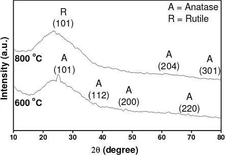
Fig. 1. X-ray diffraction (XRD) patterns of TiO2:Fe thin films as a function of annealing temperature.
Effect Of Annealing Temperature On Iron Doped Titanium Dioxide....halaman 70
change to be rutile at ~ 600 C, our findings indicate that the presence of dopants will increase the tranformation temperature. Or in the other words, the transformation temperature depends significantly on the presence of dopants.
Regarding to the XRD patterns, lattice parameter value and the structure of the crystal in the TiO2:Fe thin films which has tetragonal structure could be calculated by using the Cohen method. Calculated values of the lattices parameters of the TiO2:Fe thin films are summarised in Table 1.
Table 1. Calculated lattice parameters of TiO2:Fe thin films.
Annealing
temperature ( C) a=b ( Ǻ ) c ( Ǻ )
600 6.526 22.015
800 5.583 25.777
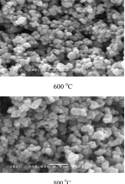
Table 1 shows that the crystal structure also depends on the annealing temperature. It seems that increasing annealing temperature reduces the a=b and increases the c parameter of the tetragonal crystal structure of TiO2:Fe thin films. Furthermore, it will be related to the surface morphologies of TiO2:Fe thin films (Fig. 2)
600 oC
800 oC
Effect Of Annealing Temperature On Iron Doped Titanium Dioxide....halaman 71
These SEM micrographs show that TiO2:Fe thin films have homogeneous grain size of about 100 nm. An alteration on crystal structure occurs due to a different annealing temperatures. Moreover, porosity analyses of the SEM micrographs by M. Rosi’s procedure 6 proceed porosity value for the annealing temperature of 600 C and 800 C are 37.7 and 39.8, respectively. It is supposed that the growth mode of film formation 7 converts from layer or Frank-van der Merwe to be the layer plus island or Stranski-Krastanov due to increasing the annealing temperature. This transformation causes the alteration of crystal structure and porosity of the thin films.
CONCLUSION
Increasing the annealing temperature of the iron doped titanium dioxide thin films prepared by spin coating method causes changes in crystal structure and morphology of the films. Porosity value could be calculated well by M. Rosi’s procedure. Our experiment results indicated that the rutile crystal orientation appears due to increasing annealing temperature of the thin films. Regarding to the application on gas sensor films increasing annealing temperature provide a superior capability due to the increasing of porosity value of TiO2:Fe thin films.
ACKNOWLEDGEMENT
The authors gratefully acknowledge the financial support of the Indonesian Ministry of National Education (Kementerian Pendidikan Nasional Republik Indonesia). The authors also wish to thank Rakhmat Randhy Prathama for his contributions in this research.
REFERENCES
1 A. Ruiz, G. Dezanneau, J. Arbiol, A. Cornet, J. R. Morante., Thin Solid Films, 436, 90 (2003).
2 E. Sotter, X. Vilanova, E. Llobet, M. Stankova, X. Correig, J. Optoelectron. Adv. Mater., 7 (3), 1395 (2005).
3 A. R. Bally, E. N. Korobeinikova, P. E. Schmid, F. Levy and F Bussy, J. Phys. D: Appl. Phys. 31, 1149 (1998).
4 D. Mardare, G. I. Rusu, J. Optoelectron. Adv. Mater., 6 (1), 333 (2004).
5 A. Nakaruk, C. Y. Lin, D. S. Perera, C. C. Sorrel, J. Sol-Gel Sci. Technol., 55, 328 (2010).
6 M. Rosi, F. D. Eljabbar, U. Fauzi, M. Abdullah dan Khairurrijal, Journal Nanosains dan Nanoteknologi, Vol. Edisi Khusus (Agustus), 29 (2009).