DI-(2-ETHYLHEXYL)PHTHALATE AND PYRANON DERIVATED FROM ENDOPHYTIC FUNGI
Penicillium
sp THE LEAVE OF KUNYIT PUTIH (Curcuma zedoaria)
Muharni
1,*, Fitrya
2, Milanti Okta Ruliza
1, Dwi Anjar Susanti
1, and Elfita
1 1Department of Chemistry, Faculty of Mathematics and Natural Sciences , Sriwijaya University, Jl. Raya Palembang Prabumulih Km 32, Indralaya, Ogan Ilir, South Sumatera 30662, Indonesia
2
Department of Pharmacy, Faculty of Mathematics and Natural Sciences , Sriwijaya University, Jl. Raya Palembang Prabumulih Km 32, Indralaya, Ogan Ilir, South Sumatera 30662, Indonesia
Received October 24, 2013; Accepted July 18, 2014
ABSTRACT
Two compounds from cultivation of the endophytic fungi Penicillium sp of leaves of kunyit putih (Curcuma zedoaria have been isolated. The endophytic fungus was cultivated on 5 L of Potatos Dextrose Broth (PDB) medium at room temperature (no shaking) for 3 weeks. The cultures were extracted with ethyl acetate to afford 3.0 g of residue after removal of the solvent under reduced pressure. The extract was separated and purified by silica gel column chromatography (CC) and afforded two pure compounds as colorless oily liquid (compound1) and yellow crystal (compound 2). The structure of these compounds were characterized by detailed UV, IR, and NMR spectroscopic analysis and compound 1 as well as comparison with the reported data. Base on spectra analysis the compound 1 was determined as Di-(2-ethylhexyl)phthalate and compound 2 as 5-(4’-ethoxy-2’-hydroxy-5’-methyl-2’,3’-dihydrofuran-3’-il (hydroxy) methyl-4-isopropyl-3-methyl-2-pyran-2-on). Compound1is not new compound, but it is new for endophytic fungus from C. zeodoria and compound2is new compound.
Keywords:endophytic fungi; Penicillium sp; Curcuma zedoaria
ABSTRAK
Telah dilakukan isolasi dua senyawa dari kultifat jamur endofitik Penicillium sp dari daun kunyit putih (Curcuma zedoaria. Jamur endofit dikultur dalam 5 L medium Potatos Dextrose Broth (PDB) pada suhu kamar (keadaan statis) selama 3 minggu. Kultur kemudian diekstraksi dengan etil asetat dan dipekatkan dengan rotary evaporator sehingga didapatkan ekstrak pekat etil asetat 3,0 g. Ekstrak dipisahkan dan dimurnikan dengan kromatografi kolom menggunakan fasa diam silika gel dan didapatkan dua senyawa murni berupa cairan minyak bening (senyawa 1) dan kristal kuning (senyawa2). Struktur senyawa hasil isolasi ditentukan berdasarkan analisis data spektroskopi UV, IR, dan NMR, dan senyawa1 juga dikonfirmasi dengan membandingkan data yang telah dilaporkan. Berdasarkan analisis data spektroskopi disimpulkan senyawa 1 adalah Di-(2-ethylhexyl)phthalate dan senyawa 2 adalah 5-(4’-etoksi-2’-hidroksi-5’-metil-2’,3’-dihidrofuran-3’-il (hidroksi) metil-4-isopropil-3-metil-2-piran-2-on). Senyawa 1 bukan merupakan senyawa baru, tetapi untuk pertama kalinya ditemukan pada jamur endofitik pada C. zeodoria dan senyawa2merupakan senyawa baru.
Kata Kunci:jamur endofitik; Penicillium sp; Curcuma zedoaria
INTRODUCTION
Endophytic microorganisms that redise in the tissues of living plants and may produce secondary metabolites of biologically active [1]. Novel antibiotics, antimycotics, immunosuppressants, anticancer compound are only a few examples of what has been found after the isolation, culture and purification and characterization of some choice endophytes in the recent past. Isolation of their bioactive secondary metabolites of endophytic fungus from plant could be
selected mainly something on ethonobotanical history [2].
Indo. J. Chem., 2014, 14 (3), 290 - 296
291
In our research of endophytic fungus, many bioactive compounds and new compounds were isolated [8-9]. In this paper we reported the isolation and structural identification one known compound namely Di-(2-ethylhexyl)phthalate (1) and one new compound as 5-(4’-ethoxy-2’-hydroxy-5’-methyl-2’,3’-dihydrofuran-3’-il (hydroxy) methyl-4-isopropyl-3-methyl-2-pyran-2-on) (2) of Penicillium sp from the leaves of C. zedoaria.
Penicillium species isolated as endophytic usually be found in plants zingiberaceae [10] and meliaceae family, although in marine organisms, three meroterpenes preaustinoids, A, B, A1, A2, and B1 have been reported to be isolated from Penicillium sp associated with the Melia azedarach [11]. Penicillium commune from the semi-mangrove plant Hibiscus tiliaceus, have been isolated one new compound 1-O-(2,4-dihydroxy-6-methylbenzoyl)-glycerol along with thirteen known products including 1-O-acetylglycerol, N-acetyl tryptophan, 3-indolylacetic acid methyl ester, 1-(2,4-dihydroxy-3,5-dimethylphenyl)ethanone, 2-(2,5-dihydroxy phenyl)acetid acid, (4R,5S)-5-hydroxyhexan-4-olide,
thymidine, uracil, thymine, ergosterol, β-sitosterol, β-daucosterol, and ergosta-7,22-dien-3β,5α,6β-triol [12].
EXPERIMENTAL SECTION
Materials
The leaves of kunyit putih were collected on May 2013 from the Indralaya, Ogan Ilir, South Sumatra. Material for isolation endophytic fungi: ethanol 70%, NaOCl, chloramphenicol, potato dextrose broth (PDB), potato dextrose agar (PDA), silica gel 60 (70-230 mesh), thin layer chromatography (TLC) from Merck (Art.5554) silica gel 60 F254,n-hexane, ethyl acetate, and methanol.
The organic solvents were used from distilled technical grade.
Instrumentation
The apparatus in the research were counter colony, autoclave, incubator, water bath, microscope, magnetic hotplate, UV lamp, column chromatography and generally apparatus in organic and microbiology laboratory, melting point was determined using Fisher John Apparatus. UV spectra were determined with Varian Conc 100 instrument. IR spectra were determined on FTIR-Perkin Elmer-Spectrum One and NMR spectra were recorded at 500 MHz (1H) and 125 MHz (13C) on JEOL JNM ECA-500 spectrometer,
UV light at λ 254 nm and 365 nm.
Procedure
Isolation of endophytic fungus
The leaves sample was washed before it was processed and surface sterilized in 70% ethanol for 3 min and 0.5% NaOCl for 1 min and rinsed thoroughly with sterile distilled water. The segment sample placed on petri-plates containing potato dextrose agar medium (PDA) (200 g potato, 20 g dextrose, and 15 g agar in 1 L of H2O, supplemented with 100 mg/L of
chloramphenicol to suppress bacterial growth). The plates were incubated at 25 ± 2 °C until fungus growth appeared. The plant segments were observed once a day for the growth of endophytic fungus. Colony fungus showed difference characteristic furthermore to pure with the plated segments were immediately transferred into new PDA plates and then subcultured until pure cultures were obtained [13].
Identification of the endophyte
The endophytic fungal strain was identified by the morphological method. The morphological examination was performed by scrutinizing the fungal culture, the mechanism of spore production, and the characteristics of the spores. All experiments and observations were repeated at twice [14].
Cultivation of pure fungal strain
The purified fungus (a small park) was transferred under sterile conditions to the PDB medium. For chemical investigations, the fungal strains were static cultivated into 15 flasks (1 L each) containing 400 mL of PDB medium for 3 weeks at room temperature [12-14].
Extraction, isolation, purification, and structure elucidation
Fig 1.Isolation of the compounds from ethyl acetate extract ofPenicilliumsp from the leaves ofC. zedoaria
Tablel 1.1H-NMR (500 MHz) and13C-NMR (125 MHz), spectral data of compound1. recorded in CD3OD
Hppm (H, multiplicity,Jin Hz) C(ppm) Carbon
no. 1 1* 1 1*
1 0.89 (3H,t) 0.84 (3H,t, 4.3) 14.1 14.1
2 1.30 (2H,m) 1.23 - 1.44 (2H,m) 23.1 24.8
3 1.29 (2H,m) 1,23 - 1.44 (2H,m) 29.0 22.7
4 1.38 (2H,m) 1.23 - 1.44 (2H, m) 23.8 29.5
5 1.67 (1H,m) 2.60 (1H,m) 38.8 40.8
6 4.21 (2H,m) 4,15 (2H,m) 68.1 65.2
7 167.8 171.1
8 1.34 (2H,m) 2.30 (2H,dq, 4.3) 30.4 29.7
9 0.91 (3H,t) 0.93 (3H,t, 4.3) 11.0 20.8
10 132.5 124.8
11 7.69 (1H,dd, 5.9 – 3.3) 6.96 (1H,dd, 6.3 – 2.2) 128.8 119.0 12 7,51 (1H,dd, 5,9 – 3.3) 7.11(1H,dd, 6.3 – 2.2) 130.9 132.6
1*[19]
of spectroscopic analysis including UV, IR, 1H-NMR,
13
C-NMR, DEPT, HMQC, HMBC, and COSY.
RESULT AND DISCUSSION
The fungus strain was identified as Penicillium sp.
Penicillium species isolated as endophytes were obtained from several plant species such as, Melia azedarach [15-16]. Zingiberaceae family [14] meliaceae family, although in marine organisms as the semi-mangrove plant Hibiscus tiliaceus [17]. Fungus
Penicillium spafter that cultivated on 5 L of PDB medium and then extracted with ethyl acetate to afford 3.0 g of residue. The extract (3.0 g) was separated by column chromatography to yield compound 1 (20 mg) and compound 2 (10 mg). The isolation of the compounds
from ethyl acetate extract of Penicillium sp from the leaves ofC. zedoariadescribed in Fig. 1.
Compound 1 was obtained as colorless oil liquid.
The UV spectra of 1 exhibited absorption at λmax nm :
206, 225, and 274. The bathochromic shift in addition of NaOH showed there is no wave length shift, it can concluded that there was no phenolic group. The IR spectrum showed the functional group such as carbonyl ester (1722 cm-1), C=C aromatic (1598–1462 cm-1), C-O (1273 cm-1), C-H aromatic (3070 cm-1), and C-H aliphatic (2927–2860 cm-1). The
1
H-NMR data (Table 1) disclosed the presence of two
protons as AB spin system at δH 7.69 (1H, dd, 5.9 &
3.3 Hz) and 7.51 (1H, dd, 5.9 & 3.3 Hz) that characteristic for aromatic proton at ortho substituted
ring. The proton signal at δH 4.21 ppm (2H, m) is
Indo. J. Chem., 2014, 14 (3), 290 - 296
293
Table 2.1H-NMR (500 MHz) and13C-NMR (125 MHz), spectral data of compound2recorded in CDCl3
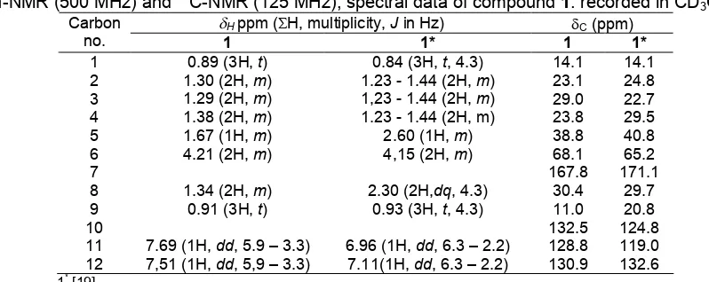
Carbon no. Cppm DEPT Hppm (H, multiplicity,Jin Hz) HMBC COSY
2 183.4 C
3 123.1 C
4 139.1 C
5 107.4 C
6 162.8 CH 8.23 (1H,s) 81.7, 107.4, 139.1
7 34.6 CH 2.98 (1H,q,J= 7.15 Hz) 107.4, 123.1, 139.1 1.22
8 18.5 CH3 1.22 (3H,d,J= 7.15 Hz) 139.1, 34.6, 81.7
9 18.2 CH3 1.34 (3H,d,J= 7.15 Hz) 34.6
10 9.5 CH3 2.01 (3H,s) 123.1, 139.1, 183.8
2’ 174.5 C
3’ 100.3 C
4’ 171.2 C
5’ 177.2 C
6’ 60.4 CH2 4.11 (2H,q,J= 7.15 Hz) 171.2 1.25
7’ 14.2 CH3 1.25 (3H,t) 60.4
8’ 21.1 CH3 2.04 (3H,s) 171.2
9’ 81.7 CH 4.77 (1H,q,J= 7.15 Hz) 139.1, 162.8 1.34
Fig 2.The1H-NMR spectrum of compound2
alcohol group. Furthermore, the presence proton signal
at δH 1.67 ppm (1H, m) for proton methine, signal at
1.2–1.4 ppm for four methilene group, and signal at 0.89 and 0.91 as pair of multiplate (3H, m) for two methyl groups.
The 13C-NMR spectrum of compound 1 (Table 1), confirming the symmetry of the molecule, exhibited the expected 12 carbon resonance. DEPT spectrum showed to two quaterner, three methane, five methylene carbons, and two methyl groups. These spectroscopic data, by comparison of 1H and 13C-NMR data to those
published in literature [19] and showed similarity, in conclusion compound 1 was identified as Di-(2-ethylhexyl)phthalate (DEHP). DEHP (compound 1) is a well known synthetic plasticizes, so already reported to be present in Calotropis gigantean [15],
Alchornea cordifolia [16], and Aloe vera [17]. The effective presence of compound1in endophytic fungus
Penicillium sp of leave C. zedoaria, not as a contaminant from solvents and endophytic fungus
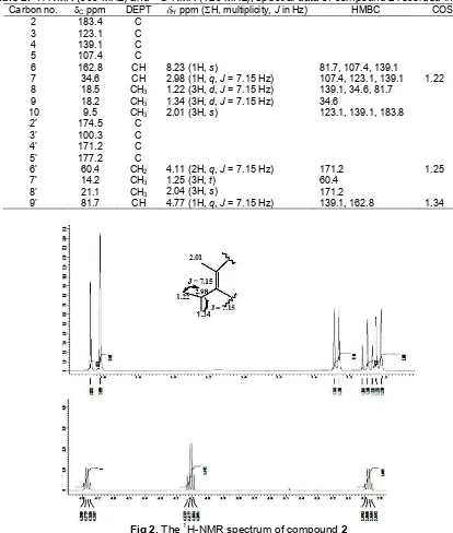
Fig 3.Spectrum13C-NMR compound2
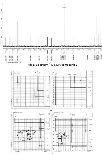
Fig 4. HMQC correlation of proton at δH 1.23–8.23 ppm (A) and HMBC correlation of proton at δH1.23–2.98 ppm (B)
compound2
Compound2was obtained as a yellow crystal, mp. 171-172 °C. The Spectra UV (MeOH) of 2 exhibited
absorption at λmax nm: 213, 253, and 321. The
bathochromic shift in addition of NaOH exhibited
absorption at λmax nm: 213, 253, and 321. Base on
Spectroscopic data UV indicate this compound was no
phenolic group. The IR spectra (KBr) showed νmaxcm -1
: 3466.08 (OH), 2980.02 and 2935.66 (C-H aliphatic), 1625.99 (conjugated C=O), 1579.70; 1521.12; 1438.90 (C=C conjugation), and 1180.44 (C-O ether). 1H-NMR
(DMSO, 500 MHz) δH ppm and 13
C-NMR (DMSO,
Indo. J. Chem., 2014, 14 (3), 290 - 296
295
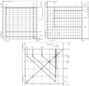
Fig 5.HMBC correlation of proton at 4.01–8.23 ppm and COSY correlation compound2
Fig 6.HMBC (A), and COSY (B)correlations and δ-assignment of compound 2
The1H-NMR spectrum, (Fig. 2) showed signal two
methyl doublet at δH1.22 and 1.34 ppm (3H,d, 7.15 Hz)
and signal methine quartet at δH 2.98 ppm (1H, q,
7.15 Hz) and one methine singlet at δH 8.23 (1H,s). At
spectrum also showed signal for methyl triplet at
δH 1.25 ppm (3H, t, 7.15 Hz), and two methyl singlet at
δH2.01 and 2.04 ppm, (3H,s) and one signal methylene
quartet at δH4.11 ppm (2H,q, 7.15 Hz).
The 13C-NMR (Fig. 3), DEPT 135 spectrum, and HMQC spectrum (Fig. 4) showed 17 signal consist that nine signal as C sp2 and 8 signal as C sp3. Analysis spectrum DEPT 135 showed 8 signal C quarternary at
δC100.3; 107.4; 123.1; 139.1; 171.2; 174.5; 177.2 and 183.8 ppm, 5 signal methyls carbon at δC 9.5; 14.2; 18.2; 18.5 and 21.1 ppm, 3 signal methines carbon at
methylene carbon at δC 60.4 ppm. Signal carbon at
δC 183.4 ppm indicated these compound have C=O carbonyl.
NMR 2D analysis for HMQC spectrum (Fig. 4)
showed the proton at δH 1.34 ppm correlation to carbon
at δH 18.2 ppm and proton at δH1.22 ppm correlation to
carbon at δC 18.5. HMBC spectrum showed correlation
from proton at δH 1.22 and 1.34 ppm to carbon at δC 34.6 and 139.1 ppm. Proton at δH 1.22 also
correlation to carbon at δC 18.2 ppm and proton at
δH 1.34 ppm showed correlation with carbon at δC 18.5. This data to indicated that proton δH 1.22 and
1.34 ppm bound to carbon fasten carbon δC 34.6 ppm.
Proton at 1.25 ppm correlation to carbon at δC60.4 ppm.
Further HMBC spectrum showed correlation proton at δH
2.01 (3H,s) to carbon at δC123.1; 139.1 and 183.8 ppm,
and correlation proton at δH 2.04 ppm to carbon at
δC 171.2 ppm. Proton at δH 2.01 and 2.04 (3H, s) at
HMQC spectrum showed fastened with carbon at
δC9.5 and 21.1 ppm.
Proton at δH4.11 correlation to carbon atδC171.2,
proton at δH 4.77 ppm showed correlation to carbon at
δC 139.1; 162.8 ppm, while proton at δH 8.23 ppm to
correlation to carbon at δC 81.7; 107.4 and 139.1 ppm. Analysis of 1H–1H COSY spectrum (Fig. 5) also to
indication of two proton spin system corresponding at δH
1.22 with proton at δH 2.98 ppm. And proton at δH1.25 to
correlation to proton at 4.11 ppm. HMBC and COSY
correlation and δ-assignment of compound showed Fig.
6. These spectroscopic data, therefore suggested that compound 2 is 5-(4’-ethoxy-2’-hydroxy-5’-methyl-2’,3’-dihydrofuran-3’-il (hydroxy) methyl-4-isopropyl-3-methyl-2-pyran-2-on).
Compound1is not new compound, but it is new for endophytic fungus from C. zedoaria and base on Dictionary Natural Products data base, 5-(4’-ethoxy-2’-hydroxy-5’-methyl-2’,3’-dihydrofuran-3’-il (hydroxy) methyl-4-isopropyl-3-methyl-2-pyran-2-on) (2) is new compound. Exploration of secondary metabolites research needs to be done in order to get the profile of organic compounds produced by endophytic fungus of
C. zedoaria.
CONCLUSION
Two compounds have been isolated from the endophytic fungus Penicillium sp from the leaves of kunyit putih (C. zedoaria). Based on spectroscopic analysis and comparison data to those published in literature compound 1 was identified as Di-(2-ethylhexyl)phthalate and compound 2 as
5-(4’-ethoxy-2’-hydroxy-5’-methyl-2’,3’-dihydrofuran-3’-il (hydroxy) methyl-4-isopropyl-3-methyl-2-pyran-2-on).
ACKNOWLEDGEMENT
The authors are statement grateful to the Directorate General of Higher Education which research grant Fundamental 2013 was supported this research.
REFERENCES
1. Strobel, G., Daisy, B., and Castillo, U., 2005,Plant Pathol. J., 4 (2), 161–176.
2. Premjanu, N., and Jayanthy, C., 2012,Int. J. Inst. Pharm. Life Sci.,2 (1), 135–162.
3. Lakshmi, S., Padmaja, G., and Remani, P., 2011,
Int. J. Med. Chem., 2011, 1–13.
4. Saikia, N., and Nath, S.C., 2003,J. Econ. Taxon. Bot., 27, 430–433.
5. Jang, M.K., Sohn, D.H., and Ryu, J-H., 2001,
Planta Med., 67 (6), 550–552.
6. Wilson, B., Abraham, G., Manju, V.S., Mathew, M., Vimala, B., Sundaresan, S., and Nambisan, B., 2005,J. Ethnopharmacol., 99 (1), 147–151.
7. Bugno, A., Nicoletti, M.A., Almodóvar, A.A.B., Pereira, T.C., and Auricchio, M.T., 2007, Braz. J. Microbiol., 28, 440–445.
8. Elfita, Muharni, Munawar, Legasari, L., and Darwati, 2011,Indo. J. Chem., 11 (1), 53–58. 9. Elfita, Muharni, Munawar, and Aryani, S., 2012,
Indo. J. Chem., 12 (2), 195–200.
10. Xu, L., Zhou, J., Zhao, J., Li, X., and Wang, J., 2008,Lett. Appl. Microbiol., 46 (1), 68–72.
11. dos Santos, R.M.G., and Rodrigues-Fo, E., 2003,
Z. Naturforsch., 58c, 663–669.
12. Yan, H-J., Gao, S-S., Li, C-S., Li, X-M., and Wang, B-G., 2010,Molecules, 15 (5), 3270–3275.
13. Barik, B.P., Tayung, K., Jagadev, P.N., and Dutta, S.K., 2010,Eur. J. Biol. Sci., 2 (1), 8–16.
14. Guo, L., Wu, J-Z., Han, T., Cao, T., Rahman, K., and Qin, L-P., 2008,Molecules,13 (9), 2114–2125. 15. Habib, M.R., and Karim, M.R., 2009, Micobiology,
37 (1), 31–36.
16. Mavar-Manga, H., Haddad, M., Pieters, L., Baccelli, C., Penge, A., and Quetin-Leclercq, J., 2008, J. Ethnopharmacol., 115 (1), 25–29.